PGHN
Original Article
Parenteral Nutrition-Associated Cholestasis in Very Low Birth Weight Infants: A Single Center Experience
Ah-Young Kim*, Ryoung-Kyoung Lim*, Young-Mi Han*, Kyung-Hee Park*, and Shin-Yun Byun*
,†*Department of Pediatrics, Pusan National University School of Medicine, †Research Institute for Convergence of Biomedical Science and Technology, Pusan National University Yangsan Hospital, Yangsan, Korea
Purpose: Parenteral nutrition (PN)-associated cholestasis (PNAC) is one of the most common complications in very low birth weight infants (VLBWIs). The aim of this study is to evaluate the risk factors of PNAC in VBLWIs.
Methods: We retrospectively reviewed the medical records of 322 VLBWIs admitted to the neonatal intensive care unit of our hospital from July 1, 2009 to December 31, 2013. We excluded 72 dead infants; 6 infants were transferred to another hospital, and 57 infants were transferred to our hospital at 2 weeks after birth. The infants were divided into the cholestasis and the non-cholestasis groups. PNAC was defined as a direct bilirubin level of ≥2.0 mg/dL in infants administered with PN for ≥2 weeks.
Results: A total of 187 VLBWI were enrolled in this study; of these, 46 infants developed PNAC. Multivariate logistic regression analysis showed that the risk factors of PNAC in VLBWI were longer duration of antimicrobial use (odds ratio [OR] 4.49, 95% confidence interval [95% CI] 4.42-4.58), longer duration of PN (OR 2.68, 95% CI 2.41-3.00), long-term lack of enteral nutrition (OR 2.89, 95% CI 2.43-3.37), occurrence of necrotizing enterocolitis (OR 2.40, 95% CI 2.16-2.83), and gastrointestinal operation (OR 2.19, 95% CI 2.03-2.58).
Conclusion: The results of this study suggest that shorter PN, aggressive enteral nutrition, and appropriate anti- microbial use are important strategies in preventing PNAC.
Key Words: Parenteral nutrition, Cholestasis, Very low birth weight infant
Received:August 13, 2015, Revised:September 14, 2015, Accepted:September 15, 2015
Corresponding author: Shin-Yun Byun, Department of Pediatrics, Pusan National University Yangsan Hospital, 20 Geumo-ro, Mulgeum-eup, Yangsan 50612, Korea. Tel: +82-55-360-2180, Fax: +82-55-360-2181, E-mail: [email protected]
Copyright ⓒ 2016 by The Korean Society of Pediatric Gastroenterology, Hepatology and Nutrition
This is an openaccess article distributed under the terms of the Creative Commons Attribution NonCommercial License (http://creativecommons.org/licenses/by-nc/4.0/) which permits unrestricted noncommercial use, distribution, and reproduction in any medium, provided the original work is properly cited.
INTRODUCTION
Providing parenteral nutrition (PN) in the neo- natal intensive care unit (NICU) is an important means of supplying nutrients to promote the growth of premature infants with a birth weight ≤1,500 g.
Due to the risk of necrotizing enterocolitis (NEC) and feeding intolerance caused by the impaired gas- trointestinal (GI) motility of very low birth weight infants (VLBWIs), many neonatologists hesitate about accelerating the rate of feeding of very-low-birth weight infants. For this reason, VLBWIs are given
nutrients through PN. However, administrating PN for a long time may induce complications, the most common of which is PN-associated cholestasis (PNAC).
The risk factors of PNAC include low birth weight, prematurity, PN duration, absence of enteral nu- trition (EN), quality or quantity of amino acid in- take, cumulative amount of lipid infusion, toxicity of trace elements, GI operation, shock, and sepsis [1,2].
To help in the care of VLBWIs in the future, this study aims to describe the risk factors of PNAC in VLBWIs at our NICU.
MATERIALS AND METHODS
Patients and criteria
All VLBWIs who received PN for ≥14 days and were admitted to our children’s hospital’s NICU be- tween July 1, 2009 and December 31, 2013 were in- cluded in the study. This was a retrospective study, with follow-up until discharge. We excluded dead in- fants, infants with major congenital anomaly, infants who were transferred to another hospital, and those who were transferred to our hospital at 2 weeks after birth. The infants were divided into the cholestasis and the non-cholestasis groups. PNAC was defined as an elevation of conjugated bilirubin to ≥2.0 mg/dL in at least one laboratory test during hospitalization.
This study was accredited by the Institutional Review Board of the Pusan National University Yangsan Hospital (study no. 05-2014-010).
Characteristics of PN and enteral nutrition PN was started within the first day of life for all VLBWIs. Dextrose infusion was started at 6 mg/kg/min and increased to a maximum of 10-12 mg/kg/min and adjusted according to the blood sugar level, calo- rie intake, and clinical conditions. Administration of amino acids (10% Primene; Baxter, Newbury, UK) was started at birth and increased from 2 g/kg/day to a maximum of 4 g/kg/day at each stage. Fish oil-based lipids (20% SMOFLipid; Fresenius Kabi AG, Graz, Austria) were started at 0.5 g/kg/day and increased daily by 0.5 g/kg/day to a maximum of 3 g/kg/day.
Lipids were stopped when EN reached calorie 100
mL/kg/day. Trace elements (Furtman; JW Life Science, Dangjin, Korea) were mixed according to the body weight at each PN administration after the first day of life.
When the infants were clinically stable, EN was started through a nasogastric or orogastric tube as soon as possible. Feeding of VLBWIs was started with breast milk or premature infant formula. EN was started at 10-30 mL/kg/day. The feeding volume was increased by 10-30 mL/kg/day according to ges- tational age (GA); the goal feeding volume was 150 mL/kg/day. Ursodeoxycholic acid was administered at 15 mg/kg/day for the cholestasis group until the conjugated bilirubin level recovered to <2.0 mg/dL.
Risk factors and definitions
Small for GA was defined as a birth body weight (BBW) below the 10th percentile for GA on a fetal growth chart. The diagnosis of respiratory distress syndrome was based on chest radiograph results to- gether with clinical symptoms. In our NICU, infants with hemodynamically significant patent ductus ar- teriosus (PDA) were treated with indomethacin or ibuprofen before ligation. The infants received fluco- nazole prophylaxis of 3 mg/kg/dose through intra- venous infusion twice a week for 6 weeks. Antimi- crobials included antibiotics and antifungal agents.
Congenital hypothyroidism was confirmed with blood thyroid function tests; infants with hypo- thyroidism were administered with levothyroxine.
NEC was defined as Bell’s stage ≥2. The diagnosis of sepsis was made based on positive blood cultures in association with the symptoms of systemic inflam- matory response syndrome, such as fever, hypother- mia, hypotension, and unstable vital signs. Broncho- pulmonary dysplasia (BPD) was defined as oxygen supplementation requirement at 36 weeks post- menstrual age. Intraventricular hemorrhage was de- fined as stage ≥3. Retinopathy of prematurity (ROP) was defined as stage ≥2 plus. Neurosensory hearing loss was confirmed with an auditory brainstem re- sponse test. Skeletal fracture was confirmed with ra- diographic examination. Near-full EN was defined as an enteral tolerance of 120 mL/kg/day. Full EN
was defined as enteral tolerance of at least 150 mL/kg/day.
Infectious causes of cholestasis (hepatitis A, hep- atitis B, hepatitis C, toxoplasmosis, rubella, herpes simplex virus, and cytomegalovirus) were excluded in this study. We investigated a-1 antitrypsin, alpha fetoprotein, gamma glutamyl transpeptidase, and the possibility of surgical causes of conjugated hy- perbilirubinemia (such as biliary atresia, choledochal cyst on abdominal ultrasonography, and hepatobiliary scintigraphy); there were no abnormalities in our VLBWIs.
Statistical analyses
The Statistical Package for Social Sciences version 21.0 (IBM Co., Armonk, NY, USA) software was used for data analysis. Categorical variables were com- pared between the two groups by using Fisher’s ex- act test and Pearson’s chi-square test. Continuous variables were compared between the two groups by using Student’s t-test. Due to the differences in GA and BBW between the two groups, these two factors were corrected with an analysis of covariance (AN- COVA). Logistic regression analysis was performed to eliminate interactions between predicted factors of PNAC; the role of specific factors that may affect PNAC was defined. A probability p-value of <0.05 was used to establish statistical significance.
RESULTS
A total of 322 VLBWIs were admitted to our hospi- tal during the study period. Of these, 135 infants were excluded: 72 infants who died before discharge, 6 infants who were transferred to another hospital, and 57 infants who were transferred to our hospital at 2 weeks after birth. Thus, a total of 187 infants were enrolled in this study and 46 of these had a di- agnosis of cholestasis.
Perinatal characteristics and neonatal demo- graphic findings
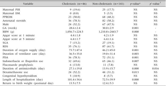
GA and BBW were significantly lower in the cho- lestasis group (28.6±2.4 vs. 30.0±2.9, 1,056.7±224.5
vs. 1,210.8±2,443.7, p<0.05). The durations of oxy- gen supply and mechanical ventilator care were sig- nificantly longer in the cholestasis group (73.7±47.4 vs. 46.2±45.0, 36.5±35.0 vs. 19.0±31.4, p<0.05).
The prevalence rate of PDA, the rate of medication treatment for PDA, and the frequency of dexame- thasone administration were higher in the choles- tasis group (78.3% vs. 51.1%, 69.6% vs. 46.1%, 52.2%
vs. 31.2%, p<0.05). The duration of antimicrobial administration and the length of hospitalization were longer in the cholestasis group (27.1±19.8 vs.
13.9±15.7, 101.4±34.6 vs. 72.5±39.9, p<0.05). To eliminate the influences of GA and BBW, we used an ANCOVA test to regulate the statistically significant results. After this regulation, only the duration of an- timicrobial administration was significantly asso- ciated with PNAC (Table 1).
Factors associated with PN, EF, and nutritional data
The durations of administration of PN, amino acid, lipid, and trace elements were longer in the cho- lestasis group (55.0±23.8 vs. 29.0±16.3, 55.0±23.8 vs.
29.0±16.3, 48.0±20.4 vs. 24.3±15.9, 43.6±18.3 vs.
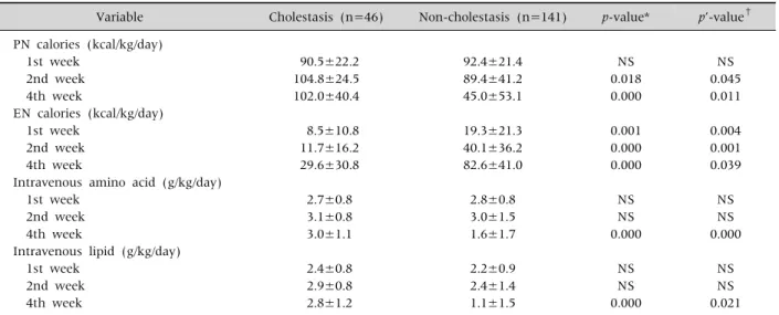
27.3±16.4, p<0.05). In this group, delayed time to initiate feeding, delayed time to near-full EN, de- layed time to full EN, and a longer duration of fasting were also observed (6.2±6.7 vs. 3.2±3.4, 52.2±20.3 vs. 29.9±14.8, 56.7±24.4 vs. 32.8±16.1, 18.7±11.0 vs. 6.8±6.8, p<0.05). Other significant differences of the cholestasis group were higher PN calories at the second and fourth weeks of life; lower EN calo- ries at the first, second, and fourth weeks of life; and higher intravenous amino acid and lipid admin- istration at the fourth week of life (104.8±24.5 vs.
89.4±41.2, 102.0±40.4 vs. 45.0±53.1, 8.5±10.8 vs.
19.3±21.3, 11.7±16.2 vs. 40.1±36.2, 29.6±30.8 vs.
82.6±41.0, 3.0±1.1 vs. 1.6±1.7, 2.8±1.2 vs. 1.1±1.5, p<0.05). After adjusting for the influences of GA and BBW by using the ANCOVA test, all of the above-mentioned statistical results were significantly associated with PNAC, except for the duration of trace element administration (Tables 2 and 3).
Table 1. Differences of Perinatal Characteristics and Neonatal Demographic Findings between the Cholestasis Group and the Non-Cholestasis Group
Variable Cholestasis (n=46) Non-cholestasis (n=141) p-value* p’-value† Maternal PIH
Maternal DM PPROM
Antenatal steroids Male
GA (week) BBW (g)
Apgar score at 1 minute Apgar score at 5 minute SGA
RDS
Duration of oxygen supply (day) Duration of ventilator care (day) PDA
Indomethacin or Ibuprofen use Fluconazole prophylaxis Duration of antimicrobials (day) Dexamethasone use
Congenital hypothyroidism Length of hospitalization (day) Return to birth weight (postnatal day)
9 (19.6) 0 (0.0) 23 (50.0) 36 (78.3) 24 (52.2) 28.6±2.4 1,056.7±224.5
4.4±1.8 6.4±1.7 7 (15.2) 35 (76.1) 73.7±47.4 36.5±35.0 36 (78.3) 32 (69.6) 6 (13.0) 27.1±19.8
24 (52.2) 5 (10.9) 101.4±34.6
13.5±7.5
25 (17.7) 5 (3.5) 68 (48.2) 82 (58.2) 67 (47.5) 30.0±2.9 1,210.8±2443.7
4.2±1.9 6.4±1.6 27 (19.1) 87 (61.7) 46.2±45.0 19.0±31.4 72 (51.1) 65 (46.1) 11 (7.8) 13.9±15.7 44 (31.2)
8 (5.7) 72.5±39.9 12.4±5.3
NS NS NS NS NS 0.001 0.000 NS NS NS NS 0.001 0.003 0.001 0.007 NS 0.000 0.013 NS 0.000
NS
NS NS NS NS NS - - NS NS NS NS NS NS NS NS NS 0.016
NS NS NS NS Values are presented as number (%) or mean±standard deviation.
*Statistics were analyzed by t-test and chi-square test. †ANCOVA (analysis of covariance) test to adjust for GA and birth body weight.
PIH: pregnancy-induced hypertension, DM: diabetes mellitus, PPROM: premature preterm rupture of membrane, GA: gestational age, BBW: birth body weight, SGA: small for GA, RDS: respiratory distress syndrome, PDA: patent ductus arteriosus, NS: nonspecific.
Table 2. Comparison of Factors Associated with Parenteral Nutrition and Enteral Nutrition between the Cholestasis Group and the Non-Cholestasis Group
Variable Cholestasis (n=46) Non-cholestasis (n=141) p-value* p’-value† PN duration (day)
Amino acid duration (day) Lipid start (postnatal day) Lipid duration (day)
Trace elements duration (day)
Time to initiate feeding (postnatal day) Time to near full EN (postnatal day) Time to full EN (postnatal day) Duration of fasting (day)
55.0±23.8 55.0±23.8 2.4±1.0 48.0±20.4 43.6±18.3 6.2±6.7 52.2±20.3 56.7±24.4 18.7±11.0
29.0±16.3 29.0±16.3 2.8±1.3 24.3±15.9 27.3±16.4 3.2±3.4 29.9±14.8 32.8±16.1 6.8±6.8
0.000 0.000 NS 0.000 0.000 0.006 0.000 0.000 0.000
0.000 0.000 NS 0.001
NS 0.000 0.000 0.000 0.000 Values are presented as mean±standard deviation.
*Statistics were analyzed by t-test. †ANCOVA (analysis of covariance) test to adjust for gestational age and birth body weight.
PN: parenteral nutrition, EN: enteral nutrition, NS: nonspecific.
Postnatal complications
The prevalence of NEC, BPD, and ROP, and the rates of GI operation and skeletal fracture were high- er in the cholestasis group (15.2% vs. 0.7%, 60.9% vs.
42.6%, 30.4% vs. 12.1%, 28.3% vs. 3.5%, 6.5% vs.
0.0%, p<0.05). NEC, GI operation, and skeletal frac- ture were significantly associated with PNAC after adjustment for GA and BBW (Table 4).
Table 3.Comparison of Nutritional Data between the Cholestasis Group and the Non-Cholestasis Group
Variable Cholestasis (n=46) Non-cholestasis (n=141) p-value* p’-value† PN calories (kcal/kg/day)
1st week 2nd week 4th week
EN calories (kcal/kg/day) 1st week
2nd week 4th week
Intravenous amino acid (g/kg/day) 1st week
2nd week 4th week
Intravenous lipid (g/kg/day) 1st week
2nd week 4th week
90.5±22.2 104.8±24.5 102.0±40.4
8.5±10.8 11.7±16.2 29.6±30.8
2.7±0.8 3.1±0.8 3.0±1.1
2.4±0.8 2.9±0.8 2.8±1.2
92.4±21.4 89.4±41.2 45.0±53.1
19.3±21.3 40.1±36.2 82.6±41.0
2.8±0.8 3.0±1.5 1.6±1.7
2.2±0.9 2.4±1.4 1.1±1.5
NS 0.018 0.000
0.001 0.000 0.000
NS NS 0.000
NS NS 0.000
NS 0.045 0.011
0.004 0.001 0.039
NS NS 0.000
NS NS 0.021 Values are presented as mean±standard deviation.
*Statistics were analyzed by t-test. †ANCOVA (analysis of covariance) test to adjust for gestational age and birth body weight.
PN: parenteral nutrition, EN: enteral nutrition, NS: nonspecific.
Table 4.Comparison of Postnatal Complications between the Cholestasis Group and the Non-Cholestasis Group
Variable Cholestasis (n=46) Non-cholestasis (n=141) p-value* p’-value† NEC (≥stage 2)
GI operation Sepsis BPD
IVH (≥grade 3) ROP (≥stage plus) PVL
Neurosensory hearing loss Skeletal fracture
7 (15.2) 13 (28.3) 13 (28.3) 28 (60.9) 5 (10.9) 14 (30.4) 3 (6.5) 0 (0.0) 3 (6.5)
1 (0.7) 5 (3.5) 22 (15.6) 60 (42.6) 6 (4.3) 17 (12.1) 10 (7.1)
5 (3.5) 0 (0.0)
0.000 0.000 NS 0.041
NS 0.006
NS NS 0.001
0.000 0.000 NS NS NS NS NS NS 0.001 Values are presented as number (%).
*Statistics were analyzed by chi-square test. †ANCOVA (analysis of covariance) test to adjust for gestational age and birth body weight.
NEC: necrotizing enterocolitis, GI: gastrointestinal, BPD: bronchopulmonary dysplasia, IVH: intravascular hemorrhage, ROP:
retinopathy of prematurity, PVL: preventricular leukomalacia, NS: nonspecific.
Laboratory findings
The maximum levels of alanine aminotrans- ferase (202.4±176.0, p<0.05), aspartate amino- transferase (252.3±192.7, p<0.05), alkaline phos- phatase (2,297.1±697.4, p<0.05), and total bilir- ubin (10.6±4.1, p<0.05) were higher in the choles- tasis group than in the control group. The maximum direct bilirubin (DB) level of the cholestasis group was 6.8 mg/dL. The time to cholestasis development was 52.4 days; the mean time to peak DB was 70.5
days. Cholestasis persisted for 63.3 days; the peak prothrombin time international normalized ratio was 1.4 (Table 5).
Independent predictive factors
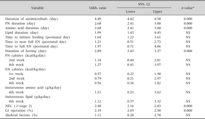
A multivariate logistic regression analysis was per- formed on the statistically significant results to eval- uate the independent predictive factors of PNAC.
The results of this analysis suggested that the factors that significantly increased the risk of PNAC were
Table 5.Comparison of Laboratory Findings between the Cholestasis Group and the Non-Cholestasis Group
Variable Cholestasis (n=46) Non-cholestasis (n=141) p-value*
ALT (IU/L) AST (IU/L) ALP (IU/L) TB (mg/dL) Peak DB (mg/dL) Time to cholestasis (day) Time to peak DB (day) Duration of cholestasis (day) Peak PT INR
202.4±176.0 252.3±192.7 2,297.1±697.4
10.6±4.1 6.8 52.4 70.5 63.3 1.4
37.0±34.8 74.9±131.6 1,625.3±748.9 8.7±2.7
0.000 0.000 0.000 0.004
Values are presented as mean±standard deviation or mean only.
*Statistics were analyzed by t-test.
ALT: alanine aminotransferase, AST: aspartate aminotrasferase, ALP: alkaline phosphatase, TB: total bilirubin, DB: direct bilirubin, PT INR: prothrombin time international normalized ratio.
Table 6.Logistic Regression Analysis for the Predictive Factors of Parenteral Nutrition-Associated Cholestasis
Variable Odds ratio 95% CI
p-value*
Lower Upper
Duration of antimicrobials (day) PN duration (day)
Amino acid duration (day) Lipid duration (day)
Time to initiate feeding (postnatal day) Time to near full EN (postnatal day) Time to full EN (postnatal day) Duration of fasting (day) PN calories (kcal/kg/day) 2nd week
4th week
EN calories (kcal/kg/day) 1st week
2nd week 4th week
Intravenous amino acid (g/kg/day) 4th week
Intravenous lipid (g/kg/day) 4th week
NEC (≥stage 2) GI operation (%) Skeletal facture (%)
4.49 2.68 2.68 1.99 1.64 1.21 1.97 2.89
1.14 1.27
0.57 0.79 0.56
1.21
1.12 2.40 2.19 1.11
4.42 2.41 2.41 1.43 1.22 0.51 0.71 2.43
0.44 0.41
0.22 0.21 0.36
0.23
0.37 2.16 2.03 0.20
4.58 3.00 3.00 4.45 3.61 2.73 4.06 3.37
2.81 3.97
1.98 2.97 1.82
3.62
3.32 2.83 2.58 2.78
0.000 0.000 0.000 NS NS NS NS 0.000
NS NS
NS NS NS
NS
NS 0.000 0.000 NS
*Statistics were analyzed by logistic regression analysis.
CI: confidence interval, PN: parenteral nutrition, EN: enteral nutrition, NEC: necrotizing enterocolitis, GI: gastrointestinal, NS:
nonspecific.
the duration of antimicrobial use (odds ratio [OR]
4.49, 95% confidence interval [95% CI] 4.42-4.58), PN (OR 2.68, 95% CI 2.41-3.00), amino acids (OR 2.68, 95% CI 2.41-3.00), and fasting (OR 2.89, 95% CI 2.43-3.37), the occurrence of NEC (OR 2.40, 95% CI
2.16-2.83), and GI operation (OR 2.19, 95% CI 2.03-2.58) (Table 6).
DISCUSSION
Although PNAC has been studied for a long time, its definite etiology is still unknown. The patho- genesis is considered to be multifactorial. Owing to a combination of immature bile acid synthetic and transport processes, bile flow in newborns, especially preterm infants, is low. The development of hep- atocyte transporters is not sufficiently complete and the plasma bile acid levels in these infants are higher than the normal adult range; thus, the immaturity of bile flow and retention promotes the development of cholestasis [3]. Lack of EN also plays a role in the de- velopment of cholestasis owing to reduced secretion of intestinal hormones and destruction of the enter- ohepatic circulation [4]. Preterm infants are also vul- nerable to infections and intestinal problems such as NEC; moreover, these infants need to receive pro- longed PN which can aggravate cholestasis [5].
Some studies suggested that PN components, such as amino acid, cause cholestasis. In 2006, Choi et al.
[6] reported that the frequency of PNAC in VLBWIs could be decreased by adjusting the composition of amino acid mixtures in PN. Also, ω-6 fatty acids, phytosterols in soybean oil, and trace elements such as copper and manganese, acting as toxicants, can lead to cholestasis [7,8].
In our study, the duration of antimicrobial admin- istration was an independent risk factor for PNAC.
Almost all VLBWIs in our NICU received anti- microbials such as ampicillin, penicillin, aminogly- coside, cephalosporin, vancomycin, meropenem, flu- conazole, amphotericin B, and caspofungin. Brown et al. suggested that antimicrobials were an im- portant cause of drug-induced liver injury due to hepatocellular, cholestatic, or mixed reactions [9].
Especially, the cholestasis condition resulted from interference with hepatocyte canalicular efflux sys- tems for bile salts, organic anions, and phospholipids due to the inhibition of the bile salt export pump [10]. The incidence of drug-induced liver injury is low and the prognosis is generally good; however, VLBWIs are exposed to numerous risk factors of cho- lestasis, such as prolonged PN and lack of EN, sepsis,
hypoxia, and their immature biliary excretion [4]. To reduce antimicrobial-related cholestasis, anti- microbials should be used properly and quickly stop- ped when the infection is solved.
Previous studies reported that, in addition to pro- longed PN, sepsis should be considered another cause of cholestasis. The mechanism of sepsis-asso- ciated cholestasis was direct hepatotoxicity due to endotoxin production by bacteria overgrowing in the intestine, especially, gram-negative bacteria [11]. By contrast, gram-positive bacteremia, such as strepto- coccal and staphylococcal infections, was not com- mon cause of liver abnormalities in neonates. However, infection with Listeria monocytogenes always pro- voked liver abnormalities, such as hepatitis and cho- lestasis [12]. Also, who had intestinal failure and re- ceived prolonged PN could have complications, such as catheter related sepsis which could had intestinal bacterial overgrowth may cause bacterial trans- location and following cholestasis [13]. However, in our study, sepsis was not a risk factor of PNAC. This was probably, the greater part of sepsis were gram-pos- itive bacterial infections, such as staphylococcus aur- eus and staphylococcus epidermidis in our NICU.
The use of fluconazole prophylaxis has been an empirical treatment method for VLBWIs in many NICUs; however, due to the lack of long-term out- comes, potential toxicities, including hepatotoxicity, remain a concern. Aghai et al. [14] reported that, when used in appropriate dose and for a targeted short-term period, fluconazole prophylaxis was ef- fective in preventing fungal infection and was not associated with an increased incidence of cholestasis.
Congruently with these findings, Kaufman et al.
[15] reported, frequency of cholestasis was no differ- ence between preterm infants who had fluconazole prophylaxis and controls. Similarly to these findings, fluconazole prophylaxis did not increase the in- cidence of cholestasis in our study.
In numerous previous studies, the duration of PN was associated with PNAC [16-18]. Prolonged sup- ply of PN leads to hyperinsulinemia, which converts glucose into fat, resulting in hepatic fatty infiltration and leading to cholestasis [19]. In our NICU, we
started PN and amino acid at the day of birth; there- fore, the duration of amino acid administration cor- responded to the duration of PN. Similarly to the other studies, the duration of PN was independently associated with PNAC in our study.
According to a recent study, minimal PN caloric intake of 60 kcal/kg/d should be given to prevent ca- tabolism in preterm infants and PN calories could be increasing to up to 90-108 kcal/kg/d which was ap- propriated to maintain body temperature [20].
Messing et al. [21] reported that excessive parenteral administration of protein, lipid, and calories induce PNAC. Also, Lloyd et al. [22] reported that increasing parenteral energy is a risk factor of cholestasis.
Excessive calories due to carbohydrate and lipid overload could lead to hepatic steatosis and choles- tasis [5]. Furthermore, increasing the intravenous amino acid causes direct damage to the hepatic cel- lular canalicular membrane, leading to cholestasis [23]. However, our data analysis demonstrated that the PN calories and the amount of amino acid and lipid were not independent predictive factors of PNAC. As in Kang et al., the amount of amino acid was not an independent risk factor of cholestasis in this study [24].
There is no confirmed link between amino acid components of PN and cholestasis. Several studies reported that some amino acids, such as tryptophan, methionine, and phenylalanine, cause hepatocellular toxicity; however, taurine increases bile acid ex- cretion and glutamine prevents gut atrophy [25,26].
We did not investigate the amino acid composition, although our NICU used amino acids (10% Primene), including taurine and glutamine. Further research is needed to evaluate the relation between amino acid components and cholestasis.
Early EN in preterm infants can lead to early full EN. Also, early EN promotes maturation of bowel motility, prevents intestinal atrophy, and reduces the PN duration and the incidence of cholestasis [27]. For these reasons, early EN and reduction of the time to full EN are excellent strategies for pre- venting PNAC in VLBWIs. However, congruently with Loomis et al. [28], the time to initiate EN and
time to full EN were not established to be statistically independent risk factors of PNAC in our study.
Long-term fasting has been suggested to be an im- portant risk factor of cholestasis. Lack of EN can re- duce the secretion of intestinal hormones, such as gastrin and cholecystokinin, and disturb bile acid secretions. Also, lack of EN can reduce the enter- ohepatic circulation of bile acid [4]. Moreover, de- creased enterohepatic circulation leads to bacterial overgrowth, which, in turn, can inhibit bile acid se- cretion, leading to cholestasis [29]. Also, lack of EN can lead to prolonged PN and its associated compli- cations, such as cholestasis. The length of enteral rest was also an independent risk factor of PNAC in our study.
Christensen et al. [17] reported that NICU pa- tients who had undergone intestinal surgery because of NEC, gastroschisis, or jejunal atresia had a high risk of PNAC. We also found an association between GI operation and PNAC, which seems to be related to the prolonged lack of EN after GI operation. Similarly to our results, a previous study suggested that NEC altered the hepatobiliary function, leading to biliary stasis. Degenerated hepatocytes could make the liver vulnerable to damage from PN; thus, NEC contrib- utes to PNAC [30].
In conclusion, longer duration of antimicrobial administration, longer duration of PN, long-term lack of EN, occurrence of NEC, and GI operation were established to be the risk factors of PNAC in VLBWIs. In other words, aggressive EN and short- ened duration of PN, as well as minimal use of anti- microbials, will be helpful in preventing PNAC.
However, this study has some limitations, includ- ing its being a short-term study (5 years, from the be- ginning of the PN) and its retrospective nature. Also, postnatal complications can be greater in the choles- tasis group, as these infants are sicker than those in the non-cholestatic group. Consequently, a lon- ger-term study and a study that would compare in- fants with a similar health status are needed to more comprehensively evaluate the risk factors of PNAC in VLBWIs.
ACKNOWLEDGEMENTS
This study was supported by 2014 Research Grant from Pusan National University Yangsan Hospital.
REFERENCES
1. Jolin-Dahel K, Ferretti E, Montiveros C, Grenon R, Barrowman N, Jimenez-Rivera C. Parenteral nu- trition-induced cholestasis in neonates: where does the problem lie? Gastroenterol Res Pract 2013;2013:163632.
2. Hsieh MH, Pai W, Tseng HI, Yang SN, Lu CC, Chen HL.
Parenteral nutrition-associated cholestasis in pre- mature babies: risk factors and predictors. Pediatr Neonatol 2009;50:202-7.
3. McKiernan PJ. Neonatal cholestasis. Semin Neonatol 2002;7:153-65.
4. Hofmann AF. Defective biliary secretion during total parenteral nutrition: probable mechanisms and possi- ble solutions. J Pediatr Gastroenterol Nutr 1995;20:
376-90.
5. Chen CY, Chen HL. The risk factors of parenteral nu- trition-associated cholestasis in preterm infants.
Pediatr Neonatol 2009;50:181-3.
6. Choi JS, Bae YJ, Lee YA. Comparison of total paren- teral nutrition-associated cholestasis according to ami- no acid mixtures in very low birth weight infants.
Korean J Pediatr 2006;49:972-6.
7. Whitington PF. Cholestasis associated with total pa- renteral nutrition in infants. Hepatology 1985;5:693-6.
8. Cheung HM, Lam HS, Tam YH, Lee KH, Ng PC. Rescue treatment of infants with intestinal failure and paren- teral nutrition-associated cholestasis (PNAC) using a parenteral fish-oil-based lipid. Clin Nutr 2009;28:
209-12.
9. Brown SJ, Desmond PV. Hepatotoxicity of antimic- robial agents. Semin Liver Dis 2002;22:157-67.
10. Kullak-Ublick GA, Stieger B, Hagenbuch B, Meier PJ.
Hepatic transport of bile salts. Semin Liver Dis 2000;20:273-92.
11. Yoon SJ, Kim CS, Lee SL. Clinical findings of sepsis-as- sociated cholestasis in the neonates. Korean J Pediatr 2004;47:380-5.
12. Vargas V, Alemán C, de Torres I, Castells L, Gavaldá J, Margarit C, et al. Listeria monocytogenes-associated acute hepatitis in a liver transplant recipient. Liver 1998;18:213-5.
13. Goulet OJ. Intestinal failure-associated liver disease and the use of fish oil-based lipid emulsions. World Rev Nutr Diet 2015;112:90-114.
14. Aghai ZH, Mudduluru M, Nakhla TA, Amendolia B, Longo D, Kemble N, et al. Fluconazole prophylaxis in extremely low birth weight infants: association with cholestasis. J Perinatol 2006;26:550-5.
15. Kaufman DA, Morris A, Gurka MJ, Kapik B, Hether- ington S. Fluconazole prophylaxis in preterm infants:
a multicenter case-controlled analysis of efficacy and safety. Early Hum Dev 2014;90 Suppl 1:S87-90.
16. Drongowski RA, Coran AG. An analysis of factors con- tributing to the development of total parenteral nu- trition-induced cholestasis. JPEN J Parenter Enteral Nutr 1989;13:586-9.
17. Christensen RD, Henry E, Wiedmeier SE, Burnett J, Lambert DK. Identifying patients, on the first day of life, at high-risk of developing parenteral nutrition- as- sociated liver disease. J Perinatol 2007;27:284-90.
18. Kim H, Lee JJ, Park SJ, Kim AR, Kim KS, Pi SY. Total parenteral nutrition-associated cholestasis in very low birth weight infants. J Korean Soc Neonatol 2001;8:
200-5.
19. Jeon GW, Choi CW, Hwang JH, Gu SH, Kim YJ, Lee JH, et al. The risk factors of cholestasis in very low birth weight infants. J Korean Soc Neonatol 2005;12:63-9.
20. Bolisetty S, Osborn D, Sinn J, Lui K; Australasian Neonatal Parenteral Nutrition Consensus Group.
Standardised neonatal parenteral nutrition formula- tions - an Australasian group consensus 2012. BMC Pediatr 2014;14:48.
21. Messing B, Colombel JF, Heresbach D, Chazouillères O, Galian A. Chronic cholestasis and macronutrient ex- cess in patients treated with prolonged parenteral nutrition. Nutrition 1992;8:30-6.
22. Lloyd DA, Zabron AA, Gabe SM. Chronic biochemical cholestasis in patients receiving home parenteral nu- trition: prevalence and predisposing factors. Aliment Pharmacol Ther 2008;27:552-60.
23. Vileisis RA, Inwood RJ, Hunt CE. Prospective con- trolled study of parenteral nutrition-associated choles- tatic jaundice: effect of protein intake. J Pediatr 1980;96:893-7.
24. Kang SJ, Park EK, Park HK, Kim CR, Seol IJ. Effect of low versus high parenteral amino acid supplementa- tion on liver unctions in premature infants. Korean J Perinatol 2010;21:266-72.
25. Dorvil NP, Yousef IM, Tuchweber B, Roy CC. Taurine prevents cholestasis induced by lithocholic acid sulfate in guinea pigs. Am J Clin Nutr 1983;37:221-32.
26. Moran JM, Salas J, Botello F, Macià E, Climent V.
Taurine and cholestasis associated to TPN. Experimen- tal study in rabbit model. Pediatr Surg Int 2005;21:
786-92.
27. Lee HY, Lee GY, Kim MJ, Jeon GW, Shim JW, Chang YS, et al. The effect of early enteral trophic feeding with- in 24 hours after birth in extremely low birth weight in- fants of 26 weeks and less, and birth weight below 1,000 g. J Korean Soc Neonatol 2007;14:59-65.
28. Loomis T, Byham-Gray L, Ziegler J, Parrott JS. Impact of standardized feeding guidelines on enteral nutrition administration, growth outcomes, metabolic bone dis- ease, and cholestasis in the NICU. J Pediatr Gastroen-
terol Nutr 2014;59:93-8.
29. Park KP, Kim SY, Kim HM. Total parenteral nu- trition-associated cholestasis in premature infants. J Korean Pediatr Soc 2003;46:17-23.
30. Veenstra M, Danielson L, Brownie E, Saba M, Natar- ajan G, Klein M. Enteral nutrition and total parenteral nutrition components in the course of total parenteral nutrition-associated cholestasis in neonatal necrotiz- ing enterocolitis. Surgery 2014;156:578-83.