• 검색 결과가 없습니다.

Der p 1 Inhibits Spontaneous Neutrophil Apoptosis by Cytokine Secretion of Normal and Allergic Lymphocytes

N/A
N/A
Protected

Academic year: 2021

Share "Der p 1 Inhibits Spontaneous Neutrophil Apoptosis by Cytokine Secretion of Normal and Allergic Lymphocytes"

Copied!
7
0
0

로드 중.... (전체 텍스트 보기)

전체 글

(1)

pISSN 1738-3544 eISSN 2288-1662

Der p 1 Inhibits Spontaneous Neutrophil Apoptosis by Cytokine Secretion of Normal and Allergic Lymphocytes

In Sik Kim 1,2 , Na Rae Lee 1 , and Ji-Sook Lee 3

1

Department of Biomedical Laboratory Science, School of Medicine, Eulji University, Daejeon 34824, Korea

2

Department of Senior Healthcare, BK21 Plus Program, Graduate School, Eulji University, Daejeon 34824, Korea

3

Department of Clinical Laboratory Science, Wonkwang Health Science University, Iksan 54538, Korea

Der p 1에 의한 정상인과 알레르기 환자의 림프구에서 사이토카인 분비를 통한 자발적인 호중구 세포고사 억제

김인식 1,2 , 이나래 1 , 이지숙 3

1

대전 을지대학교 임상병리학과,

2

대전을지대학교 BK21 플러스 프로그램 시니어 헬스케어학과,

3

원광보건대학교 임상병리학과

Dermatophagoides pteronissinus (DP) is one of the house dust mites (HDM) associated with allergic diseases. The cysteine protease Der p 1 from DP is a powerful allergen. The pathogenic mechanism of allergy is involved in cytokine secretion of lymphocytes and spontaneous apoptosis of neutrophils. In this study, we investigated whether Der p 1 induces cytokine secretion of lymphocytes and if the release of cytokines is associated with regulation of neutrophil apoptosis. In normal and allergic subjects, Der p 1 increased IL-6, IL-8, MCP-1, and GM-CSF release in a time-dependent course. Supernatants collected from normal and allergic neutrophils after Der p 1 stimulation suppressed the apoptosis of normal and allergic neutrophils, although Der p 1 alone has no effect on neutrophils. Der p 1 suppressed neutrophil apoptosis in coculture of normal neutrophils with normal lymphocytes. Der p 1 more strongly suppressed apoptosis of allergic neutrophils cocultured with allergic lymphocytes than normal neutrophils cocultured with normal lymphocytes. In summary, Der p 1 increases the secretion of cytokines, which has anti-apoptotic effects on neutrophils of normal and allergic subjects. These results will contribute to elucidate the pathogenic mechanism of allergic diseases.

Keywords: Allergy, Cytokine, Der p 1, Lymphocyte, Neutrophil apoptosis

Corresponding author: Ji-Sook Lee Department of Clinical Laboratory Science, Wonkwang Health Science University, Iksan 54538, Korea

Tel: 82-63-840-1216 E-mail: [email protected]

This is an Open Access article distributed under the terms of the Creative Commons Attribution Non-Commercial License (http://creativecommons.org/licenses/by-nc/4.0) which permits unrestricted non-commercial use, distribution, and reproduction in any medium, provided the original work is properly cited.

Copyright © 2015 The Korean Society for Clinical Laboratory Science. All rights reserved.

Received: October 7, 2014 Revised: October 23, 2014 Accepted: October 26, 2014

Introduction

Allergic diseases such as asthma, allergic rhinitis, and allergic conjunctivitis are caused by genetic, environmental, and immunologic factors (Holgate, 2008; Rondón et al., 2012;

Kim et al ., 2014). House dust mite (HDM) plays as an impor- tant allergen in the pathogenic mechanism of allergic diseases

by cytokine release and IgE synthesis of lymphocytes (Thomas

et al ., 2002; Kim et al ., 2013; Kang et al ., 2014). Der p 1, a

cysteine protease, is included in HDM and shows protease

activity. Activation of Der p 1 induces cleavage of cell surface

proteins, which favors allergen exposure to a body. Der p 1

also increases IgE generation in B lymphocytes, CD25 expres-

sion in T lymphocytes (Schulz et al ., 1998; Ghaemmaghami et

(2)

causes in pathogenesis of allergic diseases. HDM induces secretion of IL-6, IL-8/CXCL8, MCP-1/CCL2 and GM-CSF in monocytes and neutrophils, and these cytokines are involved in suppression of neutrophil apoptosis (Lee et al ., 2008; Yange et al ., 2012). HDM induces secretion of Th2 cytokines (Thomas et al ., 2002). Based on the above results, we examined whether Der p 1 activates release of cytokines involved in neutrophil survival in lymphocytes, which alter neutrophil apoptosis.

Materials and Methods

1. Normal and allergic subjects

Allergy patients, including allergic asthma and allergic rhinitis subjects, were recruited from Eulji University Hos- pital. Allergic patients had mild to severe symptoms of the disease. Allergic status was based on the presence of positive results of a skin prick test (≥2+), multiple allergen simul- taneous test (MAST) (≥ class 2), or measurement of specific HDM IgE using the Pharmacia Unicap 100 system for common allergens (≥100 IU/ml). The normal subjects had normal lung function with no history of asthma or allergic rhinitis, and did not require medication. This study was approved by the Institutional Review Board of Eulji University for normal volunteers and the Institutional Review Board of Eulji Uni- versity Hospital for allergic patients. All participants in this study gave their written informed consent.

2. Isolation of lymphocytes and neutrophils and cell culture

Human lymphocytes and neutrophils were isolated from the heparinized peripheral blood of healthy persons and allergic subjects using Ficoll-Hypaque (Amersham Phamacia biotechnology, Buckinghamshire, U.K.) gradient centrifuga- tion. CD16 microbeads magnetic cell sorting kit and a mono- cyte isolation kit II (Miltenyi Biotec, Bergisch Gladbach, Germany) were used for neutrophil and lymphocyte isolation, respectively. The cells were washed after hypotonic lysis to

yielded greater than 97% neutrophil purity and more than 99% lymphocyte purity as assessed by counting the cells using a cytospin system.

3. 3-(4,5-dimethylthiazol-2-yl)-2,5-diphenyl tetrazollum bromide (MTT) assay

MTT assay was performed to determine cell viability using the cell proliferation kit (Roche, Penzberg, Germany). Lym- phocytes in 100 L of the culture medium were plated into a 96-well culture plate. Der p 1 (INDOOR biotechnologies, Charlottesville, VA, USA) was added to each well. The plate was then incubated for 24 h at 37 o C in a CO 2 incubator. 10 L of MTT solution was added in each well. After incubation of the plate at 37 o C for 4 hrs, 100 L of solubilization solution was added to each well. After 24 hr incubation, the absor- bance was measured using an ELISA reader (Bio-Tek In- struments, Winooski, VT) at 550 nm.

4. Enzyme-linked immunosorbent assay (ELISA)

The concentrations of IL-6, IL-8, GM-CSF, and MCP-1 in a cell supernatant were measured with a sandwich enzyme- linked immunosorbent assay (ELISA) using OptEIA TM Set human IL-6, IL-8, GM-CSF, and MCP-1 (BD Biosciences, San Diego, CA, USA) according to the manufacturer's instructions.

5. Detection of apoptosis

An annexin V–fluorescein isothiocyanate (FITC) apoptosis

detection kit (BD Biosciences, San Diego, CA, USA) was used

for the detection of neutrophil apoptosis. Isolated neutrophils

were treated with DP, and then incubated with FITC-labeled

annexin V and propidium iodide (PI) for 15 min at room

temperature. Apoptotic neutrophils were analyzed using a

FACSCalibur flow cytometer with the CellQuest software (BD

bioscience) and reported as the percentage of cells showing

annexin V+/PI- and annexin V+/PI+.

(3)

Fig. 1. Derp 1 has no toxicity in lym- phocytes. Lymphocytes isolated from peripheral blood of normal (A) and allergic subjects (B) were incubated in the absence or presence of Der p 1 at the indicated concentrations for 24 h., after which the survival rate was measured by MTT assay.

Fig. 2. Der p 1 increases the release of IL-6, IL-8, MCP-1, and GM-CSF in a time-dependent course in normal and allergic lymphocytes. Normal (A) and allergic (B) lymphocytes were in- cubated with 10 g/mL Der p 1 for the indicated time. The supernatant was collected and analyzed by ELISA.

Data are expressed as the means±

S.E.M. * p <0.05 indicates a signifi-

cant difference between the control

and Der p 1-treated groups.

(4)

Fig. 3. Cytokine release by Der p 1 inhibits spontaneous apoptosis of normal and allergic neutrophils. Nor- mal and allergic lymphocytes were incubated with and without 10 g/mL of Der p 1 for 24 h. The supernatant (Sup) was collected and added to fresh neutrophils isolated from the peripheral blood of normal (A) and allergic subjects (B). Neutrophils apo- ptosis was analyzed by measuring the binding of annexin V-FITC and PI.

Data are presented relative to the control, which was set at 100% of the means±S.E.M. * p <0.05 and ** p < 0.01 indicate a significant difference between the media and supernatant- treated groups.

comparisons and one-way ANOVA for comparison of more than two groups. All analyses were conducted using the SPSS statistical software package (Version 10.0, Chicago, IL), and a p value <0.05 was considered to indicate statistical signifi- cance.

Results

1. Der p 1 induces the secretion of IL-6, IL-8, MCP-1, and GM-CSF in normal and allergic lymphocytes Prior to investigating the effect of Der p 1 on cytokine secretion of normal and allergic lymphocytes, we examined the effects of Der p 1on cytotoxicity of lymphocytes. Der p 1 at 2 g/mL, 5 g/mL and 10 g/mL was not effective on the survival of normal and allergic lymphocytes (Fig. 1). Der p 1 significantly increased the secretion of IL-6, IL-8, MCP-1 and GM-CSF in a time-dependent course ( p <0.05) (Fig. 2). These results indicate that cytokine secretion induced by Der p 1

spontaneous apoptosis of normal and allergic neutrophils

Since cytokines secreted by Der p 1 are associated with neutrophil survival, we examined whether the molecules secreted by Der p 1 delay spontaneous neutrophil apoptosis.

We first collected supernatant after Der p 1 stimulation in

normal and allergic lymphocytes, then used this supernatant

to treat normal and allergic neutrophils. As shown in Fig. 3,

Der p 1 alone has no effect on normal neutrophil apoptosis

and inhibited allergic neutrophil apoptosis without statistical

significance. The Der p 1-treated supernatant of normal and

allergic lymphocytes significantly inhibited the spontaneous

apoptosis of normal and allergic neutrophils relative to the

control supernatant ( p <0.05). Control supernatants of normal

and allergic neutrophils were significantly effective on apop-

tosis of allergic neutrophils ( p <0.05). As shown in Fig. 5A, the

anti-apoptotic effect of Der p 1-treated supernatants on

allergic neutrophils was stronger than on normal neutrophils.

(5)

Fig. 4. Der p 1 is effective on neutrophils in coculture with lymphocytes (A) Normal neutrophils or neutrophils and lymphocytes (1:1 ratio) were incubated for 24 h in the absence and presence of Der p 1 (10 g/mL). (B) Allergic neutrophils or neutrophils and lymphocytes (1:1 ratio) were incubated for 24 h in the absence and presence of Der p 1 (10 g/mL). Apoptosis was analyzed by measuring the binding of annexin V-FITC and PI. Data are expressed as the means±S.E.M and are presented relative to the control, which was set at 100%. * p <0.05 indicates a significant difference between the neutrophils and neutrophils/lymphocytes groups.

Fig. 5. Comparison between normal and allergic subjects. (A) Data from figure 3 were reconstituted to compare normal with allergic subjects. (B) Data from figure 4were reconstituted to compare normal with allergic subjects..

These results indicate that the cytokines secreted by Der p 1 inhibited neutrophil apoptosis.

3. Der p 1 has anti-apoptotic effects on neutrophils in coculture with lymphocytes

Since secretory molecules of lymphocytes after exposure to Der p 1 are related to neutrophil survival, we examined whether neutrophil apoptosis is altered in coculture with lymphocytes. As shown in Fig. 4, neutrophil apoptosis was weakly inhibited by coculture of neutrophil with lympho- cytes. Der p 1 inhibited apoptosis of normal and allergic neu- trophils in the presence of lymphocytes. As shown in Fig. 5B, the anti-apoptotic effect of Der p 1 on allergic neutrophils in the coculture with lymphocytes was stronger than on normal neutrophils.

Discussion

HDM, one of the essential allergens, induces the patho-

genic mechanism of allergic diseases. HDM contains a variety

of proteins such as Der p 1, Der p 2, and Der p 3 (Thomas,

2010; Rondón et al ., 2012; Lee et al ., 2014; Kim and Lee,

2015).We previously demonstrated that allergic diseases are

related to regulation of neutrophil apoptosis and HDM, and

that HDM inhibits the spontaneous apoptosis of neutrophils

via TLR4/Lyn/PI3K/Akt/ERK/NF-B pathway (Kim et al .,

2014; Kim et al ., 2015). In this study, we investigated that Der

p 1 regulates neutrophil apoptosis by increasing cytokine

secretion of lymphocytes.

(6)

tosis of normal and allergic neutrophils in the coculture with lymphocytes (Fig. 4). Type 2 cytokines such as IL-4, IL-5, and IL-9 is involved in allergy and IL-17 is considered as unexpec- ted key molecule of allergy (Allen et al ., 2015; Wynn, 2015). It has been recently reported that IL-6, IL-8, and MCP-1 are important in pathogenesis of allergy (Lee et al ., 2008; Yang et al ., 2012). IL-6, IL-8, MCP-1, and GM-CSF function as survival factors of neutrophils. In addition, IL-6 is an essential cyto- kine in the acute phase of inflammation. IL-8 and MCP-1 chemoattract both monocytes and neutrophils. GM-CSF differentiates myeloid progenitor cells into mature neutro- phils in bone marrow (Rossi and Zlotnik, 2000; Gabay, 2006;

Manz, 2014). Therefore, Der p 1 can affect a variety of immune responses including chemotaxis and hematopoiesis as well as regulation of neutrophil apoptosis.

Because it is important that normal and allergic subjects are different, we evaluated the different aspects between normal and allergic patients. As shown in Fig. 5, the anti-apoptotic effects of Der p 1-treated normal and allergy supernatants on allergic neutrophils were stronger than on normal neutro- phils. The anti-apoptotic effect of Der p 1 on allergic neutro- phils cocultured with lymphocytes was stronger than on normal neutrophils. These results indicate that Der p 1 dif- ferently affect neutrophil regulation in normal and allergic states. Further study is needed to unveil the exact reasons on the difference between normal and allergic subjects.

Acknowledgements: This paper was supported by Won- kwang Health Science University in 2015.

Funding: None

Conflict of interest: None

요 약

Dermatophagoides pteronissinus (DP)는 알레르기 질환과 연 관이 있는 집먼지 진드기 중 하나이다. 집먼지 진드기에 의해 생성 되는 Cystein 단백질분해효소(Derp-1)가 강력한 알레르겐으로 작

기 환자의 림프구에서, Derp-1에 의해 IL-6, IL-8, MCP-1그리고 GM- CSF의 분비가 증가됨을 보였다. Derp-1이 단독으로 호중구 에 영향을 주는 것은 아니지만, Derp-1으로 호중구를 자극한 뒤 모 은 상층액이 호중구의 세포고사를 억제시킴을 확인하였다. 정상인 의 호중구와 림프구를 co-culture하여 Derp-1을 처리한 결과 호 중구의 세포고사가 억제되었고, 이보다 알레르기 환자에서 시행되 어진 것이 그 효과가 더 크게 나타났다. 즉, Derp-1은 사이토카인 의 분비를 증가시키고, 이로 인해 정상인과 알레르기 환자의 호중 구의 세포고사를 억제시킨다. 이를 통해 알레르기 질환의 병인기전 을 밝히는데 유용한 자료가 될 수 있을 것으로 사료된다.

References

1. Allen JE, Sutherland TE, Rückerl D. IL-17 and neutrophils: un- expected players in the type 2 immune response. Curr Opin Immunol. 2015, 34:99-106.

2. Gabay C. Interleukin-6 and chronic inflammation. Arthritis Res Ther. 2006, 8 Suppl 2:S3.

3. Ghaemmaghami AM, Robins RA, Gough L, Sewell HF, Shakib F.

Human T cell subset commitment determined by the intrinsic property of antigen: the proteolytic activity of the major mite allergen Der p 1 conditions T cells to produce more IL-4 and less IFN-g. Eur J Immunol. 2001, 31:1211–1216.

4. Ghaemmaghami AM, Gough L, Sewell HF, Shakib F. The pro- teolytic activity of the major dust mite allergen Der p 1 con- ditions dendritic cells to produce less interleukin-12: aller- gen-induced Th2 bias determined at the dendritic cell level.

Clin Exp Allergy. 2002, 32:1468–1475.

5. Holgate ST. Pathogenesis of asthma. Clin Exp Allergy. 2008, 38:872-897

6. Kang BK, Kim MA, Park SH, Lee EJ, Kim JS, Kim EJ, et al. The house dust mite allergen, Dermatophagoides pteronyssinus re- gulates the constitutive apoptosis and cytokine secretion of hu- man eosinophils. Biomed Sci Lett. 2014, 20:39-42.

7. Kim DH, Choi E, Lee JS, Lee NR, Baek SY, Gu A, et al. House dust mite allergen regulates constitutive apoptosis of normal and asthmatic neutrophils via toll-like receptor 4. PLoS One.

2015, 10:e0125983.

8. Kim EH, Lee JS, Lee NR, Baek SY, Kim EJ, Lee SJ, et al. Regula- tion of constitutive neutrophil apoptosis due to house dust mite allergen in normal and allergic rhinitis subjects. PLoS One.

2014, 9:e105814.

9. Kim IS, Kim EH, Kim DH, Kim JS, Lee JS. Effect of house dust mite and CCL2 on S100A8 and S100A9 expression in human monocytes. Biomed Sci Lett. 2013, 19: 344-347.

10. Kim IS, Lee JS. Suppressive effect of arazyme on neutrophil

(7)

apoptosis in normal and allergic subjects. Biomed Sci Lett.

2014, 20:244-249.

11. Kim IS, Lee JS. Anti-apoptotic effects of house dust mite, S100A8 and S100A9 on spontaneous apoptosis of neutrophils in coculture with immune cells and in the presence of T helper cytokines. Biomed Sci Lett. 2015, 21:122-125.

12. Lee JS, Chooi E, Yang EJ, Lee NR, Baek SY, Kim EJ, et al. Induc- tion of the neutrophil migration in normal subjects due to asth- matic bronchoalveolar lavage fluid (BALF). Biomed Sci Lett.

2014, 20:1-6.

13. Lee JS, Kim IS, Ryu JS, Yun CY. House dust mite, dermatopha- goides pteronissinus increases expression of MCP-1, IL-6, and IL-8 in human monocytic THP-1 cells. Cytokine. 2008, 42:

365-371.

14. Manz MG, Boettcher S. Emergency granulopoiesis. Nat Rev Immunol. 2014, 14:302-314.

15. Rondón C, Campo P, Togias A, Fokkens WJ, Durham SR, Powe DG, et al. Local allergic rhinitis: concept, pathophysiology, and

management. J Allergy Clin Immunol. 2012, 129:1460-1467.

16. Rossi D, Zlotnik A. The biology of chemokines and their recep- tors. Annu Rev Immunol. 2000, 18:217-242.

17. Schulz O, Sewell HF, Shakib F. Proteolytic cleavage of CD25, the a subunit of the human T cell interleukin 2 receptor, by Der p 1, a major mite allergen with cysteine protease activity. J Exp Med. 1998, 187:271–275.

18. Thomas WR, Smith WA, Hales BJ, Mills KL, O'Brien RM. Charac- terization and immunobiology of house dust mite allergens. Int Arch Allergy Immunol. 2002, 129:1-18.

19. Thomas WR, Hales BJ, Smith WA. House dust mite allergens in asthma and allergy. Trends Mol Med. 2010, 16:321-328.

20. Wynn TA. Type 2 cytokines: mechanisms and therapeutic strategies. Nat Rev Immunol. 2015, 15:271-282.

21. Yang EJ, Choi E, Ko J, Kim DH, Lee JS. Differential effect of

CCL2 on constitutive neutrophil apoptosis between normal and

asthmatic subjects. J Cell Physiol. 2012, 227:2567-2577.

수치

Fig.  1.  Derp  1  has  no  toxicity  in  lym- lym-phocytes.  Lymphocytes  isolated  from peripheral  blood  of  normal  (A)  and  allergic  subjects  (B)  were  incubated  in  the  absence  or  presence  of  Der  p  1  at  the  indicated  concentrations
Fig.  3.  Cytokine  release  by  Der  p  1  inhibits  spontaneous  apoptosis  of  normal  and  allergic  neutrophils
Fig.  5.  Comparison  between  normal  and  allergic  subjects.  (A)  Data from  figure  3  were  reconstituted  to  compare  normal  with  allergic subjects

참조

관련 문서

GDP impact of COVID-19 spread, public health response, and economic policies. Virus spread and public

After first field tests, we expect electric passenger drones or eVTOL aircraft (short for electric vertical take-off and landing) to start providing commercial mobility

Micro- and nano-sized pores were formed on the surface of the alloy using PEO and anodization methods, and the pore shape change according to the Zr

Effects of Anthriscus sylvestris Hoffmann aqueous layer (ASAL) on cell apoptosis and PARP activity in NIH/3T3 fibroblast and KB, FaDu oral cancer cells by

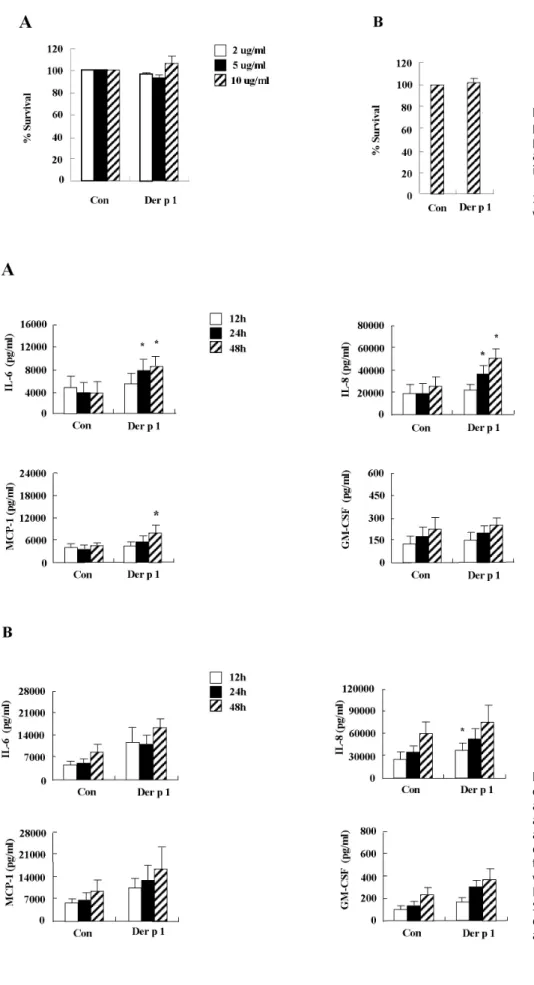
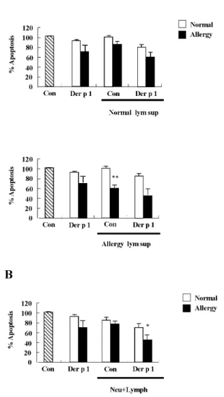
Pro-allergic cytokines were important mediators of allergic inflammation, cell recruitment and allergenic response decided to further investigate the

PI3K inhibition decreased antioxidants/GD-induced apoptosis in A549 cells, and PI3K inhibitor LY294002 had inhibitory effect on antioxidants/GD-induced caspase-3

2. No chain length effect.. 2-6 Molecular Weight Control in Linear Polymerization i) Quenching the reaction.. => Subsequent heat can change the Molecular Weight

• Equips students with fundamental knowledge in structural mechanics & structural materials for engineering.. • Pre-requisite to structural analysis/design modules