INTRODUCTION
Minoxidil, a pyrimidine derivative (2,4-diamino-6-pipe- ridino-pyrimidine-3-oxide), is an adenosine triphosphate (ATP)-sensitive potassium channel (KATPchannel) opener, and is reported to exert growth-promoting effects on follic- ular epithelial cells and to stimulate the rapid anagen induc- tion of hair cycles (1, 2). Although the mechanism of minox- idil promoting hair growth is still speculative, minoxidil was reported to mediate its effect through the growth factors- related ways, to increase blood circulation around hair folli- cles and in addition, markedly to elevate 17 -hydroxysteroid dehydrogenase activity that accelerates the conversion of tes- tosterone to weaker androgen (extensively reviewed in [3]).
Recently, it was also reported that the expression of genes encoding potassium channels and related genes was upregu- lated in hair follicle bulge stem cells (4).
Vitamin A and its active derivatives such as retinoic acid have been reported to play an important role in the growth, differentiation and maintenance of hair follicles (5, 6). Topi- cal application of all-trans retinoic acid (ATRA) is known to control hair growth cycle (7). ATRA has been suggested to stimulate the growth of sub-optimal hairs and act synergis- tically with minoxidil, producing more dense hairs than either
compound alone (8). ATRA was also reported to enhance the percutaneous absorption of minoxidil by increasing the stratum corneum permeability (9).
Recently, we have shown that minoxidil prolongs anagen stage resulting in promoting hair growth by engaging two mechanisms: the activation of Erk and Akt that enhances the survival of cultured human dermal papilla cells (DPCs) and the increase of the ratio of Bcl-2/Bax that protects cells against cell death (10). The aim of this study was to evalu- ate the possible interaction of minoxidil and ATRA on hair growth in vitro, and to investigate the effect of minoxidil in combination with ATRA on normal human epidermal kera- tinocytes (NHK) as an epithelial counterpart in addition to the suggested system in cultured dermal papilla cells (DPCs).
MATERIALS AND METHODS Drugs and reagents
Minoxidil and ATRA were purchased from Sigma (St Louis, MO, U.S.A.). Minoxidil was dissolved in 0.12 mM HCl and 1.0 mM minoxidil stock solution was stored at -20℃. ATRA was dissolved in dimethylsulfoxide (DMSO). 0.1 mM ATRA
Oh Sang Kwon, Hyun Keol Pyo, Youn Jin Oh, Ji Hyun Han, Se Rah Lee, Jin Ho Chung, Hee Chul Eun, Kyu Han Kim
Department of Dermatology, Seoul National University College of Medicine, Laboratory of Cutaneous Aging and Hair Research, Clinical Research Institute, Seoul National University Hospital, Institute of Dermatological Science, Seoul National University, Seoul, Korea
Address for correspondence Kyu Han Kim, M.D.
Department of Dermatology, Seoul National University College of Medicine, 28 Yeongeon-dong, Jongno-gu, Seoul 110-744, Korea
Tel : +82.2-2072-3643, Fax : +82.2-742-7344 E-mail : [email protected]
*This study was supported by a grant of the Korea Health 21 R&D Project, Ministry of Health & Welfare, Republic of Korea (03-PJ1-PG1-CH13-0001).
283
Promotive Effect of Minoxidil Combined with All- trans Retinoic Acid (tretinoin) on Human Hair Growth in Vitro
Minoxidil induces hair growth in male pattern baldness and prolongs the anagen phase. All-trans retinoic acid (ATRA) has been reported to act synergistically with minoxidil in vivo: they can enhance more dense hair regrowth than either compound alone. We evaluated the effect of minoxidil combined with ATRA on hair growth in vitro. The effect of co-treatment of minoxidil and ATRA on hair growth was studied in hair follicle organ culture. In cultured human dermal papilla cells (DPCs) and nor- mal human epidermal keratinocytes, the expressions of Erk, Akt, Bcl-2, Bax, P53 and P21 were evaluated by immunoblot analysis. Minoxidil plus ATRA additively pro- moted hair growth in vitro, compared with minoxidil alone. In addition, minoxidil plus ATRA elevated phosphorylated Erk, phosphorylated Akt and the ratio of Bcl-2/Bax, but decreased the expressions of P53 and P21 more effectively than by minoxidil alone. Our results suggest that minoxidil plus ATRA would additively enhance hair growth by mediating dual functions: 1) the prolongation of cell survival by activating the Erk and Akt signaling pathways, and 2) the prevention of apoptosis of DPCs and epithelial cells by increasing the ratio of Bcl-2/Bax and downregulating the ex- pressions of P53 and P21.
Key Words : Minoxidil; Tretinoin; Hair Follicle; Hair Growth; Dermal Papilla Cell; Keratinocytes
Received : 15 February 2006 Accepted : 26 July 2006
stock solution was shielded from the light with aluminum foil until required.
Anti-phosphorylated Erk-1/2 (Thr202/Tyr 204) antibody, anti-total Erk-1/2 antibody, anti-phosphorylated Akt (Ser473) antibody, and anti-total Akt antibody were purchased from Cell Signaling Technology, Inc. (Beverly, MA, U.S.A.). Anti- Bcl-2 and anti-Bax antibodies were obtained from Dako (Glo- strup, Denmark). Anti-P53 (DO7) antibody was obtained from Novocastra (Newcastle, U.K.). Anti-P21 antibody was obtained from Oncogene (San Diego, CA, U.S.A.). Anti- - actin antibody was obtained from Santa Cruz Biotech Inc.
(Santa Cruz, CA, U.S.A.).
Human hair follicle samples
Tissue samples of the occipital scalp region were obtained by excisional biopsy. Ten healthy male volunteers, aged bet- ween 20 and 35 yr, were recruited. The subjects did not have a current or prior disease and were not under medication for at least 1 month. The Institutional Review Board at the Seoul National University Hospital approved all procedures used in this study. Written informed consents were obtained from all volunteers. Tissue samples containing more than 100 hair follicles were cautiously dissected into single hair follicles.
We used hair follicles morphologically in the anagen stage only determined as described previously (11).
Hair follicle organ culture
Human scalp hair follicles were isolated and cultured in vitro as described previously (12). Briefly, dissected hair folli- cle was cut into small pieces approximately 2.5 mm in length from the bottom of dermal papilla and cultured in Williams E medium (Gibco BRL, Gaithersburg, MD) with 10 ng/mL hydrocortisone, 10 g/mL insulin, 2 mM L-glutamine, and 100 U/mL penicillin at 37℃in 5% CO2atmosphere. Minox- idil alone or in combination with ATRA was added to the culture medium. Minoxidil was used at 1 M. ATRA was used at the final concentration ranged from 1 nM to 10 nM.
A total of 180 anagen hair follicles from 3 different vol- unteers (60 follicles per subject) were cultured in triplicates under 4 different growth conditions. The data are presented as the means±SEM. In all experiments, tissue culture me- dium containing minoxidil alone or minoxidil plus ATRA was changed every other day. After culturing for 12 days, the hair growth was measured directly using an Olympus stereomicroscope with an eyepiece containing a graticule.
Culture of dermal papilla cells and normal human epider- mal keratinocytes
Human DPCs were cultured as described previously (13).
Briefly, DPCs were cultured in Dulbecco’s modified Eagle’s medium (DMEM, Gibco BRL, Gaithersburg, MD, U.S.A.)
containing 2 mM L-glutamine, 1× antibiotic antimycotic solution (1,000 units/mL of penicillin G sodium and 2.5 g/
mL of amphotericin B) and 10% fetal bovine serum (Hyclone, Logan, UT, U.S.A.) at 37℃, in a 5% CO2incubator. The fourth-passage DPCs were used.
As follicular and nonfollicular epidermal keratinocytes are known to respond similarly to minoxidil treatment (14), we used NHK of five human adult foreskins obtained from cir- cumcisions. NHK were isolated as previously described (15) and cultured in keratinocyte growth medium (Clonetics, San Diego, CA, U.S.A.) composed of MCDB 153 medium sup- plemented with epidermal growth factor (10 ng/mL), bovine pituitary extract (70 g/mL), hydrocortisone (0.5 g/mL), insulin (5 g/mL), penicillin (100 g/mL) and fungizone (0.25 g/mL). The NHK were cultured until 80% conflu- ent in tissue culture flasks (Becton Dickinson, Lincoln Park, NJ, U.S.A.) at 37℃and 5% CO2and stored in liquid nitro- gen until use. At the time of the experiments, the cells were thawed and third passage cells were used.
3-(4,5-Dimethylthiazol-2-yl)-2,5-Diphenyl Tetrazolium Bromide (MTT) assay
As a parameter to check for cytotoxic effect of minoxidil plus ATRA, cell viability was measured using the MTT assay as previously described (16). DPCs and NHK of 1.5×10⁴ cells/well were seeded into 96-well plates, cultured for 24 hr in serum-free DMEM, and treated with the vehicle (0.12 mM HCl/DMSO diluted 1:1,000 in DMEM) as control, and the various combined concentrations of ATRA (0.01-1 nM) and minoxidil (0.01-1.0 M) for 5 days. 20 L MTT solution (5 mg/mL) was added to a well, incubated for 4 hr at 37℃in the dark, removed the supernatants, added 200 L DMSO to dissolve formazan products, incubated for 30 min at room temperature, and the absorbance was measured at 570 nm using an ELISA reader. The results were expressed as the per- centage of control cells in six same culture conditions.
Western blot analysis after treatment with minoxidil alone or minoxidil plus ATRA on the dermal papilla cells and keratinocytes
For the treatment with minoxidil alone or minoxidil plus ATRA, confluent DPCs and NHK were incubated for 24 hr in serum-free DMEM and treated with the vehicle (0.12 mM HCl and/or DMSO diluted at 1:1,000 in DMEM in half), minoxidil alone (0.1 M), or 0.1 M minoxidil plus 0.1 nM ATRA for 1 hr for the evaluation of Erk and Akt, and for 24 hr for the evaluation of Bcl-2/Bax and P53/P21.
Western blot analysis was performed as follows. Briefly, proteins were extracted using the buffer containing 50 mM Tris-HCl (pH 7.4), 2 mM EDTA, 100 g/mL leupeptin, 20 g/mL aprotinin, and 100 mM NaCl. The supernatant was collected and kept at -70℃until used. Fifty microgram
of protein was loaded to a lane of 10% or 12% SDS-poly- acrylamide gel, separated by electrophoresis, and blotted onto nitrocellulose membrane. The blotted membrane was incu- bated with primary antibody (anti-total Erk polyclonal anti- body, 1:500; anti-phosphorylated Erk polyclonal antibody, 1:500; anti-total Akt polyclonal antibody, 1:500; anti-phos- phorylated Akt polyclonal antibody, 1:500; anti-Bcl-2 mon- oclonal antibody, 1:1,000; anti-Bax monoclonal antibody, 1:1,000; anti-P53 monoclonal antibody, 1:1,000; anti-P21 monoclonal antibody, 1:1,000; anti- -actin monoclonal anti- body, 1:1,000) at 4℃overnight. The membrane was incu- bated with the anti-mouse IgG-HRP conjugate (1:2,000) or the anti-rabbit IgG-HRP conjugate (1:2,000) for 1 hr at room temperature. The antibody-antigen complex was detected using the ECL system (Amersham Pharmacia Biotech; Lit- tle Chalfont, U.K.) and analyzed using a Bio-Rad GS-700 imaging densitometer (Hercules, CA, U.S.A.).
Statistical analysis
Statistical significance was determined using the ANOVA multiple comparison test and Wilcoxon-rank sum test. p value of less than 0.05 was considered statistically significant.
RESULTS
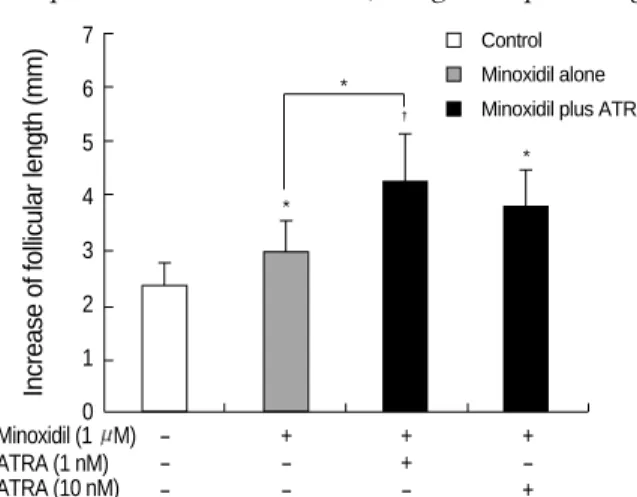
Minoxidil plus ATRA enhanced hair growth more efficiently than minoxidil alone in vitro
Compared with minoxidil alone, the growth-promoting
activity of the minoxidil plus ATRA was examined in in vitro human hair follicle culture model. Minoxidil at 1 M enhanced hair growth significantly compared with vehicle- treated control (p<0.05) (Fig. 1). The minoxidil plus ATRA (1 nM) more increased hair growth by 4.2±0.7 mm com- pared with mioxidil alone (p<0.05). Combination of the higher concentration of ATRA (10 nM) was less effective than the mixture of minoxidil plus 1 nM ATRA, although the enhanced hair growth by mixed preparation with 10 nM ATRA was at least not less than the minoxidil alone.
Minoxidil plus ATRA induced the proliferations of DPCs and NHK
Minoxidil (0.01-1 M) plus ATRA (0.01-1 nM) enhanced the proliferations of cultured DPCs and NHK. The enhance- ment was the greatest at minoxidil 0.1 M plus ATRA 0.1 nM by 189.7±42.1% (mean±SD) in DPCs and by 219.5
±57.8% in NHK.
Minoxidil plus ATRA increased the phosphorylation of Erk in DPCs and NHK more effectively than minoxidil alone
Treated with minoxidil, the phosphorylated Erk (p-Erk) level in DPCs was significantly increased to 214±21%. The minoxidil plus ATRA more significantly elevated the p-Erk level to 298±50% (p<0.05) (Fig. 2A). The NHK from five different donors was tested with minoxidil alone or minoxi- dil plus ATRA and in the experiment, it has to be mentioned that NHK from 2 donors did not respond to the treatment.
The expression of p-Erk in other three samples was slightly elevated to 106±4% by treatment of minoxidil. Minoxidil plus ATRA increased the expression of p-Erk to 207±51%
(Fig. 2B).
Increase of follicular length (mm)
7 6 5 4 3 2 1 0
- + + +
- - + -
- - - +
Fig. 1.The effect of minoxidil plus ATRA on hair growth in vitro.
Hair follicle organ culture was performed in triplicates with hair follicles obtained from the occipital scalp of three volunteers. Hair growth is enhanced by minoxidil at 1 M. Minoxidil plus ATRA promotes hair growth more significantly, especially at a concen- tration of 1 M minoxidil and 1 nM ATRA. Moreover, the increase in hair growth was significant compared with minoxidil alone. Val- ues were obtained after cumulative growth of 15 hair follicles per each growth condition for 12 days and shown as the means±SEM.
*, p<0.05; �, p<0.01.
Fig. 2.The effect of minoxidil and ATRA on the p-Erk and on total Erk in (A) DPCs and in (B) NHK. The level of p-Erk increased sig- nificantly after treatment of minoxidil, and minoxidil plus ATRA more enhanced the expression of p-Erk. The representative bands of triplicate experiments are shown. The values shown are the means
±SEM of % elevation compared with the vehicle-treated control from three different DPCs and NHK cultures. *, p<0.05, compared with the vehicle-treated control. p-Erk, phosphorylated Erk; T-Erk, total Erk; M, minoxidil.
Minoxidil (1 M) ATRA (1 nM) ATRA (10 nM)
*
*
*
�
Control Minoxidil alone Minoxidil plus ATRA
p-Erk T-Erk
CONT M M+ATRA ERK1 ERK2 ERK1 ERK2
% of control
350 300 250 200 150 100 50
0 CONT M M+ATRA p-Erk
T-Erk
*
*
A
CONT M M+ATRA ERK1 ERK2 ERK1 ERK2
% of control
300 250 200 150 100 50
0 CONT M M+ATRA p-Erk
T-Erk
B
The total Erk level was barely changed in response to mi- noxidil alone or minoxidil plus ATRA in both cells. The val- ues shown are the means±SEM of percentage increases ver- sus the control from three different batches of DPCs or NHK.
Minoxidil plus ATRA increased the phosphorylation of Akt in DPCs and NHK more efficiently than minoxidil alone
Minoxidil enhanced the level of phosphorylated Akt (p- Akt) to 126±8% in DPCs and the minoxidil plus ATRA more increased the phosphorylation of Akt to 315±8% (p<
0.05) (Fig. 3A).
The expression of p-Akt in the NHK from the three donors increased in a similar manner with DPCs by minoxidil alone and minoxidil plus ATRA to 111±10% and 284±56%, respectively (Fig. 3B). The expression of p-Akt was not ele- vated in the same two cells that were irresponsive to the treat- ment in p-Erk. Minoxidil alone and minoxidil plus ATRA had no effect on the expression of total Akt in both cells.
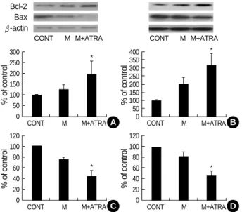
Minoxidil plus ATRA increased the Bcl-2 expression and decreased the Bax expression in DPCs and NHK more efficiently than minoxidil alone
To evaluate the possible association in changes on Bcl-2 family proteins, we investigated the effects of minoxidil alone or minoxidil plus ATRA on the expressions of Bcl-2 and Bax protein. Treated with minoxidil, the expression of Bcl-2 pro- tein was increased in DPCs to 128±18%. The increase, how- ever, was not statistically significant. Minoxidil plus ATRA
Fig. 3.The increase of p-Akt expression by minoxidil and ATRA.
(A) In DPCs, minoxidil increases p-Akt, moreover, minoxidil plus ATRA more significantly elevates the p-Akt level. (B) In NHK, mi- noxidil alone and minoxidil plus ATRA increase p-Akt expression, however, they were statistically insignificant. The blotted bands are the representative of triplicate experiments. The values are shown as the means±SEM of % elevation compared with con- trols. *, p<0.05, compared with controls treated with the vehicle.
p-Akt, phosphorylated Akt; T-Akt, total Akt; M, minoxidil.
p-Akt T-Akt
CONT M M+ATRA
% of control
400 350 300 250 200 150 100 50
0 CONT M M+ATRA p-Akt
T-Akt
*
*
A
CONT M M+ATRA
% of control
350 300 250 200 150 100 50
0 CONT M M+ATRA p-Akt
T-Akt
B
Fig. 4.The effect of minoxidil and ATRA on the expression of Bcl-2 and Bax protein in DPCs and NHK. Compared with vehicle-treat- ed control, minoxidil plus ATRA significantly increases the expres- sion of Bcl-2 in DPCs (A) and in NHK (B). (C) Minoxidil significantly decreases the expression of Bax. Minoxidil plus ATRA suppresses the expression of Bax more efficiently in DPCs. (D) Expression of Bax in NHK is also significantly decreased by minoxidil plus ATRA.
The bands are the representatives of triplicate experiments. The values are shown as the means±SEM of % elevation in compari- son with the controls. *, p<0.05 compared with vehicle-treated control. M, minoxidil.
Bcl-2 Bax -actin
CONT M M+ATRA CONT M M+ATRA
% of control
300 250 200 150 100 50 0
CONT M M+ATRA
*
A
% of control
400 350 300 250 200 150 100 50
0 CONT M M+ATRA
*
B
% of control
120 100 80 60 40 20
0 CONT M M+ATRA
*
C
% of control
120 100 80 60 40 20
0 CONT M M+ATRA
*
D
Fig. 5.The effects of minoxidil and ATRA on the expressions of P53 and P21 protein in DPCs and NHK. (A) Minoxidil alone and minoxidil plus ATRA significantly suppress the expression of P53 protein in DPCs. (B) In NHK, minoxidil plus ATRA decreases P53 expression significantly. (C) In DPCs, Minoxidil significantly dec- reases the expression of P21. Minoxidil plus ATRA reduces the expression of P21 more significantly. (D) P21 expression is sig- nificantly downregulated with minoxidil plus ATRA. The bands are the representative of triplicate experiments. *, p<0.05 com- pared with the vehicle-treated control. The data are shown as the means±SEM of % elevation compared with controls from three different DPC and NHK cultures. M, minoxidil.
p53 p21 -actin
CONT M M+ATRA CONT M M+ATRA
% of control
120 100 80 60 40 20 0
CONT M M+ATRA
* *
A
% of control
120 100 80 60 40 20 0
CONT M M+ATRA
*
B
% of control
120 100 80 60 40 20
0 CONT M M+ATRA
*
* *
C
% of control
120 100 80 60 40 20
0 CONT M M+ATRA
*
D
elevated the Bcl-2 expression to 200±73% with statistical significance (p<0.05) (Fig. 4A). For NHK, the expression of Bcl-2 protein was increased similarly; elevated Bcl-2 expres- sion to 206±38% with minoxidil alone, and with minoxi- dil plus ATRA, to 325±68% (Fig. 4B).
In contrast, minoxidil decreased the Bax protein expression significantly in DPCs, to 75±3%. Minoxidil plus ATRA more significantly downregulated the expression of Bax to 45±10% (p<0.05) (Fig. 4C). Minoxidil alone or minoxidil plus ATRA also decreased the Bax protein expression signifi- cantly in NHK to 83±7% and to 47±9%, respectively (p<0.05) (Fig. 4D).
Minoxidil plus ATRA downregulated the expressions of P53 and P21 in DPCs and NHK more efficiently than minoxidil alone
P53 plays a pivotal role in the regulation of cell growth and programmed cell death. P21 is a specific downstream target of P53 that mediates the cell cycle arrest (17). Treated in DPCs, minoxidil alone and minoxidil plus ATRA down- regulated the expression of P53 to 59±2% and to 47.5± 10%, respectively (p<0.05) (Fig. 5A). For NHK, expression patterns of P53 were also decreased significantly by minoxi- dil plus ATRA (p<0.05) (Fig. 5B).
Similarly, minoxidil decreased the expression of P21 to 65
±4% in DPCs. The minoxidil plus ATRA decreased the level of P21 more efficiently to 45±5% (p<0.05) (Fig. 5C).
In NHK, P21 expression was declined to 93±4% with minoxidil alone, and to 52±10% by minoxidil plus ATRA (p<0.05) (Fig. 5D).
DISCUSSION
All-trans retinoic acid, alone or in combination with minox- idil, has been reported to shorten the second telogen phase and lengthen the second anagen phase in the C3H mouse model (7). In this study, we demonstrated that the minoxi- dil plus ATRA additively enhances hair growth in in vitro human hair follicles, compared with minoxidil alone.
Previously, minoxidil was reported to have the concentra- tion-dependent biphasic effect on proliferation and differen- tiation; growth stimulation at low doses or anti-proliferative, pro-differentiative and partially cytotoxic effects at high doses (14). At 10 nM of ATRA co-treated with minoxidil, less elongation than at 1 nM suggested that ATRA might increase the tissue concentration of minoxidil in hair follicles in vitro.
Although ATRA is converted to other active metabolites in the dermis, the doses used in organ culture would be com- patible to the dermal concentration in vivo, because the con- centration of commercially available topical ATRA is 0.01- 0.1% and in human cadaver skin, 0.11-0.44% of ATRA is deposited in the dermis by topical application (18).
The cellular and molecular basis of ATRA mediating cell proliferation and the formation of extracellular matrix has not been delineated. ATRA at 1 nM was reported to extend the life span of human oral and epidermal keratinocytes by decreasing the expression of senescence-associated genes (p53, p21, p16) and maintaining telomerase activity, but at higher concentrations the replicative senescence was enhanced in a dose-dependent manner (19).
We demonstrated here that minoxidil plus ATRA addi- tively increased the phosphorylation of Erk in both DPCs and NHK, compared with minoxidil alone. The role of the Erk signaling pathway in cell growth has been well estab- lished (20). The diverse involvement of retinoic acid has been demonstrated in the regulation of developmental processes and the modulation of differentiation in various cellular mod- els by the MAPK pathways (21, 22). The MAPK pathway is also involved in maintaining cell survival by modulating apoptotic molecules including Bcl-2 family (23). The PI3 kinase/ Akt cascade plays a crucial role in cell survival and the prevention of apoptosis (24, 25). The crosstalk between Erk and PI3K/Akt pathway has been demonstrated to pro- long cell survival (26). Our results thus suggest that the acti- vation of Akt, an anti-apoptotic molecule, by minoxidil plus ATRA may prolong survival of DPCs and epithelial coun- terpart.
The Bcl-2 family proteins are structurally related molecules that play an essential role in the regulation of apoptosis (27).
Bcl-2 family consists of anti-apoptotic proteins such as Bcl- 2 as well as pro-apoptotic proteins such as Bad and Bax. Erk and PI3K/Akt pathways synergistically induce cell survival by Bcl-2 cascade (28). Throughout the hair cycle, dermal papilla (DP) is the only region expressing consistently Bcl-2 proteins and the Bax proteins are not detected during hair cycles (29, 30). The DP thus is considered to be resistant apoptosis. On the other hand, the downregulation of Bcl-2 expression has been shown to induce apoptosis of DPCs in tissue culture (31). The relative amount of Bcl-2 over Bax is known to determine the fate of living cells (32). Retinoid treatment has been reported to increase the ratio of mRNAs encoding bcl-2 and bax significantly (33). Our data suggest that minoxidil plus ATRA additively promote cell survival by the modulation of the ratio of Bcl-2 and Bax in DPCs and NHK.
P53 is a tumor suppressor gene that plays a pivotal role in the regulation of cell growth and cell death. The downstream transcriptional targets of P53 are the cyclin-dependent kinase inhibitor P21WAF1that mediates G1 arrest and Bax that is involved in apoptosis (17). In addition, bcl-2 gene was demon- strated to act as a transcriptional target for wild-type P53 that downregulates the endogenous Bcl-2 expression and upregulates the Bax expression (34). Moreover, in addition to transcriptional activation, P53-dependent apoptosis may be induced through a transactivation-independent means, such as by the induction of oxidative stress, the inhibition
of RNA, or the inhibition of protein synthesis (35). In this study, minoxidil plus ATRA worked together to suppress the expression of P53 and its downstream target P21, although the exact mechanism has to be elucidated in future experi- ments.
In summary, our data suggest that the enhanced hair growth by minoxidil plus ATRA in our short-term organ culture may be explained through prolonged survival of epithelial cells and DPCs, which mediate signals for follicular epithe- lium. Minoxidil plus ATRA more increased the phosphoryla- tion of Erk and Akt early 1 hr after the treatment than mino- xidil alone. The change of Bcl-2/Bax ratio, P53 and P21 were also detected later 24 hr after the treatments. Minoxidil plus ATRA could work together to prolong the survival of cul- tured DPCs and epithelial cells and to protect them from apoptosis by dual mechanisms with different kinetics: 1) the activation of Erk- and Akt-dependent pathways and 2) the increase of the ratio of Bcl-2/Bax and the suppression of the expression of P53 and its downstream target P21.
ACKNOWLEDGEMENT
We thank for excellent statistical assistance to Min Jeong Lee in Clinical Research Institute, Seoul National Universi- ty Hospital.
REFERENCES
1. Tanigaki-Obana N, Ito M. Effects of cepharanthine and minoxidil on proliferation, differentiation and keratinization of cultured cells from the murine hair apparatus. Arch Dermatol Res 1992; 284: 290-6.
2. Lee WS, Ahn HJ, Kim YH. The effect of coapplication of capsaicin and minoxidil on the murine hair growth. Korean J Dermatol 2003;
41: 451-60.
3. Messenger AG, Rundegren J. Minoxidil: mechanisms of action on hair growth. Br J Dermatol 2004; 150: 186-94.
4. Morris RJ, Liu Y, Marles L, Yang, Z, Trempus C, Li S, Lin JS, Saw- icki JA, Cotsarelis G. Capturing and profiling adult hair follicle stem cells. Nat Biotechnol 2004; 22: 411-7.
5. Bergfeld WF. Retinoids and hair growth. J Am Acad Dermatol 1998;
39: 86-9.
6. Fisher GJ, Voorhees JJ. Molecular mechanisms of retinoid actions in skin. FASEB J 1996; 10: 1002-13.
7. Bazzano G, Terezakis N, Attia H, Bazzano A, Dover R, Fenton D, Mandir N, Celleno L, Tamburro M, Jaconi S. Effect of retinoids on follicular cells. J Invest Dermatol 1993; 101: 138-42.
8. Bazzano GS, Terezakis N, Galen W. Topical tretinoin for hair growth promotion. J Am Acad Dermatol 1986; 15: 890-3.
9. Ferry JJ, Forbes KK, VanderLugt JT, Szpunar GJ. Influence of treti- noin on the percutaneous absorption of minoxidil from an aqueous topical solution. Clin Pharmacol Ther 1990; 47: 439-46.
10. Han JH, Kwon OS, Chung JH, Cho KH, Eun HC, Kim KH. Effect
of minoxidil on proliferation and apoptosis in dermal papilla cells of human hair follicle. J Dermatol Sci 2004; 34: 91-8.
11. Messenger AG. The culture of dermal papilla cells from human hair follicles. Br J Dermatol 1984; 110: 685-9.
12. Philpott MP, Green MR, Kealey T. Human hair growth in vitro. J Cell Sci 1990; 97: 463-71.
13. Randall VA, Thornton MJ, Redfern CP. Dermal papilla cells from human hair follicles express mRNA for retinoic acid receptors in culture. Ann N Y Acad Sci 1991; 642: 457-8.
14. Boyera N, Galey I, Bernard BA. Biphasic effects of minoxidil on the proliferation and differentiation of normal human keratinocytes. Skin Pharmacol 1997; 10: 206-20.
15. Boyce ST, Ham RG. Calcium-regulated differentiation of normal human epidermal keratinocytes in chemically defined clonal culture and serum-free serial culture. J Invest Dermatol 1983; 81: 33-40.
16. Mosmann T. Rapid colorimetric assay for cellular growth and sur- vival: application to proliferation and cytotoxicity assays. J Immunol Methods 1983; 65: 55-63.
17. Kim R, Tanabe K, Emi M, Uchida Y, Inoue H, Toge T. Inducing cancer cell death by targeting transcription factors. Anticancer Drugs 2003; 14: 3-11.
18. Skov MJ, Quigley JW, Bucks DA. Topical delivery system for treti- noin: research and clinical implications. J Pharm Sci 1997; 86: 1138- 43.
19. You YO, Lee G, Min BM. Retinoic acid extends the in vitro life span of normal human oral keratinocytes by decreasing p16 (INK4A) ex- pression and maintaining telomerase activity. Biochem Biophys Res Commun 2000; 268: 268-74.
20. Robinson MJ, Cobb MH. Mitogen-activated protein kinase pathways.
Curr Opin Cell Biol 1997; 9: 180-6.
21. Bost F, Caron L, Marchetti I, Dani C, Le Marchand-Brustel Y, Bi- netruy B. Retinoic acid activation of the ERK pathway is required for embryonic stem cell commitment into the adipocyte lineage. Bio- chem J 2002; 361: 621-7.
22. Chung JH, Kang S, Varani J, Lin J, Fisher GJ, Voorhees JJ. Decreased extracellular-signal-regulated kinase and increased stress-activated MAP kinase activities in aged human skin in vivo. J Invest Dermatol 2000; 115: 177-82.
23. Chang F, Steelman LS, Shelton JG, Lee JT, Navolanic PM, Blalock WL, Franklin R, McCubrey JA. Regulation of cell cycle progression and apoptosis by the Ras/Raf/MEK/ERK pathway [Review]. Int J Oncol 2003; 22: 469-80.
24. Ahmad S, Singh N, Glazer RI. Role of AKT1 in 17beta-estradiol- and insulin-like growth factor I (IGF-I)-dependent proliferation and pre- vention of apoptosis in MCF-7 breast carcinoma cells. Biochem Phar- macol 1999; 58: 425-30.
25. Tang Y, Zhou H, Chen A, Pittman RN, Field J. The Akt proto-onco- gene links Ras to Pak and cell survival signals. J Biol Chem 2000;
275: 9106-9.
26. Chang F, Lee JT, Navolanic PM, Lee JT, Shelton JG, Navolanic PM, Blalock WL, Franklin RA, McCubrey JA. Involvement of PI3K/Akt pathway in cell cycle progression, apoptosis, and neoplastic trans- formation: a target for cancer chemotherapy. Leukemia 2003; 17:
590-603.
27. Cory S, Huang DC, Adams JM. The Bcl-2 family: roles in cell sur- vival and oncogenesis. Oncogene 2003; 22: 8590-607.
28. McCubrey JA, Steelman LS, Blalock WL, Lee JT, Moye PW, Chang F, Pearce M, Shelton JG, White MK, Franklin RA, Pohnert SC. Syn- ergistic effects of pi3k/akt on abrogation of cytokine-dependency induced by oncogenic raf. Adv Enzyme Regul 2001; 41: 289-323.
29. Stenn KS, Lawrence L, Veis D, Korsmeyer S, Seiberg M. Expres- sion of the bcl-2 protooncogene in the cycling adult mouse hair fol- licle. J Invest Dermatol 1994; 103: 107-11.
30. Lindner G, Botchkarev VA, Botchkareva NV, Ling G, van der Veen C, Paus R. Analysis of apoptosis during hair follicle regression (cata- gen). Am J Pathol 1997; 151: 1601-17.
31. Ferraris C, Cooklis M, Polakowska RR, Haake AR. Induction of
apoptosis through the PKC pathway in cultured dermal papilla fibro- blasts. Exp Cell Res 1997; 234: 37-46.
32. Adams JM, Cory S. The Bcl-2 protein family: arbiters of cell sur- vival. Science 1998; 281: 1322-6.
33. Chung JJ, Cho S, Kwon YK, Kim DH, Kim K. Activation of retinoic acid receptor gamma induces proliferation of immortalized hippo- campal progenitor cells. Brain Res Mol Brain Res 2000; 83: 52-62.
34. Reed JC, Miyashita T, Takayama S, Wang HG, Sato T, Krajewski S, Aime-Sempe C, Bodrug S, Kitada S, Hanada M. BCL-2 family proteins: regulators of cell death involved in the pathogenesis of cancer and resistance to therapy. J Cell Biochem 1996; 60: 23-32.
35. Polyak K, Xia Y, Zweier JL, Kinzler KW, Vogelstein B. A model for p53-induced apoptosis. Nature 1997; 389: 300-5.