D I A B E T E S & M E T A B O L I S M J O U R N A L
This is an Open Access article distributed under the terms of the Creative Commons Attribution Non-Commercial License (http://creativecommons.org/licenses/by-nc/4.0/) which permits unrestricted non-commercial use, distribution, and reproduction in any medium, provided the original work is properly cited.
Statins Increase Mitochondrial and Peroxisomal Fatty Acid Oxidation in the Liver and Prevent Non-Alcoholic Steatohepatitis in Mice
Han-Sol Park1,*, Jung Eun Jang2,*, Myoung Seok Ko1, Sung Hoon Woo1, Bum Joong Kim1,2, Hyun Sik Kim1, Hye Sun Park1, In-Sun Park3, Eun Hee Koh2, Ki-Up Lee2
1Asan Institute for Life Sciences, 2Department of Internal Medicine, Asan Medical Center, University of Ulsan College of Medicine, Seoul,
3Department of Anatomy, Inha University School of Medicine, Incheon, Korea
Background: Non-alcoholic fatty liver disease is the most common form of chronic liver disease in industrialized countries. Re- cent studies have highlighted the association between peroxisomal dysfunction and hepatic steatosis. Peroxisomes are intracellu- lar organelles that contribute to several crucial metabolic processes, such as facilitation of mitochondrial fatty acid oxidation (FAO) and removal of reactive oxygen species through catalase or plasmalogen synthesis. Statins are known to prevent hepatic steatosis and non-alcoholic steatohepatitis (NASH), but underlying mechanisms of this prevention are largely unknown.
Methods: Seven-week-old C57BL/6J mice were given normal chow or a methionine- and choline-deficient diet (MCDD) with or without various statins, fluvastatin, pravastatin, simvastatin, atorvastatin, and rosuvastatin (15 mg/kg/day), for 6 weeks. Histo- logical lesions were analyzed by grading and staging systems of NASH. We also measured mitochondrial and peroxisomal FAO in the liver.
Results: Statin treatment prevented the development of MCDD-induced NASH. Both steatosis and inflammation or fibrosis grades were significantly improved by statins compared with MCDD-fed mice. Gene expression levels of peroxisomal prolifera- tor-activated receptor α (PPARα) were decreased by MCDD and recovered by statin treatment. MCDD-induced suppression of mitochondrial and peroxisomal FAO was restored by statins. Each statin’s effect on increasing FAO and improving NASH was independent on its effect of decreasing cholesterol levels.
Conclusion: Statins prevented NASH and increased mitochondrial and peroxisomal FAO via induction of PPARα. The ability to increase hepatic FAO is likely the major determinant of NASH prevention by statins. Improvement of peroxisomal function by statins may contribute to the prevention of NASH.
Keywords: Fatty acid oxidation; Non-alcoholic fatty liver disease; Peroxisomes; Statins
Corresponding author: Ki-Up Lee http://orcid.org/0000-0001-6233-6093 Department of Internal Medicine, Asan Medical Center, University of Ulsan College of Medicine, 88, Olympic-ro 43-gil, Songpa-gu, Seoul 05505, Korea E-mail: [email protected]
* Han-Sol Park and Jung Eun Jang contributed equally to this study as first
INTRODUCTION
Non-alcoholic fatty liver disease (NAFLD) is a clinical spec- trum of liver damage, from simple steatosis to more advanced stages, such as non-alcoholic steatohepatitis (NASH), fibrosis, or cirrhosis [1,2]. Because NAFLD is closely associated with obesity, diabetes, and cardiovascular disease, it is regarded as a representative hepatic phenotype of metabolic syndrome [3].
NAFLD is the most common cause of chronic liver disease in developed countries [4], and the prevalence in the general population ranges from 20% to 30% [5].
Statins competitively inhibit 3-hydroxy-3-methylglutaryl- coenzyme A (HMG-CoA) reductase, the rate-limiting enzyme in cholesterol synthesis, and are widely used as cholesterol- lowering drugs. The overall benefits of statins seem to be greater than what might be expected from an alteration in the http://dx.doi.org/10.4093/dmj.2016.40.5.376
pISSN 2233-6079 · eISSN 2233-6087
lipid profile alone, suggesting that statins have cholesterol-in- dependent pleiotropic effects [6]. However, the basic mecha- nism underlying these pleiotropic effects is largely unknown.
Statins prevent hepatic steatosis in animals, and suggested mechanisms include prevention of carbohydrate response ele- ment-binding protein activation [7] and the induction of per- oxisomal proliferator-activated receptor α (PPARα) [8], the master regulator of fatty acid oxidation (FAO). Statins also at- tenuate hepatic inflammatory reactions induced by angioten- sin II [9] and prevent hepatic fibrosis by inactivating hepatic stellate cells [10]. Accumulation of free cholesterol in the mi- tochondria is suggested to be a major mechanism for steato- hepatitis [11], and lowering of free cholesterol by statins may also be a mechanism of how statins prevent NASH.
It is well known that decreased mitochondrial function con- tributes to the development of hepatic steatosis [12]. Recent studies have also highlighted the association between peroxi- somal dysfunction and hepatic steatosis [13,14]. Peroxisomes are ubiquitous, single-membrane-bounded organelles and ex- ist in all eukaryotes [15]. The main metabolic functions of per- oxisomes in mammalian cells include degradation of very long chain and branched-chain fatty acids, which cannot be in- stantly oxidized in mitochondria. Peroxisomes are responsible for the biosynthesis of plasmalogen, a special class of lipids, and docosahexaenoic acid, a final elongation and desaturation product of n-3 polyunsaturated fatty acids [16]. Peroxisomes also play a critical role in maintenance of intracellular redox balance. Production of reactive oxygen species is inevitable in fuel metabolism, and peroxisomes possess several anti-oxida- tive systems, including catalase, superoxide dismutases, and peroxiredoxins [17]. Despite their importance, less attention has been paid to peroxisomes than other organelles, such as mitochondria, endoplasmic reticulum, and lysosomes.
In this study, we found that treatment with various statins ameliorated hepatic steatosis and steatohepatitis and that this was associated with increased hepatic FAO. In particular, per- oxisomal FAO, as well as mitochondrial FAO, was significantly decreased in the liver of NASH animals, and this was recov- ered by statins.
METHODS
Animals
Seven-week-old male C57BL/6N mice were purchased from Central Lab Animal Inc. (Seoul, Korea) and acclimated for 1
week prior to the experiment. Animals were housed at an am- bient temperature (22°C±1°C) on a 12-hour/12-hour light/
dark cycle with free access to water and diet. Mice were fed normal chow diet (ND; n=10), methionine- and choline-defi- cient diet (MCDD; n=10; Dyets Inc., Bethlehem, PA, USA), or MCDD with 15 mg/kg/day of each statin supplementation (n=6 to 10) for 6 weeks. At the end of experiment period, mice were fasted (5 hours) in the morning and then sacrificed.
Blood samples were collected and the livers were rapidly har- vested, quickly frozen in liquid nitrogen, and stored at –80°C.
All animal experiment protocols were approved by the Institu- tional Animal Care and Use Committee of the Asan Institute for Life Sciences, Seoul, South Korea.
Histological analysis
Liver tissue samples were fixed with 4% paraformaldehyde and embedded for 5 µm serial paraffin sections. The sections were stained with hematoxylin and eosin for evaluation of the steatosis and with the Masson’s trichrome (MT) for determi- nation of the fibrosis. The severities of the hepatic histological changes were assessed and scored in a blind manner using the NASH-Clinical Research Network scoring system [18]. Briefly, the steatosis grade was scored according to the degree of pa- renchymal involvement as follows: 0, <5%; 1, 5% to 33%; 2, 33% to 66%; and 3, >66%. The steatosis location was scored as follows: 0, zone 3 predominant; 1, zone 1 predominant; 2, azonal; and 3, panacinar. The lobular inflammation grade was scored by the numbers of the inflammation foci in the area of
×200 microscopic fields as follows: 0, no foci; 1, <2 foci per
×200 field; 2, 2 to 4 foci per ×200 field; and 3, >4 foci per
×200 field. The fibrosis stage was scored by the location and density of the fibrosis as follows: 0, none; 1, perisinusoidal or periportal fibrosis; 2, perisinusoidal and periportal fibrosis; 3, bridging fibrosis; and 4, cirrhosis.
Plasma and tissue biochemical assays
Plasma and hepatic triglyceride (TG) levels were measured us- ing the GPO-Trinder kit (Sigma-Aldrich, St. Louis, MO, USA), according to the manufacturer’s instructions. Plasma free fatty acid levels were determined using an enzymatic as- say kit (Wako Chemicals, Richmond, VA, USA). Plasma ala- nine aminotransferase (ALT) levels were measured using the IDToxTM Alanine Transaminase Endpoint Assay Kit (ID Labs Inc., London, ON, Canada).
Measurement of FAO
The rate of FAO was measured as 14CO2 generation from 14C palmitate (NEN Life Sciences, Boston, MA, USA), as previ- ously described [19]. Peroxisomal FAO was determined in the presence of inhibitors of mitochondrial oxidation, namely, an- timycin A and rotenone (final concentrations 100 and 12.5 µM, respectively) [14].
Quantitative real-time polymerase chain reaction analysis Total RNA was isolated using TRIzol reagent (Invitrogen, Carlsbad, CA, USA); 1 μg of each sample was reverse tran- scribed with random primers using the Reverse Aid M-MuLV reverse transcription kit (Fermentas, Hanover, MD, USA).
Target cDNA levels were quantified by real-time polymerase chain reaction (PCR) using gene-specific primers (Table 1) and the 7500 Fast RT-PCR system (Applied Biosystems, Foster City, CA, USA) containing SYBR green. The data were nor- malized to the levels of expression of the internal control t-box protein (Tbp) and expressed in arbitrary units.
Western blot analysis
Liver tissues were homogenized in lysis buffer and centrifuged at 13,000 rpm for 30 minutes at 4°C. Samples with equal amounts of protein (20 to 50 μg) were analyzed by Western blotting using antibodies against PPARα (#sc9000; Santa Cruz Biotechnology, Santa Cruz, CA, USA) and α-tubulin (#NB100-690; Novus Bio- logicals, Littleton, CO, USA).
Measurement of lipid peroxidation
Hepatic lipid peroxidation was assessed by measuring malo- ndialdehyde (MDA) levels using a Bioxytech MDA-586 assay kit (OxisResearch, Portland, OR, USA), according to the man- ufacturer’s instruction. MDA values were corrected to the tis- sue protein contents.
Statistical analyses
All values are presented as the mean±standard error of the mean. Statistical significance of the differences between ex- perimental groups was determined by the Student t-test or Table 1. Primer sets for real-time polymerase chain reaction
analysis
Gene Mouse primer sequences
Pparα 5´-AGAGCCCCATCTGTCCTCTC-3´
5´-ACTGGTAGTCTGCAAAACCAAA-3´
Pex7 5´-TGGTGACAGGTGCGGTTGAC-3´
5´-ATAGGAGCAGGAGGCCAGCA-3´
Cpt-1α 5´-TGGCCGCATGTCAAGCCAGA-3´
5´-AGGAGAGCAGCACCTTCAGCGA-3´
Cpt-1β 5´-TTGGTCCCGTGGCGGATGA-3´
5´-AAAGCGCTGGGCGTTCGTCT-3´
Acox1 5´-CACGCACATCTTGGATGGTAGTCCG-3´
5´-ACGCTGGCTTCGAGTGAGGAAGTTA-3´
Dbp1 5´-ACGCCCTGGCGTTTGCAGAA-3´
5´-TGGCCACTGCTTTTCCGCCT-3´
Catalase 5´-GTGGCCAACTACCAGCGTGA-3´
5´-ATCTACAGCGCACTGGACGC-3´
MnSOD 5´-GGTGGAGAACCCAAAGGAGA-3´
5´-CTTGGACTCCCACAGACACG-3´
Gpx-1 5´-CGTGCAATCAGTTCGGACAC-3´
5´-TAAAGAGCGGGTGAGCCTTC-3´
H&EMT
ND MCDD MCDD
Fluvastatin+
MCDD+ Pravastatin
MCDD+ Simvastatin
MCDD+ Atorvastatin
MCDD+ Rosuvastatin
Fig. 1. Statin treatment attenuates methionine- and choline-deficient diet (MCDD)-induced hepatic steatosis and steatohepatitis.
Representative histological images of each experimental group. H&E (×200; scale bar=100 μm), MT (×100; scale bar=50 μm).
ND, normal chow diet; MT, Masson’s trichrome.
one-way analysis of variance with the Bonferroni correction using SPSS version 18 (SPSS Inc., Chicago, IL, USA). P<0.05 was considered statistically significant.
RESULTS
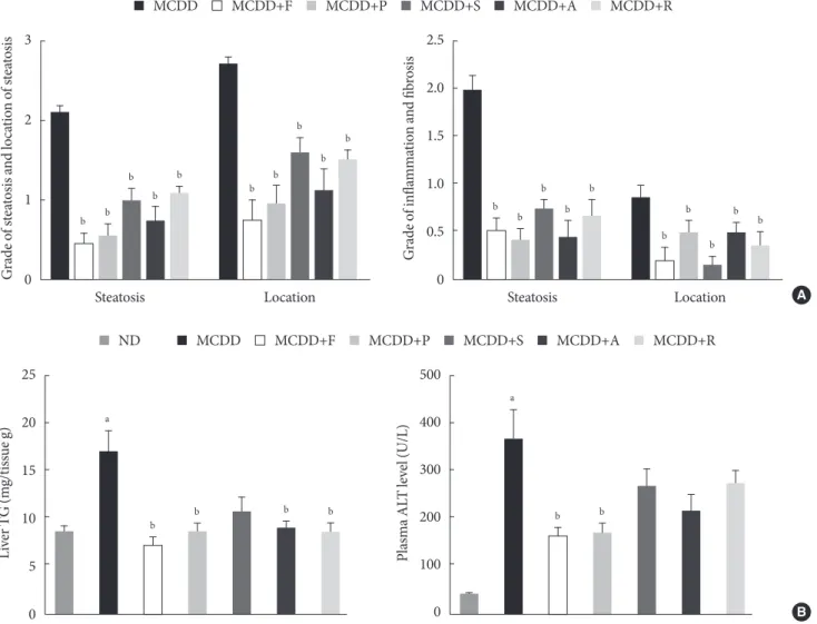
Administration of statins prevent hepatic lipid accumulation and steatohepatitis in MCDD-fed mice Of the animal models of NAFLD, the MCDD model has been used frequently as a valuable model of NASH. Consistent with previous studies [20], administration of MCDD to C57BL6/N mice for 6 weeks caused NASH and mild hepatic fibrosis (Figs.
1 and 2A). Feeding MCDD significantly increased plasma ALT levels, a specific marker of liver injury, and hepatic TG contents (Fig. 2B). The effects of different types of statins, in- cluding fluvastatin, pravastatin, simvastatin, atorvastatin, and rosuvastatin, on MCDD-induced NAFLD were examined. It is well known that the efficacy and potency of lowering plasma cholesterol are different between the types of statins. However, it is not established whether this lipid-lowering effect corre- lates with pleiotropic effects of the drugs. Because the effective dose of each statin for reducing hepatic steatosis has not been established in rodent models, we treated five statins with the same dose (15 mg/kg/day) [21]. Table 2 shows the changes in
MCDD MCDD+F MCDD+P MCDD+S MCDD+A MCDD+R
3
2
1
scaosiattef sn otiond loGs aosiattef srade o 0
Steatosis Location
b b
b b
b
b b
b b
b
2.5
2.0
1.5
1.0
0.5
0
Grade of inflammation and fibrosis
Steatosis Location
b b
b b
b
b b
b
b b
A MCDD
ND MCDD+F MCDD+P MCDD+S MCDD+A MCDD+R
25
20
15
10
5
0
Liver TG (mg/tissue g)
a
b b b b
500
400
300
200
100
0
Plasma ALT level (U/L)
a
b b
B
Fig. 2. Histologic scores and hepatic triglyceride (TG) content and plasma alanine aminotransferase (ALT) level. (A) Histologic scores for location and severity of steatosis, inflammation, and fibrosis, according to criteria of Kleiner et al. [18]. (B) Hepatic TG content and plasma ALT level in mice fed normal chow diet (ND) and methionine- and choline-deficient diet (MCDD) with or without various statins for 6 weeks. F, fluvastatin; P, pravastatin; S, simvastatin; A, atorvastatin; R, rosuvastatin. aP<0.05 com- pared with ND, bP<0.05 compared with MCDD.
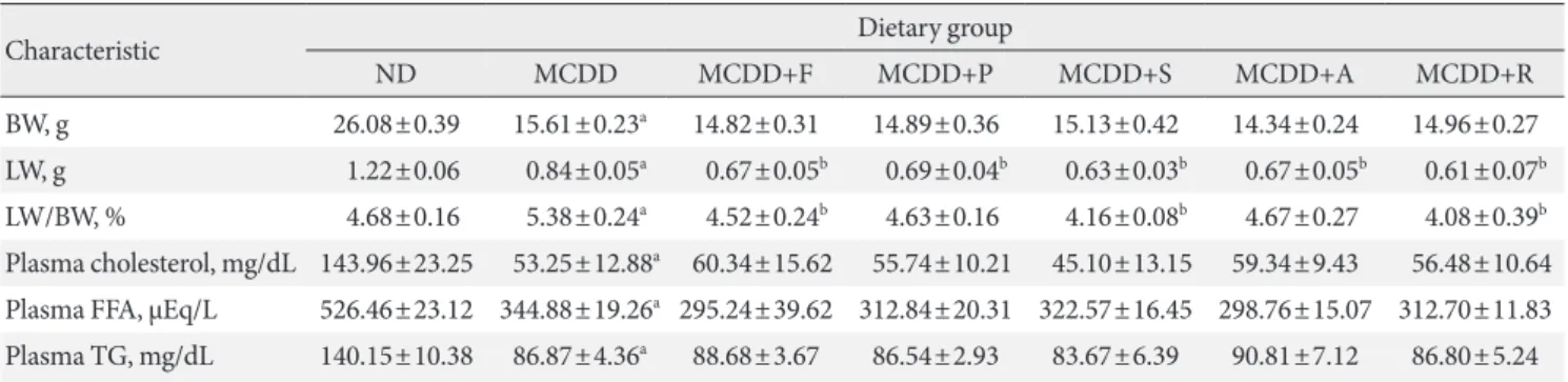
Table 2. Metabolic characteristics of mice fed methionine- and choline-deficient diet with or without statins
Characteristic Dietary group
ND MCDD MCDD+F MCDD+P MCDD+S MCDD+A MCDD+R
BW, g 26.08±0.39 15.61±0.23a 14.82±0.31 14.89±0.36 15.13±0.42 14.34±0.24 14.96±0.27 LW, g 1.22±0.06 0.84±0.05a 0.67±0.05b 0.69±0.04b 0.63±0.03b 0.67±0.05b 0.61±0.07b LW/BW, % 4.68±0.16 5.38±0.24a 4.52±0.24b 4.63±0.16 4.16±0.08b 4.67±0.27 4.08±0.39b Plasma cholesterol, mg/dL 143.96±23.25 53.25±12.88a 60.34±15.62 55.74±10.21 45.10±13.15 59.34±9.43 56.48±10.64 Plasma FFA, μEq/L 526.46±23.12 344.88±19.26a 295.24±39.62 312.84±20.31 322.57±16.45 298.76±15.07 312.70±11.83 Plasma TG, mg/dL 140.15±10.38 86.87±4.36a 88.68±3.67 86.54±2.93 83.67±6.39 90.81±7.12 86.80±5.24 Values are presented as mean±standard error of means (n=6–10).
ND, normal chow diet; MCDD, methionine- and choline-deficient diet; F, fluvastatin; P, pravastatin; S, simvastatin; A, atorvastatin; R, rosuvas- tatin; BW, body weight; LW, liver weight; FFA, free fatty acid; TG, triglyceride.
aP<0.05 between ND and MCDD groups, bP<0.05 between MCDD and MCDD with statin-treated groups.
1.5
1.0
0.5
0
PPARα/α-tubulin (arbitrary unit) b
MCDDND MCDD+F
ND MCDD MCDD+F
PPARα α-Tubulin
a
1.5
1.0
0.5
0
PPARα mRNA (fold change)
a
b b
b b b
MCDD ND MCDD+F MCDD+P MCDD+S MCDD+A MCDD+R PPARα
A B
1.5
1.0
0.5
0
Pex7 mRNA (fold change)
a b
b b
b
Pex7 2.0
1.5
1.0
0.5
0
mRNA (fold change)
a b
b b b
b b
b b
b a
b b
b
b a
b
FAO enzyme
CTP-1α
Mitochondrial FAO Peroxisomal FAO
CTP-1β ACOX1 DBP1
C D
Fig. 3. Treatment with statins recovers methionine- and choline-deficient diet (MCDD)-induced suppression of peroxisomal proliferator-activated receptor α (PPARα) and target gene expression levels in the liver. (A) mRNA expression levels and (B) pro- tein expression levels of PPARα. Expression levels of genes involved in mitochondrial and peroxisomal (C) fatty acid oxidation (FAO), and (D) peroxisomal biogenesis factor (Pex) 7. ND, normal chow diet; F, fluvastatin; P, pravastatin; S, simvastatin; A, atorvastatin; R, rosuvastatin; CPT, carnitine palmitoyltransferase; ACOX, acyl CoA oxidase; DBP, D-bifunctional protein.
aP<0.05 compared with ND, bP<0.05 compared with MCDD.
body weight, liver weight, and plasma levels of cholesterol, free fatty acid, and TG in each experimental group. Interestingly, administration of various statins with MCDD for 6 weeks failed to further reduce plasma cholesterol levels in the mice fed MCDD alone (Table 2), but most statins reduced hepatic TG levels and plasma ALT levels (Fig. 2B). Among them, fluv- astatin showed most prominent effect on reducing hepatic TG levels and plasma ALT levels. The histological analysis of statin- fed mice revealed a significant reduction in hepatic lipid accu- mulation, as well as inflammation or fibrosis (Figs. 1 and 2).
Gene expression of PPARα and enzymes responsible for hepatic FAO was decreased in MCDD and recovered by statin treatment
PPARα is a master regulator of FAO. After 6 weeks of MCDD feeding, a gene expression level of PPARα was significantly de- creased (Fig. 3A). All statins recovered PPARα mRNA levels in the liver compared with MCDD-fed mice. Western blot analysis also showed that MCDD feeding significantly decreased and fluvastatin treatment increased, respectively, protein expression of PPAR (Fig. 3B). In line with this result, the PPARα target genes encoding enzymes involved in mitochondrial FAO (carnitine-palmitoyltransferase-1α and -1β), and peroxisomal FAO (acyl-CoA oxidase-1 and D-bifunctional protein-1) were decreased by MCDD and recovered by most of statins (Fig. 3C).
In addition, MCDD significantly decreased and most statins
significantly increased the expression of peroxisomal biogenesis factor (Pex)-7, a gene encoding a protein that imports several essential enzymes into peroxisomes (Fig. 3D) [22].
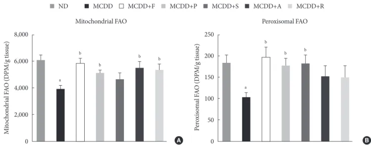
Statin restores MCDD-induced suppression of hepatic mitochondrial and peroxisomal FAO
To ensure the effect of MCDD and statins on hepatic FAO rate, we directly measured both mitochondrial and peroxisomal FAO using 14C palmitate oxidation. In agreement with previous studies [23], feeding MCDD significantly suppressed mito- chondrial FAO in the liver (Fig. 4A). Treatment with statins sig- nificantly increased mitochondrial FAO, except for simvastatin (P=0.21). Feeding MCDD also significantly decreased peroxi- somal FAO and fluvastatin, whereas pravastatin and simvas- tatin supplementation significantly increased it (Fig. 4B).
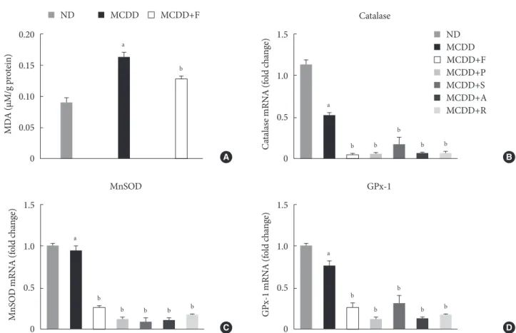
Hepatic gene expression levels of anti-oxidative enzymes was decreased in MCDD-fed mice but not recovered by statins A recent study showed that MCDD increases oxidative stress through decreased anti-oxidative capacity [24]. It was also shown that statins reduce inflammatory responses [25]. Ac- cordingly, the MDA assay showed that the lipid peroxidation levels in the liver of MCDD-fed mice were more elevated than ND-fed mice, whereas these findings were ameliorated in the fluvastatin supplementation group (Fig. 5A).
Thus, we examined the possibility that statins may increase MCDD
ND MCDD+F MCDD+P MCDD+S MCDD+A MCDD+R
250
200
150
100
50
0
Peroxisomal FAO (DPM/g tissue)
a b
b b
Peroxisomal FAO 8,000
6,000
4,000
2,000
0
Mitochondrial FAO (DPM/g tissue)
a b
b
b b
Mitochondrial FAO
A B
Fig. 4. Measurement of hepatic fatty acid oxidation (FAO) in mice fed methionine- and choline-deficient diet (MCDD) with or without statins. (A) Mitochondrial FAO. (B) Peroxisomal FAO. DPM, disintegrations per minute; ND, normal chow diet; F, fluv- astatin; P, pravastatin; S, simvastatin; A, atorvastatin; R, rosuvastatin. aP<0.05 compared with ND, bP<0.05 compared with MCDD.
the gene expression of anti-oxidant enzymes: catalase, a major peroxisomal anti-oxidant; manganese superoxide dismutase, which exists in both mitochondria and peroxisomes; and glu- tathione peroxidase (GPx)-1, a cytosolic anti-oxidant. MCDD significantly decreased gene expression levels of catalase and GPx-1. However, contrary to our expectation, statins further de- creased mRNA expressions of all of these enzymes (Fig. 5B-D).
DISCUSSION
In the present study, we showed that various statins prevented MCDD-induced hepatic steatosis and NASH and increased hepatic mitochondrial and peroxisomal FAO. Gene expression levels of PPARα and its target genes, which are responsible for mitochondrial and peroxisomal FAO, were increased by statin treatment. Our study is the first to demonstrate that statin treatment increases peroxisomal FAO to prevent NASH devel-
opment.
Interestingly, the overall preventive effect of statins was not related with its potency to reduce plasma cholesterol levels. In our study, we used same dosages of various statins because there has been no study directly comparing potency of these drugs. Interestingly, fluvastatin, known to have lower potency to reduce plasma cholesterol level than atorvastatin and rosuv- astatin, showed most prominent effects on reducing hepatic steatosis and NASH. Fluvastatin was also most effective in in- creasing mitochondrial and peroxisomal FAO, as well as ex- pression of PPARα and its downstream FAO enzymes. Thus, it is suggested that each statin’s effect on increasing FAO is inde- pendent of its effect on decreasing cholesterol level and that the ability to increase hepatic FAO is the major determinant of NASH prevention by statins. However, the molecular mecha- nism by which this kind of discrepancy occurs among various statins is presently unknown.
Fig. 5. Changes in hepatic lipid peroxidation and gene expression levels of several anti-oxidative enzymes. (A) Hepatic malondi- aldehyde (MDA) levels in mice fed methionine- and choline-deficient diet (MCDD) and fluvastatin supplementation. (B) mRNA expression levels of catalase, (C) manganese superoxide dismutase (MnSOD), and (D) glutathione peroxidase 1 (GPx-1).
ND, normal chow diet; F, fluvastatin; P, pravastatin; S, simvastatin; A, atorvastatin. aP<0.05 compared with ND, bP<0.05 com- pared with MCDD.
0.20
0.15
0.10
0.05
0
MDA (μM/g protein)
a
b
MCDD
ND MCDD+F
A
1.5
1.0
0.5
Catalase mRNA (fold change) 0
a
b b
b
b b
MCDDND MCDD+F MCDD+P MCDD+S MCDD+A MCDD+R Catalase
B
1.5
1.0
0.5
MnSOD mRNA (fold change) 0
a
b
b b b b
MnSOD
1.5
1.0
0.5
0
GPx-1 mRNA (fold change)
a
b b
b
b b
GPx-1
C D
The two-hit hypothesis is a well-known theory to explain the pathogenesis of NAFLD and its progression from steatosis to NASH [26]. The “first hit” is the accumulation of fatty acids or TGs in the liver that may increase susceptibility of the hepa- tocellular damage induced by second hits. There are several mechanisms leading to the development of hepatic steatosis:
(1) increased hepatic fatty acid uptake, (2) increased de novo lipogenesis in the liver, (3) decreased hepatic FAO, and (4) de- creased very low density lipoprotein secretion from the liver.
The “second hit’’ is a combination of inflammatory responses, oxidative stress, and mitochondrial dysfunction, which leads to hepatocellular damage and fibrosis [26].
Among them, FAO occurs mainly in mitochondria, but peroxisomes and microsomes also play a role. Peroxisomal β-oxidation is required for efficient mitochondrial β-oxidation [27]. Peroxisomal dysfunction induces functional abnormali- ties in mitochondria and consequently compromises cellular ATP production [28]. Especially when the liver is overloaded with fatty acids, the role of peroxisomal β-oxidation becomes more important because dicarboxylic acids are increased through ω-oxidation in endoplasmic reticulum [14,29]. In line with this, recent studies have highlighted the association be- tween peroxisomal dysfunction and hepatic steatosis. The up- regulation of genes, which regulates peroxisomal biogenesis and FAO in a certain strain of mice, was related with resistance to diet-induced hepatic steatosis [13]. The liver-specific Pex5-/- mice developed hepatic steatosis even though mitochondrial FAO was increased [14].
Mitochondria and peroxisomes are closely related organelles and play a critical role in the cellular energy metabolism. X- linked adrenoleukodystrophy (X-ALD) is an inherited disorder caused by mutation of the ABCD1 gene, which encodes a per- oxisomal transporter of very long chain fatty acids. The mouse model of X-ALD showed impaired oxidative phosphorylation of mitochondria and increased oxidative stress [30]. Peroxi- somal biogenesis disorder, Zellweger syndrome, is character- ized by severe neurologic deficits with multiple organ dysfunc- tions. Pex5-/- mice, a mouse model for Zellweger syndrome, caused alteration of mitochondrial morphology, changes of mitochondrial respiratory chains, and increased oxidative stress in the liver [31]. In our study, statin treatment increased both peroxisomal and mitochondrial FAO, suggesting that im- provement of peroxisomal FAO may underlie improvement of mitochondrial FAO. Taken together, improvement of peroxi- somal FAO may be the primary mechanism of NASH preven-
tion by statins. However, it should be noted that each statin showed a different level of effect on mitochondrial or peroxi- somal FAO, whereas all statins improved steatosis and NASH.
Therefore, there may be additional mechanisms of preventive effect of statins on steatosis and NASH.
Increased oxidative stress and altered anti-oxidative system play an important role in the development of NASH/NAFLD [32]. Because mitochondria and peroxisomes are major sourc- es of free radical generation, resulting in oxidative stress, main- tenance of its function is critical to prevent NAFLD. In agree- ment with previous reports [24], feeding MCDD significantly decreased gene expression levels of peroxisomal anti-oxidative enzymes, including catalase and GPx. A number of studies have demonstrated that statins act as an anti-oxidant in vari- ous tissues [33]. In line with this, we demonstrated that the MCDD-induced hepatic lipid peroxidation was suppressed by statin treatment. Previous studies have shown that treatment with various statins increased the activity of anti-oxidant en- zymes, such as catalase or SOD [34]. However, in our study, statins failed to increase gene expression levels of anti-oxidant enzymes. The reason for this discrepancy between current study and previous studies is presently unclear. We recently found that fluvastatin treatment significantly increased hepatic level of plasmalogen, which is well known to act as an endoge- nous anti-oxidant (unpublished data) [35,36]. Thus, it can be suggested that changes in anti-oxidant enzymes are not the primary reason of improvement of anti-oxidative defense function by statins.
Statins are known to cause several hepatic adverse effects, ranging from transient elevation of transaminases to acute liv- er failure [37]. However, recent studies have reported that statin-induced acute liver failure is extremely rare and may be related with idiosyncratic reaction [38]. Indeed, several recent studies showed that statins can be used safely in NASH pa- tients [39]. In addition, a recent meta-analysis showed that statins may improve serum aminotransferase levels and ultra- sound findings in NASH patients [40]. Therefore, it is suggest- ed that favorable effects of statins on liver function in animal studies can be extended to humans.
In summary, statin treatment prevented hepatic steatosis and NASH in MCDD-fed mice. Feeding MCDD for 6 weeks caused hepatic steatosis, inflammation, and early fibrosis through de- creased hepatic mitochondrial and peroxisomal FAO. Various statins exhibited significant improvement of histological scores and enhanced hepatic FAO via induction of PPARα and target
genes. Interestingly, these pleiotropic effects were not correlated with cholesterol-lowering potency of statins. Based on these data, we suggest a new possibility that improvement of peroxi- somal function by statins may contribute to the prevention of NASH.
CONFLICTS OF INTEREST
No potential conflict of interest relevant to this article was re- ported.
ACKNOWLEDGMENTS
This work was supported by the National Research Foundation of Korea (NRF) funded by the Ministry of Education, Science and Technology (2006-2005412, K.U.L.; 2012R1A1A3012626, E.H.K.). This work was also supported by the grants (2009- 006, 2013-578, 2014-006) from the Asan Institute for Life Sci- ences, Seoul, South Korea.
REFERENCES
1. Farrell GC, Larter CZ. Nonalcoholic fatty liver disease: from steatosis to cirrhosis. Hepatology 2006;43(2 Suppl 1):S99-112.
2. Williams CD, Stengel J, Asike MI, Torres DM, Shaw J, Contre- ras M, Landt CL, Harrison SA. Prevalence of nonalcoholic fatty liver disease and nonalcoholic steatohepatitis among a largely middle-aged population utilizing ultrasound and liver biopsy: a prospective study. Gastroenterology 2011;140:124-31.
3. Yoo HJ, Choi KM. Hepatokines as a link between obesity and cardiovascular diseases. Diabetes Metab J 2015;39:10-5.
4. Browning JD, Szczepaniak LS, Dobbins R, Nuremberg P, Hor- ton JD, Cohen JC, Grundy SM, Hobbs HH. Prevalence of he- patic steatosis in an urban population in the United States: im- pact of ethnicity. Hepatology 2004;40:1387-95.
5. Bellentani S, Scaglioni F, Marino M, Bedogni G. Epidemiology of non-alcoholic fatty liver disease. Dig Dis 2010;28:155-61.
6. Davignon J. Beneficial cardiovascular pleiotropic effects of statins. Circulation 2004;109(23 Suppl 1):III39-43.
7. Rodriguez-Calvo R, Barroso E, Serrano L, Coll T, Sanchez RM, Merlos M, Palomer X, Laguna JC, Vázquez-Carrera M. Atorv- astatin prevents carbohydrate response element binding pro- tein activation in the fructose-fed rat by activating protein ki- nase A. Hepatology 2009;49:106-15.
8. Landrier JF, Thomas C, Grober J, Duez H, Percevault F, Souidi
M, Linard C, Staels B, Besnard P. Statin induction of liver fatty acid-binding protein (L-FABP) gene expression is peroxisome proliferator-activated receptor-alpha-dependent. J Biol Chem 2004;279:45512-8.
9. Moreno M, Ramalho LN, Sancho-Bru P, Ruiz-Ortega M, Ram- alho F, Abraldes JG, Colmenero J, Dominguez M, Egido J, Ar- royo V, Ginès P, Bataller R. Atorvastatin attenuates angiotensin II-induced inflammatory actions in the liver. Am J Physiol Gastrointest Liver Physiol 2009;296:G147-56.
10. Trebicka J, Hennenberg M, Odenthal M, Shir K, Klein S, Gran- zow M, Vogt A, Dienes HP, Lammert F, Reichen J, Heller J, Sauerbruch T. Atorvastatin attenuates hepatic fibrosis in rats after bile duct ligation via decreased turnover of hepatic stel- late cells. J Hepatol 2010;53:702-12.
11. Mari M, Caballero F, Colell A, Morales A, Caballeria J, Fernan- dez A, Enrich C, Fernandez-Checa JC, Garcia-Ruiz C. Mito- chondrial free cholesterol loading sensitizes to TNF- and Fas- mediated steatohepatitis. Cell Metab 2006;4:185-98.
12. Grattagliano I, de Bari O, Bernardo TC, Oliveira PJ, Wang DQ, Portincasa P. Role of mitochondria in nonalcoholic fatty liver disease: from origin to propagation. Clin Biochem 2012;45:
610-8.
13. Hall D, Poussin C, Velagapudi VR, Empsen C, Joffraud M, Beckmann JS, Geerts AE, Ravussin Y, Ibberson M, Oresic M, Thorens B. Peroxisomal and microsomal lipid pathways asso- ciated with resistance to hepatic steatosis and reduced pro-in- flammatory state. J Biol Chem 2010;285:31011-23.
14. Peeters A, Swinnen JV, Van Veldhoven PP, Baes M. Hepatoste- atosis in peroxisome deficient liver despite increased β-oxidation capacity and impaired lipogenesis. Biochimie 2011;93:1828-38.
15. Imoto Y, Kuroiwa H, Yoshida Y, Ohnuma M, Fujiwara T, Yo- shida M, Nishida K, Yagisawa F, Hirooka S, Miyagishima SY, Misumi O, Kawano S, Kuroiwa T. Single-membrane-bounded peroxisome division revealed by isolation of dynamin-based machinery. Proc Natl Acad Sci U S A 2013;110:9583-8.
16. Wanders RJ, Waterham HR. Peroxisomal disorders: the single peroxisomal enzyme deficiencies. Biochim Biophys Acta 2006;
1763:1707-20.
17. Antonenkov VD, Grunau S, Ohlmeier S, Hiltunen JK. Peroxi- somes are oxidative organelles. Antioxid Redox Signal 2010;
13:525-37.
18. Kleiner DE, Brunt EM, Van Natta M, Behling C, Contos MJ, Cummings OW, Ferrell LD, Liu YC, Torbenson MS, Unalp- Arida A, Yeh M, McCullough AJ, Sanyal AJ; Nonalcoholic Ste- atohepatitis Clinical Research Network. Design and validation
of a histological scoring system for nonalcoholic fatty liver dis- ease. Hepatology 2005;41:1313-21.
19. Kim JY, Koves TR, Yu GS, Gulick T, Cortright RN, Dohm GL, Muoio DM. Evidence of a malonyl-CoA-insensitive carnitine palmitoyltransferase I activity in red skeletal muscle. Am J Physiol Endocrinol Metab 2002;282:E1014-22.
20. Caballero F, Fernandez A, Matias N, Martinez L, Fucho R, Ele- na M, Caballeria J, Morales A, Fernandez-Checa JC, Garcia- Ruiz C. Specific contribution of methionine and choline in nu- tritional nonalcoholic steatohepatitis: impact on mitochondri- al S-adenosyl-L-methionine and glutathione. J Biol Chem 2010;285:18528-36.
21. Campbell MJ, Esserman LJ, Zhou Y, Shoemaker M, Lobo M, Borman E, Baehner F, Kumar AS, Adduci K, Marx C, Petricoin EF, Liotta LA, Winters M, Benz S, Benz CC. Breast cancer growth prevention by statins. Cancer Res 2006;66:8707-14.
22. Rodrigues TA, Alencastre IS, Francisco T, Brites P, Fransen M, Grou CP, Azevedo JE. A PEX7-centered perspective on the peroxisomal targeting signal type 2-mediated protein import pathway. Mol Cell Biol 2014;34:2917-28.
23. Serviddio G, Giudetti AM, Bellanti F, Priore P, Rollo T, Tambo- rra R, Siculella L, Vendemiale G, Altomare E, Gnoni GV. Oxi- dation of hepatic carnitine palmitoyl transferase-I (CPT-I) im- pairs fatty acid beta-oxidation in rats fed a methionine-choline deficient diet. PLoS One 2011;6:e24084.
24. Jorgacevic B, Mladenovic D, Ninkovic M, Prokic V, Stankovic MN, Aleksic V, Cerovic I, Vukicevic RJ, Vucevic D, Stankovic M, Radosavljevic T. Dynamics of oxidative/nitrosative stress in mice with methionine-choline-deficient diet-induced nonal- coholic fatty liver disease. Hum Exp Toxicol 2014;33:701-9.
25. Margaritis M, Channon KM, Antoniades C. Statins as regula- tors of redox state in the vascular endothelium: beyond lipid lowering. Antioxid Redox Signal 2014;20:1198-215.
26. Day CP. Pathogenesis of steatohepatitis. Best Pract Res Clin Gastroenterol 2002;16:663-78.
27. Reddy JK, Hashimoto T. Peroxisomal beta-oxidation and per- oxisome proliferator-activated receptor alpha: an adaptive met- abolic system. Annu Rev Nutr 2001;21:193-230.
28. Islinger M, Grille S, Fahimi HD, Schrader M. The peroxisome:
an update on mysteries. Histochem Cell Biol 2012;137:547-74.
29. Van Veldhoven PP. Biochemistry and genetics of inherited dis- orders of peroxisomal fatty acid metabolism. J Lipid Res 2010;
51:2863-95.
30. Lopez-Erauskin J, Galino J, Ruiz M, Cuezva JM, Fabregat I, Cacabelos D, Boada J, Martinez J, Ferrer I, Pamplona R, Villar- roya F, Portero-Otin M, Fourcade S, Pujol A. Impaired mito- chondrial oxidative phosphorylation in the peroxisomal dis- ease X-linked adrenoleukodystrophy. Hum Mol Genet 2013;
22:3296-305.
31. Baumgart E, Vanhorebeek I, Grabenbauer M, Borgers M, De- clercq PE, Fahimi HD, Baes M. Mitochondrial alterations caused by defective peroxisomal biogenesis in a mouse model for Zellweger syndrome (PEX5 knockout mouse). Am J Pathol 2001;159:1477-94.
32. Rolo AP, Teodoro JS, Palmeira CM. Role of oxidative stress in the pathogenesis of nonalcoholic steatohepatitis. Free Radic Biol Med 2012;52:59-69.
33. Shishehbor MH, Brennan ML, Aviles RJ, Fu X, Penn MS, Spre- cher DL, Hazen SL. Statins promote potent systemic antioxi- dant effects through specific inflammatory pathways. Circula- tion 2003;108:426-31.
34. Yilmaz MI, Baykal Y, Kilic M, Sonmez A, Bulucu F, Aydin A, Sayal A, Kocar IH. Effects of statins on oxidative stress. Biol Trace Elem Res 2004;98:119-27.
35. Braverman NE, Moser AB. Functions of plasmalogen lipids in health and disease. Biochim Biophys Acta 2012;1822:1442-52.
36. Wallner S, Schmitz G. Plasmalogens the neglected regulatory and scavenging lipid species. Chem Phys Lipids 2011;164:
573-89.
37. Lewis JH. Clinical perspective: statins and the liver: harmful or helpful? Dig Dis Sci 2012;57:1754-63.
38. Bjornsson E, Jacobsen EI, Kalaitzakis E. Hepatotoxicity associ- ated with statins: reports of idiosyncratic liver injury post-mar- keting. J Hepatol 2012;56:374-80.
39. Athyros VG, Tziomalos K, Gossios TD, Griva T, Anagnostis P, Kargiotis K, Pagourelias ED, Theocharidou E, Karagiannis A, Mikhailidis DP; GREACE Study Collaborative Group. Safety and efficacy of long-term statin treatment for cardiovascular events in patients with coronary heart disease and abnormal liver tests in the Greek Atorvastatin and Coronary Heart Dis- ease Evaluation (GREACE) Study: a post-hoc analysis. Lancet 2010;376:1916-22.
40. Eslami L, Merat S, Malekzadeh R, Nasseri-Moghaddam S, Ara- min H. Statins for non-alcoholic fatty liver disease and non-al- coholic steatohepatitis. Cochrane Database Syst Rev 2013;12:
CD008623.