bloodresearch.or.kr Blood Res 2019;54:282-295.
Letters to the Editor 285
in RBC or platelet level is not evident, but the increase is resolved during follow-up. “Probable MPN” indicates that the increase in RBC or platelet level continues during fol- low-up. “Proven MPN” is diagnosed with PV or ET based on the WHO criteria. In total, 1,729 CI patients (1,003 men; 726 women) of median age 73 years (range, 19–96 yr) were reviewed. Thrombocytosis (platelets ≥450×109/L) was evident in 69 (4.0%) patients at diagnosis or during follow-up. Reactive thrombocytosis was the most common form of thrombocytosis (N=62, 3.6%). Three (0.2%) patients were considered to exhibit possible ET, and four (0.2%) had proven ET. The causes of reactive thrombocytosis (N=62 patients) included infection (N=59, 95.2%), bleeding (N=1, 1.6%), and iron-deficiency (N=1, 1.6%). Erythrocytosis was evident in 79 (4.6%) patients at diagnosis or during fol- low-up. Reactive erythrocytosis was the most common form of erythrocytosis (N=50, 2.9%), followed by possible PV (N=21, 1.2%), probable PV (N=6, 0.3%), and proven PV (N=2, 0.1%). None of the 27 patients with possible or prob- able PV underwent further investigations. Particularly, the JAK2 mutational status was not explored. Reactive eryth- rocytosis (N=50) was detected during diagnosis and fol- low-up in 28 (56.0%) and 22 (44.0%) patients, respectively, and all cases were attributable to hemoconcentration. Of the four patients with proven ET, two lacked any other predisposing factor for thrombosis. All patients with proven ET and PV exhibited multifocal CI and previously un- detected infarctions on CI diagnosis.
These results showed that many CI patients with eryth- rocytosis did not undergo further evaluation in terms of a PV diagnosis and that JAK2 mutational status should be evaluated in such patients. Stroke is a global health problem with a global lifetime risk of approximately 25% in people 25 years and older (as of 2016). People living in East Asia, Central Europe, and Eastern Europe have the highest risk of stroke [6]. In Korea, stroke accounts for roughly 1 out of every 10 deaths, and the proportion of ischemic stroke has steadily increased [7]. To effectively care for patients with PV-associated CI, hematologists should communicate well with neurologists.
Ik-Chan Song1, Yoon-Seok Choi1, Jong Wook Shin2, Hee-Jung Song2, Jei Kim2, Deog-Yeon Jo1
1Division of Hematology/Oncology, Department of Internal Medicine, 2Department of Neurology, College of Medicine, Chungnam National University, Daejeon, Korea
Correspondence to: Deog-Yeon Jo Department of Internal Medicine, Chungnam National University Hospital, 282 Munhwa-ro, Daejeon 35015, Korea
E-mail: [email protected]
Received on May 19, 2019; Revised on Jul. 21, 2019; Accepted on Aug. 13, 2019 https://doi.org/10.5045/br.2019.54.4.284
AuthorsÊ Disclosures of Potential Conflicts of Interest No potential conflicts of interest relevant to this article were reported.
REFERENCES
1. Kaifie A, Kirschner M, Wolf D, et al. Bleeding, thrombosis, and anticoagulation in myeloproliferative neoplasms (MPN): analy- sis from the German SAL-MPN-registry. J Hematol Oncol 2016;
9:18.
2. Martin K. Risk factors for and management of MPN-associated bleeding and thrombosis. Curr Hematol Malig Rep 2017;12:
389-96.
3. Enblom A, Lindskog E, Hasselbalch H, et al. High rate of abnor- mal blood values and vascular complications before diagnosis of myeloproliferative neoplasms. Eur J Intern Med 2015;26:344-7.
4. Ong E, Barraco F, Nighoghossian N, et al. Cerebrovascular events as presenting manifestations of myeloproliferative neoplasm.
Rev Neurol (Paris) 2016;172:703-8.
5. Arber DA, Orazi A, Hasserjian R, et al. The 2016 revision to the World Health Organization classification of myeloid neoplasms and acute leukemia. Blood 2016;127:2391-405.
6. GBD 2016 Lifetime Risk of Stroke Collaborators. Global, region- al, and country-specific lifetime risks of stroke, 1990 and 2016.
N Engl J Med 2018;379:2429-37.
7. Hong KS, Bang OY, Kang DW, et al. Stroke statistics in Korea:
part I. Epidemiology and risk factors: a report from the Korean Stroke Society and clinical research center for stroke. J Stroke 2013;15:2-20.
Beta-2 microglobulin as a prognostic factor of primary central nervous system lymphoma
TO THE EDITOR: Primary central nervous system lympho- ma (PCNSL) is an extra-nodal non-Hodgkin lymphoma in- volving the brain, leptomeninges, eyes, or spinal cord and no primary malignancy outside of the central nervous system (CNS). PCNSL is a rare lymphoma and accounts for only 1% of all incident lymphomas [1, 2]. Prevalence of the disease is higher in the sixth to eighth decades of life. The median age at diagnosis of PCNSL is 65 years and the in- cidence is rising in the elderly population [3, 4].
Among several previously published prognostic models, the International Extranodal Lymphoma Study Group (IELSG) and the Memorial Sloan Kettering Cancer Center (MSKCC) models are the most widely used in current prac- tice [5, 6]. Old age and poor performance status at initial diagnosis were strong poor prognostic factors commonly found in both studies. Meanwhile, serum beta-2 micro- globulin (B2MG) is a well-established prognostic factor in multiple myeloma and follicular lymphoma [7, 8]. Thus,
Blood Res2019;54:282-295. bloodresearch.or.kr
286 Letters to the Editor
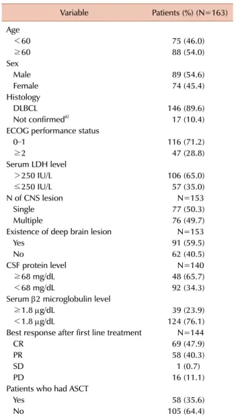
Table 1. Baseline clinical characteristics.
Variable Patients (%) (N=163) Age
<60 75 (46.0)
≥60 88 (54.0)
Sex
Male 89 (54.6)
Female 74 (45.4)
Histology
DLBCL 146 (89.6)
Not confirmeda) 17 (10.4)
ECOG performance status
0–1 116 (71.2)
≥2 47 (28.8)
Serum LDH level
>250 IU/L 106 (65.0)
≤250 IU/L 57 (35.0)
N of CNS lesion N=153
Single 77 (50.3)
Multiple 76 (49.7)
Existence of deep brain lesion N=153
Yes 91 (59.5)
No 62 (40.5)
CSF protein level N=140
≥68 mg/dL 48 (65.7)
<68 mg/dL 92 (34.3)
Serum 2 microglobulin level
≥1.8 g/dL 39 (23.9)
<1.8 g/dL 124 (76.1)
Best response after first line treatment N=144
CR 69 (47.9)
PR 58 (40.3)
SD 1 (0.7)
PD 16 (11.1)
Patients who had ASCT
Yes 58 (35.6)
No 105 (64.4)
a)Not confirmed in histology means lymphoid malignancy without established exact diagnosis among NHL subtypes.
Abbreviations: ASCT, autologous stem cell treatment; CNS, central nervous system; CR, complete response; CSF, cerebrospinal fluid;
DLBCL, diffuse large B-cell lymphoma; ECOG, Eastern Cooperative Oncology Group; LDH, lactate dehydrogenase; PD, progressive disease; PR, partial response; SD, stable disease.
the purpose of this study was to validate previously suggested prognostic factors and to evaluate prognostic value of serum B2MG level in PCNSL patients.
Methods Patients
We retrospectively analyzed the PCNSL registry data for patients treated from March 1993 to May 2017 at the Asan Medical Center in Seoul, South Korea. Variables that were extracted from the medical records and analyzed included patient demographics, Eastern Cooperative Oncology Group (ECOG) performance status, tumor characteristics, treat- ment profiles, serum lactate dehydrogenase (LDH) level (normal range <250 IU/L), serum B2MG level (normal range <2.5 g/dL), cerebrospinal fluid (CSF) total protein level, number of CNS lesions, existence of deep CNS lesions (brain stem, thalamus, basal ganglia, and cerebellum), date of disease progression, and survival status.
Statistics
Overall survival (OS) was defined as the time from the beginning of first-line therapy to death from any cause.
Univariate and multivariate analyses were performed to identify prognostic factors for OS using a Cox proportional hazards model. Survival curves were estimated by the Kaplan-Meier method and compared using log-rank tests.
A two-sided P-value less than 0.05 was considered statisti- cally significant. All statistical analyses were performed us- ing the Statistical Package for the Social Sciences (SPSS) version 21.0 (IBM Corp, Armonk, NY, USA).
Ethical approval
All procedures performed in studies involving human participants were in accordance with the ethical standards of the institutional review board and with the 1964 Helsinki declaration and its later amendments or comparable ethical standards. For this type of study, formal consent was not required (IRB 2018-1275).
Results
Clinical characteristics
In total, 163 patients were identified and included in the analysis. The median follow-up duration was 3.5 years [95% confidence interval (CI), 2.4–4.6] and median OS was 4.0 years (95% CI, 2.1–5.9). Baseline patient characteristics are summarized in Table 1. The median age was 60 years (range, 19–83), 88 patients (54.0%) were 60 years of age or older and 89 patients (54.6%) were males. All patients received high-dose methotrexate-based chemotherapy as in- itial treatment. Human immunodeficiency virus (HIV) se- rology was checked in 67 patients at the time of diagnosis and all were negative.
Prognostic factors
Univariate analysis of prognostic factors revealed that poor performance status [Eastern Cooperative Oncology Group Performance scale (ECOG PS) ≥2] and elevated se-
rum B2MG (≥1.8 g/mL) were significantly associated with shorter OS (Table 2). Old age (≥60 yr) showed borderline association in terms of poor OS [hazard ratio (HR) 1.55;
95% CI, 0.95-2.52; P=0.068]. Multivariate analysis results were consistent with HRs of 2.5 (95% CI, 1.06–3.03; P=0.001) for ECOG PS ≥2 and 1.79 (1.06–3.03; P=0.038) for elevated serum B2MG (≥1.8 g/mL). The survival curves of the two groups according to serum B2MG level are shown on Fig. 1.
Discussion
In our analysis, ECOG PS higher than 1 and elevated
bloodresearch.or.kr Blood Res 2019;54:282-295.
Letters to the Editor 287
Table 2. Univariate analysis and multivariate analysis of overall survival.
Variables Univariate Multivariate
HR (95% CI) P HR (95% CI) P
Age
<60 Reference
≥60 1.55 (0.95–2.52) 0.068
ECOG performance status
0–1 Reference Reference
≥2 2.24 (1.38–3.63) 0.001 2.30 (1.06–3.03) 0.001
Serum LDH level
≤250 IU/L Reference
>250 IU/L 1.53 (0.94–2.48) 0.089
Number of CNS lesions
Multiple Reference
Single 0.88 (0.54–1.44) 0.616
Presence of deep CNS lesion
Yes Reference
No 1.15 (0.70–1.89) 0.583
CSF protein level
≥68 mg/dL Reference
<68 mg/dL 1.18 (0.68–2.04) 0.552
Serum 2 microglobulin
<1.8 g/dL Reference Reference
≥1.8 g/dL 1.70 (1.01–2.88) 0.047 1.79 (1.06–3.03) 0.038
Abbreviations: CI, confidence interval; CNS, central nervous system; CSF, cerebrospinal fluid; ECOG, Eastern Cooperative Oncology Group;
HR, hazard ratio; LDH, lactate dehydrogenase; OS, overall survival; PFS, progression-free survival.
Fig. 1. Overall survival from the initiation of first-line treatment.
serum B2MG level were significantly predictive of poor prognosis according to univariate analysis. Most studies de- fined a cutoff level of serum B2MG level between 2.0 to 3.5 and our analysis showed 1.8 as the best cutoff level to establish a significant survival benefit [7-9]. Although the mechanism underlying the negative prognostic impact of elevated serum B2MG is unclear, a widely accepted hy- pothesis is that it is related to high tumor burden [9, 10].
Based on this hypothesis, we might explain the relatively low cutoff level of B2MG in PCNSL by reiterating that
even a small elevation of serum B2MG could reflect high tumor burden in the CNS. Our results are reliable consider- ing the large number of enrolled patients and comparable survival outcomes of our cohort with other studies [6]. In the MSKCC study, 240 patients were enrolled, and the me- dian OS and failure-free survival were 37 (95% CI, 31–42) months and 17 (95% CI, 12–21) months, respectively.
Our study has some limitations including the retrospective nature and also it is a single center study. Another limitation is that the cut-off value of B2MG level 1.8 g/dL is arbitrary.
However, the cutoff value in this study represents only a population from single center. Higher serum B2MG level could have some relationship with survival outcomes in PCNSL and further investigations via multi-center studies are needed. In conclusion, ECOG performance status and serum B2MG were associated with prognosis in PCNSL patients. Serum B2MG may have some association with prognosis of PCNSL.
Jaewon Hyung1#, Jung Yong Hong1#, Shin Kim1, Jin Sook Ryu2, Jooryung Huh3, Cheolwon Suh1
1Departments of Oncology, 2Nuclear Medicine, 3Pathology, Asan Medical Center, University of Ulsan College of
Medicine, Seoul, Korea Correspondence to: Cheolwon Suh Department of Oncology, Asan Medical Center, University
Blood Res2019;54:282-295. bloodresearch.or.kr
288 Letters to the Editor
of Ulsan College of Medicine, 88 Olympic-ro 43-gil, Songpa-gu, Seoul 05505, Korea E-mail: [email protected]
Received on Aug. 16, 2019; Revised on Oct. 16, 2019; Accepted on Nov. 1, 2019 https://doi.org/10.5045/br.2019.54.4.285
#These authors contributed equally to this work.
AuthorsÊ Disclosures of Potential Conflicts of Interest No potential conflicts of interest relevant to this article were reported.
REFERENCES
1. Haldorsen IS, Espeland A, Larsson EM. Central nervous system lymphoma: characteristic findings on traditional and advanced imaging. AJNR Am J Neuroradiol 2011;32:984-92.
2. van der Sanden GA, Schouten LJ, van Dijck JA, et al. Primary cen- tral nervous system lymphomas: incidence and survival in the Southern and Eastern Netherlands. Cancer 2002;94:1548-56.
3. Villano JL, Koshy M, Shaikh H, Dolecek TA, McCarthy BJ. Age, gender, and racial differences in incidence and survival in pri- mary CNS lymphoma. Br J Cancer 2011;105:1414-8.
4. O'Neill BP, Decker PA, Tieu C, Cerhan JR. The changing in- cidence of primary central nervous system lymphoma is driven primarily by the changing incidence in young and middle-aged men and differs from time trends in systemic diffuse large B-cell non-Hodgkin's lymphoma. Am J Hematol 2013;88:997-1000.
5. Ferreri AJ, Blay JY, Reni M, et al. Prognostic scoring system for primary CNS lymphomas: the International Extranodal Lymphoma Study Group experience. J Clin Oncol 2003;21:266-72.
6. Abrey LE, Ben-Porat L, Panageas KS, et al. Primary central nerv- ous system lymphoma: the Memorial Sloan-Kettering Cancer Center prognostic model. J Clin Oncol 2006;24:5711-5.
7. Munshi NC, Anderson KC, Bergsagel PL, et al. Consensus recom- mendations for risk stratification in multiple myeloma: report of the International Myeloma Workshop Consensus Panel 2. Blood 2011;117:4696-700.
8. Federico M, Bellei M, Marcheselli L, et al. Follicular lymphoma international prognostic index 2: a new prognostic index for fol- licular lymphoma developed by the international follicular lym- phoma prognostic factor project. J Clin Oncol 2009;27:4555-62.
9. Hagberg H, Killander A, Simonsson B. Serum beta 2-micro- globulin in malignant lymphoma. Cancer 1983;51:2220-5.
10. Shi C, Zhu Y, Su Y, Chung LW, Cheng T. Beta2-microglobulin:
emerging as a promising cancer therapeutic target. Drug Discov Today 2009;14:25-30.
Long-term response in refractory AML following azacitidine-failed MDS by salvage decitabine-bridged
allogenic transplantation
TO THE EDITOR: Myelodysplastic syndromes (MDS) are a group of heterogeneous hematological malignancies which demand personalized and risk-adapted clinical management [1]. Current therapeutic approaches are rather limited for patients unsuitable for allogeneic stem cell transplantation (SCT), the only realistic and potentially curative treatment measure that exists [1]. With regard to patients with high risk MDS, the standard of care is currently represented by treatment with hypomethylating agents (HMAs), such as decitabine and azacitidine. The latter is used as initial therapy in most cases, and induces responses in 40–50%
of treated patients [2, 3]. Obstacles to azacitidine admin- istration as well as recommendations for the optimization of treatment with this agent have been reported [2, 4].
However, despite optimal management of azacitidine treat- ment, the duration of its clinical benefit, although variable, is usually transient and almost all patients ultimately experi- ence loss of response to the drug, disease progression, and therefore very poor outcomes [1, 2, 5, 6]. After this loss of response or disease progression despite treatment, there are no standard care regimens available [5]. Rescue strategies including intensive chemotherapy (ICT) only provide minor benefits, whereas allogeneic SCT is feasible only in a minor- ity of cases. With these results in mind, especially the cata- strophic outcome of azacitidine-failed patients, typical con- cerns about decision making and clinical management in these settings can be summarized by an unusual case we observed which is reported herein. A 59-year-old woman was admitted for profound malaise due to pancytopenia on March 2015. The bone marrow (BM) and trephine biopsy revealed refractory anemia with an excess of blasts-2 (RAEB-2), remarkable multilineage dysplasia, and 18% of BM infiltrating blasts; the karyotype analysis and molecular study for typical abnormalities found in MDS were negative.
She was diagnosed as having an Inter-2 MDS, according to the International Prognostic Scoring System [7]. On the basis of the patient’s overall fitness level, and given the lack of a suitable familiar donor to proceed to immediate allogeneic SCT, we recommended therapy with azacitidine (75 mg/m2, schedule 5+2+2). Therapy was started on April 2015 without significant adverse effects. Meanwhile, a matched unrelated donor (MUD) was fruitlessly sought.
After six cycles (September 2015), a partial remission (according to Cheson’s criteria) was achieved [8]. Because of this, the same treatment was continued for another three cycles until December 2015, when a progressive pan- cytopenia unveiled progression to secondary acute myeloge- nous leukemia (AML). At the time of evolution, standard