http://www.ecevr.org/ 83
CLINICAL
EXPERIMENTAL VACCINE
RESEARCH
Foot-and-mouth disease (FMD) O, A, and Asia 1 type viruses have been circulating in Asian countries [1]. In Korea, FMD occurred once in 2000, once in 2002, and three times in 2010, making a total of five outbreaks since 2000 [2-6]. All animals susceptible to FMD were vaccinated in early 2011 to help prevent recurrence of the disease. Since that time, due to concerns about a recurrence, vaccines against three serotypes, O, A, and Asia 1, have continued to be vaccinated (O1 Manisa, A Malaysia 97, and Asia 1 Shamir) [7]. In July-August 2014, FMD occurred on three pig farms in Uiseong and Go- ryeong in Gyeongbuk province and in Hapcheon in Gyeongnam province. This paper describes the outbreak, the diagnosis, the quarantine measures for FMD under the condition of vaccination and pathogenesis, genetic analysis of the isolated virus.
The outbreak of FMD among pigs was reported in Uiseong, Gyeongbuk province on July 23, 2014; the symptoms were detaching of the hooves and vesicles. Using a lateral flow device for detecting antigens [8] by the veterinarian of regional veterinary service in Gyeongbuk, the pigs were identified as being FMD-positive in the farm site. The susceptible animals were thoroughly examined using reverse transcriptase poly- merase chain reaction and enzyme-linked immunosorbent assays (ELISAs), and FMD was confirmed (Table 1).
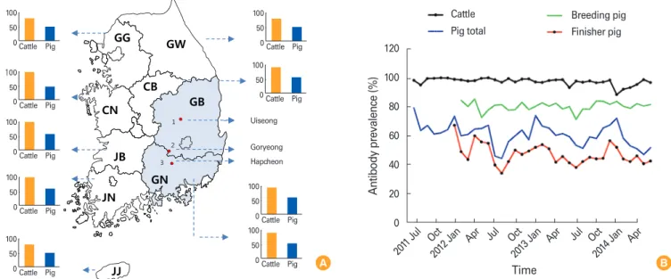
Four days later, on July 27, cases of suspected FMD were reported to have occurred on a pig infected premise (IP) in Goyrong, Gyeongbuk. The detachment of hooves and the formation of ulceration in the mouth were additionally reported 64 km away from the farm where FMD first occurred. The third report was made 10 days later on August 6 in Hapcheon, Gyeongnam, 25.7 km away from the second IP where FMD occurred;
the symptoms included vesicles in the mouth, ulcers on the bridge of the nose, the in- ability to stand up, and lack of appetite. FMD was confirmed after a laboratory diagno- sis (Fig. 1A).
At the first premise where FMD occurred, FMD case occurred in the herd where
© Korean Vaccine Society.
This is an Open Access article distributed under the terms of the Creative Commons Attribution Non-Com- mercial License (http://creativecommons.org/licenses/
by-nc/3.0) which permits unrestricted non-commercial use, distribution, and reproduction in any medium, pro- vided the original work is properly cited.
K O R E A N V A C C I N E S O C I E T Y
K O R E A N K O R E A N A C C I N E O C I E T Y V
S
Clin Exp Vaccine Res 2016;5:83-87 http://dx.doi.org/10.7774/cevr.2016.5.1.83 pISSN 2287-3651 • eISSN 2287-366X
Jong-Hyeon Park, Dongseob Tark, Kwang-Nyeong Lee, Seo-Yong Lee, Mi-Kyeong Ko, Hyang-Sim Lee, Su-Mi Kim, Young-Joon Ko, Min-Goo Seo, Ji-Eun Chun, Myoung-Heon Lee, Byounghan Kim
Animal and Plant Quarantine Agency, Anyang, Korea
Received: October 16, 2015 Revised: November 2, 2015 Accepted: December 7, 2015
Corresponding author: Jong-Hyeon Park, DVM, PhD Animal and Plant Quarantine Agency, 175 Anyang-ro, Manangu, Anyang 14089, Korea Tel: +82-31-467-1719, Fax: +82-31-463-4516 E-mail: [email protected]
No potential conflict of interest relevant to this article was reported.
This study was supported by an internal research fund provided by the Animal and Plant Quarantine Agency, and we appreciate the efforts of its staff in implementing quarantine measures to protect against FMD.
Despite nation-wide immunization with O, A, and Asia 1 type vaccines in Republic of Ko- rea, foot-and-mouth disease type O occurred again in July 2014 after three years and three months. This virus was a Mya-98 strain of the Southeast Asian topotype and was most similar to the identified type that circulated in East Asia in 2014. This was new virus with the deletion of 23 amino acids in 3A/3B1 region and low pathogenic property.
Keywords: Foot-and-mouth disease, Sequence deletion, Vaccination, Korea
Novel foot-and-mouth disease
virus in Korea, July-August 2014
vaccinations had not been implemented prior to the FMD outbreak. At the second premise where FMD case occurred, the disease occurred in some pigs that had not been vacci- nated. FMD virus (FMDV)−infected animals of a lower level were identified on this farm compared to the other farms.
The owner in third IP recognized the FMD outbreak around the area and simultaneously vaccinated to the own pigs, but it is estimated that its animals were infected with FMD before protectable immunity induced. In the process of an epidemi- ological survey, a retrospective analysis of serum collected on 16 June was positive for the non-structural protein (NSP) an- tibody around the second IP, and finally NSP-positive anti- body in an additional six farms were detected. Therefore, the
first outbreak is estimated to have been before June. Although the etiology of FMD was uncertain, there was no outbreak of FMD from April 2011 for 3 years and 3 months.
In Korea, the average antibody prevalence rate by structural protein (SP)–ELISA was 98.9% for cattle and 50.5% for pigs.
The antibody prevalence rate of cattle was much higher than the 80% immunity level, a transmission inhibition level in general [9], but in finisher pigs it was relatively low due to one shot vaccination (Fig. 1B). The antibody prevalence rate in cattle tested in Korea was high level, and therefore we esti- mated that it was difficult for field viruses to circulate. In pigs, the rate accounted for approximately 80% in breeding pigs and 40%-45% in finisher pigs. The possibility of virus infection Fig. 1. Foot-and-mouth disease (FMD) outbreak in South Korea, July 2014 and mean rate of FMD antibody prevalence in each provinces. (A) The region of three FMD cases in Gyeongbuk (GB), Gyeongnam (GN) provinces and antibody prevalence of cattle and pigs in outbreak time point. (B) Monthly FMD antibody prevalence in Korea. GG, Gyeonggi; GW, Gangwon; CN, Chungnam; CB, Chungbuk; JN, Jeonnam; JB, Jeonbuk; JJ, Jeju.
Fig. 1.
GG GW
CN CB
GB JB
JN
GN
JJ
GB
GN
1
2 3
100 50
0 Cattle Pig 100
50 0 Cattle Pig
100 50
0 Cattle Pig 100
50 0 Cattle Pig
100 50
0 Cattle Pig
100 50
0 Cattle Pig 100
50 0 Cattle Pig 100
50 0 Cattle Pig 100
50 0 Cattle Pig
Uiseong
Goryeong Hapcheon
Cattle Pig total
Breeding pig Finisher pig 120
100 80 60 40 20 0
Antibody prevalence (%)
Time 2011 Jul Oct
2012 Jan Apr Jul Oct
2013 Jan Apr Jul Oct
2014 Jan Apr B A
Table 1. Summary of diagnosis results in the FMDV-infected farm in South Korea, July-August 2014
Infected
premise Reported
date Province, region
Antigen test Antibody test
Specimen RT-PCR Antigen ELISA
LFD (No. of positive/No.
of tested)
No. of tested
SP-ELISA
(No. of positive) NSP-ELISA (No. of positive)
O A Asia1
1st 23-Jul G yeongbuk,
Uiseong ve, ts, sa + O type 2/6 16 13 7 1 1
2nd 27-Jul G yeongbuk,
Goryeong ts, sa + ND ND 8 7 7 4 2
3rd 06-Aug G yeongnam, Hapcheon
ve, ts, sa + ND 6/6 16 7 4 1 0
FMDV, foot-and-mouth disease virus; RT-PCR, reverse transcriptase polymerase chain reaction; ELISA, enzyme-linked immunosorbent assay; LFD, lateral flow device; SP, structural protein; NSP, non-structural protein; ND, not done; ve, vesicle; ts, tissue; sa, saliva.
Table 2. Similarity comparison of VP1 amino acids of the closest FMDV or reference virus to FMDV identified in South Korea, July-August 2014 Virus (infected
premise) Topotype
(lineage) Similarity (%)
Topotype-specific variation and consensus of VP1 amino acid (amino acid position) 24 28 47 58 85 96 137 138 139 140 141 142 153 158 180 198 194 205 212
O/SKR/01/2014 (1st) SEA (Mya-98) Identity T H S P D T T G G S L P P P V A V L S
O/SKR/03/2014 (3rd) SEA (Mya-98) 100 - - - - - - - - - - - - - - - - - - -
O/GZ-MT/CHA/2013 SEA (Mya-98) 97.65 I - - - - A - - - - - - - - M - - - -
O/HKN/7/2010 SEA (Mya-98) 97.18 V - - - - A A - - - - - Q - M - - - -
O/KOR/1/2010 SEA (Mya-98) 96.71 V - - - - A A - - - - - Q - M - - - -
O/JPN/1/2010 SEA (Mya-98) 96.71 V - - S - A A - - - - - - - M - - - -
O/Andong/SKR/2010 SEA (Mya-98) 96.71 V - - S - A A - - - - - Q - M - - - -
O/PAK/44/2000 ME-SA (PanAsia-2) 92.02 V Q Q S N - G E S - T T Q A M E I I L
O/SKR/2000 ME-SA (PanAsia) 91.11 V Q Q S - - G E S P V T Q T M E I I F
O/SKR/2002 ME-SA (PanAsia) 88.73 V Q Q S N A G E S P V T Q T M E I I L
O1 Manisa ME-SA 90.14 V Q Q S N A G D - T V A Q A M Q I I L
FMDV, foot-and-mouth disease virus.
is higher among finisher pigs than breeding pigs (Fig. 1B).
Pigs with clinical symptoms were culled and pigs without clinical symptoms on farms where FMD occurred were vacci- nated emergently. The movement of livestock on the farms
that tested positive was restricted for 3 weeks and additional quarantine measures, such as disinfection, were taken. Addi- tional tests and careful examinations of the environment or other risk factors were made until the risk disappeared.
Fig. 2. Phylogenetic tree and genetic analysis using the VP1 genes of foot-and-mouth disease viruses (FMDVs) identified in South Korea and Asian countries during foot-and-mouth disease (FMD) outbreaks in the same periods. (A) A phylogenetic tree was inferred using the neighbor-joining method. The black dots represent cases of FMD reported in Korea, July-August 2014. The percentage of the repli- cate tree, in which the associated taxa clustered together in the bootstrap test (1,000 replicates), is shown next to the branches. The analysis was conducted using MEGA 6. (B) Deletion of 23 amino ac- ids in 3A/3B region of FMDV occurred in Korea. Rectangle box rep- resents the deleted region in 3A/3B region. Arabic numbers show nucleotide position of O1 Manisa strain (AY593823).
HKN/19/2010 JQ070305 HKN/20/2010, HM229661 O/NC/CHA/2010 HQ652080 O/GSLX/2010, JQ900581 O/VN/YB10/2010 HQ260720
O/VN/YB09/2010 HQ260719 O/VN/YB08/2010, HQ260718 O/MY/CHA/2010 HQ652079 O/VN/LC169/2009 HM055510
O/GZ/CHA/2010,JN998086 MYA/5/2009, KF112880 O/CHN/Mya98/33-P JQ973889 SKR/4/2010, KF112886 O/KOR/1/2010 HM143846
HKN/2/2011 KJ606979 HKN15/2010, KF112889
HKN/7/2010 JQ070303 HKN/9/2010 JQ070304
JPN/1/2010,KF112885
O/TZ/CHA/2010 HQ652081 O/CHA/31/2010 JF792356
O/BY/CHA/2010,JN998085 O/Yangju/SKR/2010, KF501487
DRK/31/2011, KF112888 SKR/12/2010 KC438373 SKR/5/2010, KF112887 RUS/Jul-2010 JQ070329
RUS/Jul 2010,KF112883 O/GZ-MT/CHA/2013 KJ646655
O/JC/SKR/2014 Dec
O/SKR/01/2014 Jul
O/SKR/03/2014 Aug 99
98
99
96 72
93 93 83
78 58
69 33 55
42 19 9
26
52
99
55 50 72 56
21
39
25 21
22 18
0.005
B
A
3A
According to the results by full-length or VP1 sequence analysis of the FMDV, the outbreaks were of O type, SEA topotype, and Mya-98 lineage and were classified as the vi- ruses most similar to those that occurred in East Asia in 2014 (Fig. 2A). According to the similarity analysis of the VP1 nu- cleotide, they were different by 3.13% from O/Primorskiy/
RUS/2014 identified to be the viruses closest to a nucleotide sequence reported thus far (World Reference Laboratory for FMD), by 3.44% from O/GZ-MT/CHA/2013 in China, and by 4.23% from O/HKN/13/2010 in Hong Kong (Table 2, Fig. 2A).
Surprisingly, we found a deletion of 69 nucleotides (23 ami- no acids [aa]) in 3A/3B1 region of the virus (5 aa of 3A region and 18 aa of 3B1 region) by genome analysis (Fig. 2B). This vi- rus is a natural variant from the field. Experimentally, the re- duced single-VPg viruses produced a mild disease in swine, indicating that the VPg copy number is an important determi-
nant of host range and virulence. However, the deletion of in- dividual 3B proteins in FMDV cause clinical disease in cattle [10,11].
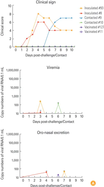
For the pathogenesis test, the footpads of pigs were inocu- lated with the FMDV (103.0 TCID50/0.1 mL) that occurred in first case in 2014, and a contact group was established at the same time point. A vaccination group was inoculated with commercial trivalent vaccine (O1 Manisa, A Malaysia 97, and Asia 1 Shamir) 5 days prior to the contact-challenge. Next, FMD specific clinical symptoms were observed to identify the transmission of FMD. According to the experimental re- sults, the pigs that were inoculated started to show lameness, erosions at the coronary band, and vesicles in their hooves, and the one of contact group began to have the same symp- toms 6 days after the start of the experiment. However, the one contacted and two vaccinated pigs did not appear any
100 80 60 40 20 -ELISSPA (Percent inhibition, %) 0
-5 0 1 2 3 4 5 6 7 8 9 10 Days post-challenge/Contact
SP-ELISA for type O
Inoculated/contact Vaccinated 3.5
3.0 2.5 2.0 s (log)Neutralizing titer 1.5
-5 0 1 2 3 4 5 6 7 8 9 10 21 Days post-challenge/Contact
VN antibody titer
Inoculated/contact Vaccinated
Inoculated #93 Inoculated #8 Contacted #9 Contacted #10 Vacinated #127 Vacinated #11 10
8 6 4 2 0
Clinical score
0 1 2 3 4 5 6 7 8 9 10 Days post-challenge/Contact
Clinical sign
Inoculated #93 Inoculated #8 Contacted #9 Contacted #10 Vacinated #127 Vacinated #11
1,000,000 100,000 10,000 1,000 100 s of viral RNA/0.1 mLCopy number 10
0 1 2 3 4 5 6 7 8 9 10 Days post-challenge/Contact
Viremia
1,000,000 100,000 10,000 1,000 100 s of viral RNA/0.1 mLCopy number 10
0 1 2 3 4 5 6 7 8 9 10 Days post-challenge/Contact
Oro-nasal excretion
Fig. 3. Pathogenesis and transmission after virus challenge and con- tact or vaccination in pigs. (A) Clinical score, viremia, and virus excre- tion during 10 days post-infection (vaccinated animals was inoculated with commercial trivalent vaccine before 5 days of contact chal- lenge). (B) Kinetics of virus neutralizing (VN) antibody titers against O/Andong/SKR/2010 and structural protein (SP) enzyme-linked immu- nosorbent assay (ELISA) antibody for type O virus after challenge and contact.
B
A
symptoms until 21 days of cohabiting, the ending time point of the experiment. In the animals with clinical signs during the experiment, viremia and virus excretion were identified, but in the vaccination group, small amount of virus after con- tact was only detected (Fig. 3A). The SP antibody was pro- duced in 4-5 days post-inoculation in inoculated animals and in 8 days post-contact in the contact pigs (Fig. 3B). Therefore, it was proven that even though the immunized period was short after emergency vaccination, this vaccination was shown the protective capacity with no clinical symptom, vi- rus excretion. In the field, decreases in transmission and the protection capability were identified when high potency vac- cination (>6 PD50) carried out although result of vaccine matching of the isolated virus with vaccine strain, O1 Manisa was identified comparatively low as r1 value of 0.14. The O1 Manisa vaccine was able to protect against O/Andong/
SKR/2010 [4] and O/SKR/01/2014, which was verified in the field.
The outbreak of FMD in Korea that has been determined to be closely related to the occurrence of FMD in neighboring countries is considered to have been affected from viruses of adjacent nations by genetic analysis in this case, too. Howev- er, the direct cause of this inflow has not been verified. This, O/SKR/01/2014, was new virus with the deletion of 23 amino acids in 3A/3B1 region and low pathogenic property.
ORCID
Jong-Hyeon Park http://orcid.org/0000-0003-0825-8121 Dongseob Tark http://orcid.org/0000-0001-7499-4253 Kwang-Nyeong Lee http://orcid.org/0000-0002-8267-3983 Seo-Yong Lee http://orcid.org/0000-0001-9596-6075 Mi-Kyeong Ko http://orcid.org/0000-0002-9714-4232 Hyang-Sim Lee http://orcid.org/0000-0002-3393-8295 Su-Mi Kim http://orcid.org/0000-0002-9242-2731 Young-Joon Ko http://orcid.org/0000-0002-3436-8175 Min-Goo Seo http://orcid.org/0000-0003-1752-5105 Ji-Eun Chun http://orcid.org/0000-0003-3844-8567 Myoung-Heon Lee http://orcid.org/0000-0001-5143-5125 Byounghan Kim http://orcid.org/0000-0002-7252-7763
References
1. Knowles NJ, He J, Shang Y, et al. Southeast Asian foot-and- mouth disease viruses in Eastern Asia. Emerg Infect Dis 2012;18:499-501.
2. Park JH, Park JY, Kim YJ, et al. Vaccination as a control measure during the outbreak of foot-and-mouth disease in 2000 in Korea. Dev Biol (Basel) 2004;119:63-70.
3. Wee SH, Park JY, Joo YS, Lee JH, An SH. Control measures implemented during the 2002 foot-and-mouth disease outbreak in the Republic of Korea. Vet Rec 2004;154:598- 600.
4. Park JH, Lee KN, Ko YJ, et al. Control of foot-and-mouth disease during 2010-2011 epidemic, South Korea. Emerg Infect Dis 2013;19:655-9.
5. Park JH, Lee KN, Ko YJ, et al. Diagnosis and control measures of the 2010 outbreak of foot-and-mouth disease A type in the Republic of Korea. Transbound Emerg Dis 2013;60:188-92.
6. Park JH, Lee KN, Ko YJ, et al. Outbreaks and diagnosis of foot-and-mouth disease serotype O in the Republic of Ko- rea, April-June 2010. Transbound Emerg Dis 2014;61:277-84.
7. Lee HS, Lee NH, Seo MG, et al. Serological responses after vaccination of growing pigs with foot-and-mouth disease trivalent (type O, A and Asia1) vaccine. Vet Microbiol 2013;
164:239-45.
8. Oem JK, Ferris NP, Lee KN, Joo YS, Hyun BH, Park JH. Sim- ple and rapid lateral-flow assay for the detection of foot-and- mouth disease virus. Clin Vaccine Immunol 2009;16:1660-4.
9. Doel TR. FMD vaccines. Virus Res 2003;91:81-99.
10. Pacheco JM, Henry TM, O’Donnell VK, Gregory JB, Ma- son PW. Role of nonstructural proteins 3A and 3B in host range and pathogenicity of foot-and-mouth disease virus.
J Virol 2003;77:13017-27.
11. Pacheco JM, Piccone ME, Rieder E, Pauszek SJ, Borca MV, Rodriguez LL. Domain disruptions of individual 3B pro- teins of foot-and-mouth disease virus do not alter growth in cell culture or virulence in cattle. Virology 2010;405:149- 56.