bloodresearch.or.kr Blood Res 2014;49:196-207.
Letters to the Editor 203
mal abnormalities in utero, 5 years before leukaemia. Br J Haematol 2004;126:307-12.
3. Northup JK, Gadre SA, Ge Y, Lockhart LH, Velagaleti GV. Do cy- togenetic abnormalities precede morphologic abnormalities in a developing malignant condition? Eur J Haematol 2007;78:
152-6.
4. Wong KF, Kwong YL. Isochromosome 8q is a marker of secon- dary acute myeloid leukemia. Cancer Genet Cytogenet 2000;
120:171-3.
5. Mitelman F, Johansson B, Mertens F. Mitelman database of chro- mosome aberrations and gene fusions in cancer. Bethesda, MD:
National Cancer Institute, 2014. (Accessed March 10, 2014, at http://cgap.nci.nih.gov/Chromosomes/Mitelman)
6. Jackson A, Carrara P, Duke V, et al. Deletion of 6q16-q21 in hu- man lymphoid malignancies: a mapping and deletion analysis.
Cancer Res 2000;60:2775-9.
7. Dalsass A, Mestichelli F, Ruggieri M, et al. 6q deletion detected by fluorescence in situ hybridization using bacterial artificial chromosome in chronic lymphocytic leukemia. Eur J Haematol 2013;91:10-9.
8. Betts D. i(8)(q10) in acute myeloid leukaemia. Atlas Genet Cytogenet Oncol Haematol 2007;11:245-6.
9. Devitt KA, Lunde JH, Lewis MR. New onset pancytopenia in adults: a review of underlying pathologies and their associated clinical and laboratory findings. Leuk Lymphoma 2014;55:
1099-105.
10. Kelly K, Murphy P. Aplastic anaemia preceding acute lympho- blastic leukaemia in an adult with isolated deletion of chromo- some 9q. Leuk Res 2008;32:1936-8.
11. Li Q, Chen Z, You Y, Zou P. Transient pancytopenia preceding acute lymphoblastic leukemia with positive Philadelphia chromosome. Leuk Res 2008;32:1317-20.
T-cell large granular lymphocytic leukemia: 4 cases
TO THE EDITOR: T-cell large granular lymphocytic leuke- mia (T-LGL) is a rare clonal hematological disorder charac- terized by peripheral blood and bone marrow lymphocytic infiltration with large granular lymphocytes (LGLs), spleno- megaly, and cytopenia, of which neutropenia is most com- mon [1]. T-LGL is characterized by persistent increases in LGLs ranging from 2×10⁹/L to 20×109/L on peripheral blood in the absence of a reactive cause [2]. The exact pathogenesis is unknown, but is believed to result from clonal expansion of mature postthymic T cells [3]. T-LGL typically presents in the sixth decade of life, with an equal male to female ratio [4]. Approximately 40% of patients have an associated autoimmune disorder, most commonly rheumatoid arthritis (RA), pure red cell aplasia, or immune thrombocytopenia [5]. Approximately one third of patients are asymptomatic when routine blood counts reveal cytopenia and LGL, which
leads to diagnosis. The symptoms, if present, are related to cytopenia [6].
Bone marrow aspirate may be required to confirm the diagnosis, especially in those with low absolute numbers of circulating LGLs. Patients with T-LGL have a median survival of more than 10 years [7]. The most common in- dications for treatment are cytopenia, recurrent infection, pure red cell aplasia, progressive splenomegaly, and B symp- toms [8]. The reported incidence of T-LGL is 2–5% of the chronic lymphoproliferative disorders (LPDs) in North America and about 6% in Asia [9]. The incidence and preva- lence is not known in Pakistan. In this case series, we de- scribe the laboratory findings and clinical courses of 4 pa- tients diagnosed with T-LGL in a tertiary care hospital.
CASES
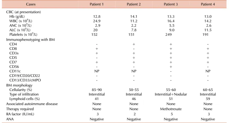
The first case was a 24-year-old man who presented with lymphocytosis in November 2010. His physical examination was unremarkable and showed no hepatosplenomegaly or lymphadenopathy. A complete blood cell (CBC) count re- vealed lymphocytosis with the presence of LGLs (Table 1). Based on bone marrow morphological and im- munophenotypic assessments, the patient was diagnosed with T-LGL (Table 1). His follow-up visits showed persistent lymphocytosis with a declining platelet count. On his fol- low-up in December 2013, CBC revealed hemoglobin (Hb), 13.5 g/dL; white blood cell (WBC) count, 17.9×10⁹/L with an absolute lymphocyte count (ALC) of 11.4×10⁹/L; and platelets, 93×109/L.
The second case was a 61-year-old man who was referred in November 2010 for assessment of persistent lymphocy- tosis lasting for about 7 months. His physical examination was unremarkable. CBC showed lymphocytosis, whereas blood smear, bone marrow morphology, and immuno- phenotyping were consistent with T-LGL (Table 1). He re- mained asymptomatic throughout the disease course with- out any treatment. His CBC in August 2013 revealed Hb, 14 g/dL; WBC, 7.6×10⁹/L with an ALC of 5.3×10⁹/L; and platelets, 100×109/L.
The third case was a 61-year-old man who was diagnosed with hairy cell leukemia (HCL) in 1992 after morphological findings from peripheral smear/bone marrow and under- went splenectomy as the sole treatment for HCL. He was then lost to follow-up until 1998, when he presented with recurrent chest infections. His physical examination was unremarkable, whereas CBC revealed lymphocytosis and LGLs on peripheral smear (Table 1). Subsequent examination of bone marrow morphology and immunophenotyping was consistent with T-LGL (Table 1). During follow-up visits, he continued to have chest infections and was started on low-dose oral methotrexate 5 mg once daily for 7 days in a 4-week cycle. He responded well to treatment and the repeated infections resolved. He is still on the regimen and is asymptomatic. His most recent follow-up CBC re- vealed Hb, 12.5 g/dL; WBC, 19.5×10⁹/L with an ALC of 12×109/L; and platelets, 181×109/L.
Blood Res2014;49:196-207. bloodresearch.or.kr
204 Letters to the Editor
Table 1. Laboratory findings of the patients with T-LGL.
Cases Patient 1 Patient 2 Patient 3 Patient 4
CBC (at presentation) Hb (g/dL)
WBC (x 109/L) ANC (x 109/L) ALC (x 109/L) Platelets (x 109/L)
12.824.9 202.9 152
14.111.2 2.27.8 151
13.316.4 5.59.0 249
13.014.2 11.52.6 191 Immunophenotyping with BM
CD4CD8 CD3sCD5 CD7CD56 CD11c
CD19/CD20/CD22 CD13/CD33/cMPO
+- +- +- NP --
+- ++ +- NP --
++ ++ ++ - --
+- ++ +- NP --
BM morphology Cellularity (%) Type of infiltration Lymphoid cells (%)
85–90 Interstitial
41
50–55 Interstitial
46
55–60 Interstitial+Nodular
51
60–65 Interstitial
59
Associated autoimmune disease None None None None
Therapy required None None Methotrexate None
RA factor (IU/mL) 3 2 5 3
ANA Negative Negative Negative Negative
Abbreviations: T-LGL, T-cell large granular lymphocytic leukemia; CBC, complete blood count; WBC, white blood cell; ANC, absolute neutrophil count; ALC, absolute lymphocyte count; BM, bone marrow; NP, not performed; RA, rheumatoid arthritis; ANA, anti-neutrophil antibody.
The forth case was a 62-year-old woman who presented with persistent lymphocytosis for 10 months in 2010. Her physical examination was unremarkable. She had lichen sclerosis that was diagnosed in 1996 and was treated with topical steroids. Peripheral smear, bone marrow examina- tion, and immunophenotyping were consistent with CD8+
T-LGL (Table 1). In the most recent follow-up visit, she was well and a CBC revealed Hb, 11.7 g/dL; WBC, 7.6×10⁹/L with an ALC of 4.3×10⁹/L; and platelets, 242×109/L.
DISCUSSION
This case series illustrates the clinicopathological features of T-LGL, which was first described in 1977 as a syndrome displaying increased circulating LGLs and chronic neu- tropenia [10]. Approximately 40% of cases are asymptomatic and are discovered incidentally through persistent neu- tropenia, asymptomatic lymphocytosis, or as an associated phenomenon with autoimmune disorders such as RA.
Peripheral smear examination is very helpful in such cases.
In our clinical experience of more than 15 years in a tertiary care hospital where patients are referred from all regions of the country, we have seen 4 T-LGL cases. T-LGL is not only rare, it can also be easily missed if peripheral smears are not reviewed, especially in patients with neutropenia. In these cases, the median age at presentation was 61 years, which is comparable with the literature [11].
Only 1 of our patients was female.
Natural killer cell lymphoproliferative disorder was ex- cluded in these patients on the basis of positivity for surface CD3 and characteristic morphology of LGLs. The most com- mon presentation was persistent lymphocytosis, with an LGL count ranging from 7.8×10⁹/L to 20×109/L [12]. None of our patients presented with neutropenia or splenomegaly, as has been reported in the literature [1, 5]. There was no clinical or serological evidence of RA or other auto- immune disorder.
Case 3 had various interesting issues. First, he had HCL, for which he had a splenectomy. A literature review revealed sporadic cases of T-LGL with other B-cell lymphoprolifer- ative disorders, including chronic lymphocytic leukemia, HCL, and multiple myeloma [13]. In most cases in the liter- ature, there were simultaneous occurrences of B-cell lym- phoproliferative disorders and T-LGL [13], whereas because our patient was lost to follow-up, it could not be clearly delineated that the HCL transformed to T-LGL or if it was a second malignancy. Peripheral smear and bone marrow showed LGL only, and flow cytometry revealed expression of all T-cell markers but not B-cell markers and CD11c.
Second, increased numbers of LGLs have been found in splenectomized patients [14]. LGL clonality has been sup- ported by flow cytometry results, and, in our case, clinical behavior. Third, recurrent infections requiring treatment can be related to CD56 expression associated with a relatively aggressive clinical course [15].
bloodresearch.or.kr Blood Res 2014;49:196-207.
Letters to the Editor 205
Currently, there is no standard treatment for patients with T-LGL. For asymptomatic T-LGL patients with an in- dolent course, a wait-and-see approach can be considered [11]. In our series, all 4 patients are alive and stable, with a slowly declining trend of platelets during the more than 3-year follow-up. T-LGL clonality could not be confirmed by a T-cell receptor gene rearrangement in our cases because our facility lacks this capacity. The clinical features and laboratory findings of the T-LGL patients in our study were similar to that reported in the literature. It remains unclear if the incidence is truly low or the disease has been under- diagnosed because most cases are asymptomatic on presen- tation. This raises the importance of reviewing the periph- eral smears in asymptomatic patients who have persistent lymphocytosis or neutropenia. Systematic long-term fol- low-up studies need to be performed.
Anila Rashid, Mohammad Khurshid, Arsalan Ahmed Department of Pathology and Microbiology, The Aga Khan
University Hospital, Karachi, Pakistan Correspondence to: Anila Rashid Department of Pathology and Microbiology, The Aga Khan University Hospital, P.O. Box 3500, Stadium Road, Karachi, Pakistan
E-mail: [email protected]
Received on May 2, 2014; Revised on Jun. 25, 2014; Accepted on Aug. 25, 2014 http://dx.doi.org/10.5045/br.2014.49.3.203
AuthorsÊ Disclosures of Potential Conflicts of Interest No potential conflicts of interest relevant to this article were reported.
REFERENCES
1. Aribi A, Huh Y, Keating M, et al. T-cell large granular lympho- cytic (T-LGL) leukemia: experience in a single institution over 8 years. Leuk Res 2007;31:939-45.
2. Chan WC, Foucar K, Morice WG, Catovsky D. T cell large gran- ular lymphocytic leukemia. In: Swerdlow SH, Campo E, Harris NL, et al, eds. WHO classification of tumours of haematopoietic and lymphoid tissues. 4th ed. Lyon, France: IARC Press, 2008:272-3.
3. Dearden C. Large granular lymphocytic leukaemia pathogenesis and management. Br J Haematol 2011;152:273-83.
4. Swerdlow SH, Campo E, Harris NL, et al, eds. WHO classification of tumours of haematopoietic and lymphoid tissues. 4th ed. Lyon, France: IARC Press, 2008.
5. Bareau B, Rey J, Hamidou M, et al. Analysis of a French cohort of patients with large granular lymphocyte leukemia: a report on 229 cases. Haematologica 2010;95:1534-41.
6. Loughran TP Jr. Clonal diseases of large granular lymphocytes.
Blood 1993;82:1-14.
7. Lamy T, Loughran TP. Large granular lymphocyte leukemia.
Cancer Control 1998;5:25-33.
8. Lamy T, Loughran TP Jr. Clinical features of large granular lym-
phocyte leukemia. Semin Hematol 2003;40:185-95.
9. Yamamoto JF, Goodman MT. Patterns of leukemia incidence in the United States by subtype and demographic characteristics, 1997-2002. Cancer Causes Control 2008;19:379-90.
10. McKenna RW, Parkin J, Kersey JH, Gajl-Peczalska KJ, Peterson L, Brunning RD. Chronic lymphoproliferative disorder with un- usual clinical, morphologic, ultrastructural and membrane sur- face marker characteristics. Am J Med 1977;62:588-96.
11. Zhang D, Loughran TP Jr. Large granular lymphocytic leukemia:
molecular pathogenesis, clinical manifestations, and treatment.
Hematology Am Soc Hematol Educ Program 2012;2012:652-9.
12. Kondoh K, Morimoto M, Keino D, et al. T-cell large granular lym- phocyte leukemia in a child with anemia and Crohn's disease.
Pediatr Int 2013;55:111-4.
13. Lamy T, Loughran TP Jr. Clinical features of large granular lym- phocyte leukemia. Semin Hematol 2003;40:185-95.
14. Papadaki T, Stamatopoulos K, Kosmas C, et al. Clonal T-large granular lymphocyte proliferations associated with clonal B cell lymphoproliferative disorders: report of eight cases. Leukemia 2002;16:2167-9.
15. Kelemen E, Gergely P, Lehoczky D, Triska E, Demeter J, Vargha P. Permanent large granular lymphocytosis in the blood of sple- nectomized individuals without concomitant increase of in vitro natural killer cell cytotoxicity. Clin Exp Immunol 1986;63:696- 702.
Second case of postpartum acquired hemophilia A in a Korean female
TO THE EDITOR: Acquired hemophilia A (AHA) is a very rare condition with an incidence of about 1.5 per million of the general population per year [1]. It develops in middle age due to autoantibody production directly against the factor VIII (FVIII) and occurs in both sexes equally. The underlying conditions associated with AHA are other auto- immune diseases, malignancy, drugs and pregnancy.
However, about half of AHA has no specific cause, thus they are called idiopathic. Postpartum AHA is a special category with distinct clinical manifestation contributing to 7-21% of patients with AHA [2-4]. In Korea, the first case of postpartum AHA in a 40 years old female has been reported by Lee et al. [5]. To the best of our knowledge, the present case report is the second one in Korea.
A previously healthy 18 years old female had several minor bruises on hands and feet at 5 months after delivery.
There was no specific complication immediately after delivery. She previously didn’t take any drugs which could result in AHA and her family history was nonspecific. The hemorrhagic symptoms were aggravated during the process of time and she visited a private medical doctor at 8 months after delivery. At the medical clinic, she was only supple- mented with iron to treat her iron deficiency anemia.