Arthroscopy-assisted anterior cruciate ligament (ACL) reconstruction by means of bone-patellar-bone (BTB)
autografting is an increasingly popular procedure for the stabillization of ACL-deficient knee (1, 2). The central one-third of the patellar tendon and bony plugs are ex- cised together to increase strength, durability and elas- ticity (3, 4) and the donor site is allowed to heal by pri- mary intensification. The outcome of ACL reconstruc- tion, using patellar tendon, is usually excellent, but sev- eral authors have reported certain complications.
Among these, quadriceps weakness, flexion contracture and patello-femoral pain are common (5, 6).
Infrapatellar contracture syndrome and patellar tendon
MR Imaging Findings of Patellar Tendon after Anterior Cruciate Ligament Reconstruction with Bone-Tendon-Bone Autograft
1Jin Hyoung Kim, M.D., Hyoung Seuk Kim, M.D.2, Hyoung Rae Kim, M.D., Baek Hyun Kim, M.D., Hae Young Seol, M.D., In Ho Cha, M.D., Chang hee Lee, M.D.3, Hong Cheol Im, M.D.4
Purpose: To evaluate the postoperative changes occurring in the patellar tendon after re- construction of the anterior cruciate ligament (ACL) using the central one-third of the patellar tendon together with patellar and tibial bony plugs.
Materials and Methods: Ten patients with ACL injury underwent sagittal and coronal T1- weighted MR imaging of both postoperative and normal knee joints. In all cases, recon- struction of the ACL was performed using the central one-third of the patellar tendon, to- gether with patellar and tibial bony plugs. During the follow-up period of 6-27 months, patient were clinically stable. We compared the length, signal intensity and contour of both patellar tendons, as seen on MR images.
Results: No defects was found in harvested patellar tendons, and MR images showed high signal intensity within harvested tendons in six of the ten patients. In seven of ten, patellar tendons had irregular margins and were poorly delineated from adjacent tissue.
The mean length of patellar tendons was 44.2±2.9 mm in normal knee and 43.9±3.1 mm in postoperative knee, while their mean thickness in postoperative knee, measured at mid-portion, averaged 4.3±1.2 mm. There were no statistically significant differences (p>0.05). The greatest mean thickness of patellar tendon was 6.9±1.2 mm and 4.3±0.5 mm in normal and postoperative knee, respectively. Thus, on average, postoperative patellar tendon was 161% thicker than normal tendon (p<0.05).
Conclusion: In clinically stable patients, patellar tendons after graft harvesting had a high- er signal intensity, worse-defined margins and a greater thickness than normal. We sug- gest that these are the normal postoperative findings.
Index words : Knee, MR Knee, surgery Knee, injuries
1Department of Diagnostic Radiology, Korea University College of Medicine
2Department of Diagnostic Radiology, Inje University Ilsan Paik Hospital
3Department of Diagnostic Radiology, Konkuk University Chungju Hospital
4Department of Orthopaedic Surgery, Korea University College of Medicine
Received July 12, 2001; Accepted October 4, 2001
Address reprint requests to : Hae Young Seol, M.D., Department of Diagnostic Radiology, Korea University Guro Hospital, 80 Guro-dong, Guro-ku, Seoul 152-050, Korea.
Tel. 82-2-818-6784 Fax. 82-2-818-9282 E-mail: [email protected]
rupture, although rare, have also been reported (1, 7, 8).
Many studies have evaluated the morphologic changes occurring in grafted ACL, but only a few have used US or MR to determine those occurring in residual patellar tendon. Using the contralateral knee as a con- trol, Coupens et al. (9) found that the cross-sectional area of the donor site was, on average, 53 percent larger, and that signal intensity, which was initially high, returned to normal throughout the ligament within eighteen months. A study by Proctor et al. (10), using a gout mod- el, reported that as seen on T1-weighted images, the patellar tendon after harvesting was thicker than that on the control side, and its signal intensity was slightly higher.
The purpose of our study is to evaluate the postopera- tive morphological changes occurring in the patellar ten- don and seen at MRI after reconstruction of the ACL with mid one-third BTB autograft in clinically stable subjects.
Materials and Methods
We assessed ten patients who had undergone arthro- scopic-assisted ACL reconstruction by means of mid one-third BTB autograft between March 1992 and November 1996 at our institute and were followed up for a mean period of 16.7 (6-27) months. All patients were male aged 20-38 (average, 26) yesars, and the caus- es of ACL injury were sports injury (n=6), traffic acci- dent (n=3), or a fall-down (n=1).
In all cases, surgery was performed by one operator. A 10 mm strip of central patellar tendon with patellar bone and tibial tuberosity was obtained in each patient, and arthroscopically-guided reconstruction involving a single incision was performed. The remaining patellar tendon, including the tendon itself and the paratendon, was loosely sutured using absorbable suture material.
After surgery, the knee joint was fixed with a long leg splint. Continuous passive movement and isotonic movement of knee joint muscles were started on post- surgical day 1, and the range of joint motion was contin- uously increased. Partial weight bearing movement was permitted two weeks after surgery, and total weight- bearing and active movement, two weeks later.
All patients were stable during the follow-up period.
During a visit to the orthopedic outpatient department, a KT 1000 arthrometer was used to measure the differ- ence in stability between the knee joints, and this was 2.71 mm and 2.64 mm in postoperative and normal
knee, respectively. This result was not statistically sig- nificant (p>0.05). Clinical outcome was estimated using the Lysholm scoring scale and was, on average, 90.1 points. The score, on a scale of 0 to 100, is knee function following ACL reconstruction. A high score indicates a greater return to normal function, and the department of orthopedic surgery at our institution, considers that scores of above 80 indicate normal functioning of the knee.
Using magnetom 63 SP 1.5T scanner(Siemens, Erlangen, Germany), we obtained MR images of both postoperative and contralateral normal knees. In all pa- tients, the standard examination consisted of contiguous 3mm images in coronal and sagittal planes. A three-di- mensional T1-weighted turbo spin-echo sequence with a repetition of 630 ms and effective time-to-echo of 15 ms was used, and the field of view was 16 cm, with a 256×
256 matrix. On MR images, lengths, thicknesses and sig- nal intensities of harvested and normal patellar tendons were estimated. Length was measured from the lower pole of the patellar bone to the highest portion of tibial tuberosity, while thickness was determined at the mid and thickest portions of the tendon. The signal intensi- ties of harvested and normal tendon were compared.
Any defect in the tendon donor site was carefully exam- ined, and tendon contours were recorded.
All measurements were acquired by three radiologists working independently, the average of these three sets of data being regarded as estimated data. The estimated lengths and thicknesses of both patellar tendons were statistically analyzed using the paired t test, and we also determined whether any correlation existed between follow-up period and ratio of the thickness of postopera- tive to nonoperative patellar tendon, measured at the thickest portion of the tendon and calculated using Pearson’s correlation coefficient (R).
Results
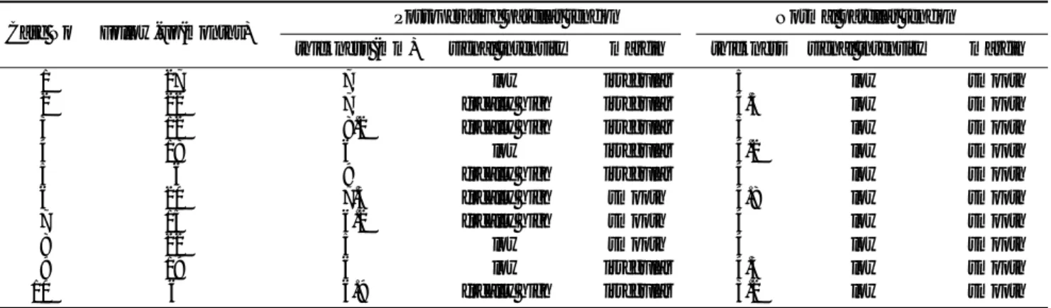
MR images (Table 1) revealed no definite defect in harvested patellar tendons, but six of ten cases showed focal areas of higher signal intensity within the harvest- ed tendon compared with normal patellar tendon (Fig.
1). In seven of ten cases, donor patellar tendons had ir- regular margins and were poorly delineated from adja- cent tissues. All patellar tendons with focal high signal intensity had irregular margins, and the signal intensity of all normal patellar tendons was uniformly low, with well-defined margins.
In Table 2, the dimensions of postoperative patellar tendon are compared with those of nonoperative ten- dons. In normal and postoperative knees, respectively, average length was 44.2±2.9 mm, and 43.9±3.1 mm, and average thickness at mid portion was 4.3±0.5 mm and 4.5±1.2 mm with no statistically significant differ- ence (p>0.05). The greatest thickness , however, aver- aged 4.3 mm±0.5 mm in the normal knee and 6.9±
1.2mm in postoperative knee (Fig. 2), and postoperative tendon was on average 161 percent thicker than normal tendon (p<0.05).
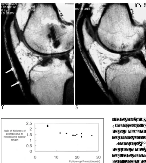
Figure 3 plots follow-up period against the ratio of the greatest thickness of postoperative to nonoperative patellar tendon. Pearson’s correlation coefficient (R) for this graph was -0.8799.
Discussion
Rupture of the ACL is a devastating knee injury, and in most cases, the correction of disability due to anterior instability and the prevention of secondary degenerative
Table 2. Average Length and Thickness of Postoperative and Non-operative Patellar Tendons
Postoperative Nonoperative p value PT(mm) PT(mm)
Length 44.2±2.9 43.9±3.1 0.4
Thickness at
center of PT 04.5±1.2 04.3±0.5 0.1 Thickness at the
thickest portion of PT 06.9±1.2 04.3±0.5 0.001 (PT; patellar tendon)
A B
Fig. 1. Focal high signal intensity (ar- rows) is seen on mid-portion of postop- erative patellar tendon (A) comparing with the contralateral normal patellar tendon (B) on T1 weighted sagittal im- ages.
Table 1. MR Finding of Postoperative and Non-postoperative Patellar Tendons in Patients with ACL Reconstruction with Central one- third of Patellar Tendon
Case No Follow-up(months) Postoperative patellar tendon Normal patellar tendon thickness (mm) signal intensity margin thickness signal intensity margin
01 27 7.0 low irregular 5.0 low smooth
02 22 7.0 focally high irregular 4.5 low smooth
03 12 8.2 focally high irregular 5.0 low smooth
04 18 6.0 low irregular 4.2 low smooth
05 06 9.0 focally high irregular 4.0 low smooth
06 20 7.3 focally high smooth 4.8 low smooth
07 15 6.2 focally high smooth 4.0 low smooth
08 22 5.0 low smooth 4.0 low smooth
09 19 6.0 low irregular 4.3 low smooth
10 6 6.9 focally high irregular 3.2 low smooth
meniscoid change requires surgical intervention (11).
The patellar tendon, semitendinous muscle, the iliotibial tract, achilles tendon, brachial muscles and deep femoral fascia are used as donor material in ACL recon- struction. The central one-third of the patellar tendon is commonly used in this way because of its biomechani- cal advantages, strength at procurement, ease with which each end of the graft can be fixed to bone, and the quality of this osseous fixation compared with other tis- sues. These benefits, however, must be weighed against the risks of harvesting the autograft (4).
MR imaging very cleared demonstrates the morpho- logic details of the knee, and has proven highly accurate in diagnosing tendon disease such as inflammation, as well as partial and complete tears of the patellar tendon (12-15).
This study provides a quantitative description of the changes revealed by MR imaging at the donor site of the
central one-third of the patellar after graft harvesting.
Reports have also described the MRI or ultrasound find- ing of morphologic change occuring at the donor site of the central patellar tendon.
Proctor et al. (10) performed an experimental study us- ing a gout model. They obtained a 6mm-wide strip from the central portion of the patellar tendon, together with patellar and tibial bony plugs, and after 21 months, the repair tissue that formed in the defect was characterized in terms of its structural, material, histological and ultra- structural properties. Thus, the reported focal area of in- creased signal intensity in harvested patellar tendon and the thickness of donor tendon was, on average, 198 per- cent greater than in normal tendon. Histologically, cen- tral tissue demonstrated a consistent pattern of well-de- fined, longitudinally-oriented collagen fascicles (10).
Our study involving clinically stable patients showed similar results. The greatest thickness of harvested patellar tendon was 161 percent more than that of nor- mal patellar tendon. Six of ten cases showed focal high signal intensity and irregular margins within harvested patellar tendon, but its length, width, and thickness at mid-portion showed no significant change.
Several studies have reported an absence of morpho- logical difference between postoperative and nonopera- tive patellar tendons. Meisterling et al. studied fourteen patients who had undergone arthroscopic ACL recon- struction using the central one-third of patellar tendon followed by bilateral MRI of the knee at least 22 months after surgery. They reported no statistically significant Fig. 3. Plot diagram show negative correlation between follow-
up period and ration of thickness of postoperative patellar ten- don.
A B
Fig. 2. Postoperative patellar tendon (arrows) on mid-portion shows more thicker (A) than contralateral normal patellar tendon (B) on T1 weighted sag- ittal images.
differences in the length, width and thickness of postop- erative and nonoperative patellar tendons, results which indicate that ACL reconstruction using the central one- third of the patellar tendon does not affect its morpholo- gy (7).
We believe that this difference between the two stud- ies is due to the time lag between surgery and MRI. In our study, the average follow-up period was 16.7 (6-27) months, compared with at least 22 months in Meisterling’s study. Kiss et al. performed ultrasound studies of postoperative patellar tendon healing, ran- domly dividing 20 patients into four groups and studing them at 3, 6, 9 and 12 months postoperatively. The size of tendon defect diminished progressively from mean 109 mm2at 3 months to mean 23 mm2at 12 months (16).
Coupens et al. performed magnetic resonance imaging at 6 weeks and 4, 6, 9, and 18 months after ACL recon- struction using the central one-third of the patellar ten- don in 20 patients, and the thickness, length and width of donor patellar tendon were measured. No significant change was seen in the length or width of harvested patellar tendon, but its thickness increased in all cases.
The rate of increase in thickness was highest six weeks after surgery, decreasing subsequently (9). Nixon et al.
performed a similar study. In 14 cases, the MR images from six weeks to two years after ACL reconstruction showed that the size of the defect and the signal intensi- ty of the central one-third of harvested patellar tendon decreased with time since surgery, and at two years, the defect was indistinguishable from normal tendon (17).
In our study, harvested patellar tendons may have been thicker than normal tendons because of the relatively short follow-up period.
The correlation coefficient between follow-up period and ratio of thickness of postoperative patellar tendon to that of nonoperative patellar tendon measured at the thickest portion was -0.87994 (p=0.0008), and despite the small sample size, strong correlation is thus evident.
In rare cases, rupture of the patellar tendon or patellar tendinosis(jumper’s knee) has occurred as a complica- tion after ACL reconstruction using the central one-third of the patellar tendon. In a case of patellar tendinosis, Kiss et al. reported that no observed ultrasound features distinguished the case from asymptomatic cases (16). In another study, persistent diffuse thickening of donor patellar tendon correlated with clinical symptoms of patellar tendinitis after ACL reconstruction (18), find- ings which were also noted in our patients. It is on the basis of clinical features alone, that complications aris-
ing in patellar tendon grafting are detected.
In conclusion, increased thickness and focal high sig- nal intensity of harvested patellar tendon were revealed by MR imaging in clinically stable patients, and these findings should be regarded as normal postoperative findings.
References
1. Bonamo JJ, Krinick RM, Sporn AA. Rupture of the patellar liga- ment after use of its central third for anterior cruciate ligament re- construction. J Bone Joint Surg Am 1984;66:1294-1297
2. Breituss H, Frolich R, Povacz P, Resch H, Wicker A. The tendon defect after anterior cruciate ligament reconstruction using the mid third patellar tendon - a problem for the patellofemoral joint?
Knee Surg Sports Traumatol Arthrosc 1996;3:194-198
3. Lambert KL. Vasculized patellar tendon graft with rigid internal fixation for anterior cruciate ligament insufficiency. Clin Orthop 1983;172:85
4. Noyes FR, Butler DL, Paulos LE, Grood ES. Intra-articular cruciate reconstruction. I: perspectives on graft strength, vascularization, and immediate motion after replacement. Clin Orthop 1983;172:
71-77
5. Indelicato P, Bittar E, Prevot T. Clinical comparison of free dried and fresh frozen patellar tendon allograft for anterior cruciate liga- ment reconstruction of the knee. Am J Sports Med 1990;18:335-342 6. Jackson D, Schaefer P. Cyclops syndrome - loss of extension fol-
lowing intra-articular anterior cruciate ligament reconstruction.
Arthroscopy 1990;6:171-178
7. Meisterling RC, Wadsworth T, Adrill R, Griffiths H, Lane-Larsen CL. Morphologic changes in the human patellar tendon after bone- tendon-bone anterior cruciate ligament reconstruction. Clin Orthop 1993;289:208-212
8. Marumoto JM, Mitsunaga MM, Richardson AB, Medoff RJ, Mayfield GW. Late patellar tendon ruptures after removal of the central third for anterior cruciate ligament reconstruction. Am J Sports Med 1996;24:698-702
9. Coupens SD, Yates CK, Sheldon C, Ward C. Magnetic resonance imaging evaluation of the patellar tendon autograft with press fit femoral fixation. Am J Sports Med 1992;200:332-335
10. Proctor CS, Jackson DW, Simon TM. Characterization of the re- paired tissue after removal of the central one-third of the patellar tendon. J Bone Joint Surg Am 1997;79:997-1006
11. Macdaniel WJ, Dameron TB. Untreated ruptures of the anterior cruciate ligament: a follow-up. J Bone Joint Surg Am 1980;62:696- 705
12. Beltran J, Noto AAM, Herman LJ, Lubbers LM. Tendons: High- field-strength, surface coil MR imaging. Radiology 1997;162:735- 740
13. Bonde D, Quinn SF, Murray WT, et al. Magnetic resonance im- ages of chronic patellar tendinitis. Skeletal Radiol 1998;17:24-28 14. Davies SG, BAudouin CJ, King JB, perry GD. Ultrasound, comput-
ed tomography, and magnetic resonance imaging in patellar ten- dinitis. Clin Radiol 1991;43:52-56
15. Ehman RL. Berquist TH. Magnetic resonance imaging of muscu- loskeletal trauma. Radiol Clin North Am 1986;24:291-319
16. Kiss ZS, KLellaway DP, Cook JL, Khan KM. Postoperative patellar tendon healing: An ultrasound study. Austral Radiol 1998;42:28-32 17. Nixon RG, SeGall GK, Sax SL, Cain TE, Tullos HS. Reconstitution
of the patellar tendon donor site after graft harvest. Clin Orthop
1995;317:162-171
18. Gillogly SD, Schaefer RA, Rak KM, et al. Accuracy of Magnetic Resonance Imaging in Assessment of Patellar Tendon Autograft
Anterior Cruciate Ligament Reconstruction. Presentation to the American Academy of Orthopedic Surgeons Annual Meeting, Anaheim, California, 1991
대한방사선의학회지 2002;46:67-72
자가 골 - 슬개건 - 골을 이용한 전방십자인대 재건술 후 슬개건의 자기공명영상 소견1
1고려의대 진단방사선과, 2인제의대 일산백병원 진단방사선과
3건국의대 충주병원 진단방사선과, 4고려의대 정형외과
김진형・김형석2・김형래・김백현・설혜영・차인호・이창희3・임홍철4
목적: 중앙부 슬개건 이식편을 이용한 외상성 전방십자인대 파열 복구 수술 후 슬관절 자기공명영상을 이용하여 수술한 슬개건과 정상 슬개건의 소견을 비교하여 보았다.
대상과 방법: 외상성 전방십자인대 파열 소견을 보인 10명에서 슬개건의 중앙부분에서 이식편을 얻어 전방십자인대 복
구 수술을 시행한 환자들을 중 임상적으로 증상을 보이지 않았던 10명의 환자들을 대상으로 하였으며, 수술 후 6-27 개월(평균 16.7개월) 후 반대편 정상 슬관절과 같이 양쪽 슬관절에서 자기공명영상을 시행하였다. 자기공명영상은 시 상면과 관상면상으로 시행하였고, 슬개건의 길이, 중앙부와 가장 두꺼운 부위에서의 두께 및 신호강도를 측정하여 양쪽 슬관절을 서로 비교하였다.
결과: 수술한 슬관절에서 슬개건 내에 국소적인 고신호 강도가 6명(60%)에서 보였으며, 7명(70%)에서는 슬개건의 경 계가 불분명하였다. 길이는 건측 슬개건에서는 44.2 mm±2.9 mm이었고, 수술한 슬개건에서는 43.9 mm±3.1 mm이 었다. 중앙부위에서의 두께는 양측에서 통계학적으로 유의한 차이를 발견할 수 없었다(p>0.05). 가장 두꺼운 부분에 서의 두께는 건측이 4.3 mm±0.5 mm이고 수술한 쪽의 슬개건은 6.9 mm±1.2 mm로 측정되어 통계학적으로 유의하 게 수술한 쪽의 슬개건이 건측 슬개건보다 두꺼웠다(161%).
결론: 슬개건을 이용하여 전방십자인대 복구 수술을 받은 환자에서 시행한 슬관절 자기 공명영상에서 이식편을 얻은 슬 개건은 정상 슬개건과 비교하여 경계가 불분명하며, 국소적인 고신호강도를 보이고, 두께가 두껍게 보였고, 이는 정상 수술 후 자기공명영상 소견으로 생각된다.