JCS
Journal of Chest SurgeryClinical Research Outcomes after Extracorporeal Membrane Oxygenation in
Neonates with Congenital Diaphragmatic Hernia: A Single- Center Experience
Wooseok Choi, M.D. 1,* , Won Chul Cho, M.D., Ph.D. 2,* , Eun Seok Choi, M.D., Ph.D. 1 , Tae-Jin Yun, M.D., Ph.D. 1 , Chun Soo Park, M.D., Ph.D. 1
1
Division of Pediatric Cardiac Surgery, Department of Thoracic and Cardiovascular Surgery, Asan Medical Center, University of Ulsan College of Medicine, Seoul;
2
Department of Thoracic and Cardiovascular Surgery, Gangneung Asan Hospital, University of Ulsan College of Medicine, Gangneung, Korea
ARTICLE INFO Received May 11, 2021 Revised September 6, 2021 Accepted September 8, 2021 Corresponding author Chun Soo Park Tel 82-2-3010-3583 Fax 82-2-3010-6966 E-mail [email protected] ORCID
https://orcid.org/0000-0001-8718-8904
*
These authors equally contributed to this work.
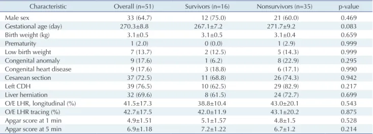
Background: Congenital diaphragmatic hernia (CDH) is a rare disease often requiring mechanical ventilation after birth. In severe cases, extracorporeal membrane oxygenation (ECMO) may be needed. This study analyzed the outcomes of patients with CDH treated with ECMO and investigated factors related to in-hospital mortality.
Methods: Among 254 newborns diagnosed with CDH between 2008 and 2020, 51 pa- tients needed ECMO support. At Asan Medical Center, a multidisciplinary team approach has been applied for managing newborns with CDH since 2018. Outcomes were com- pared between hospital survivors and nonsurvivors.
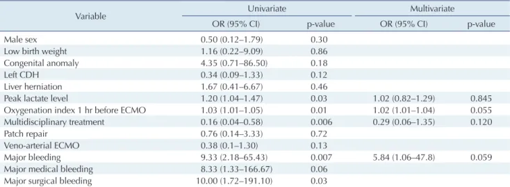
Results: ECMO was established at a median of 17 hours after birth. The mean birth weight was 3.1±0.5 kg. Twenty-three patients (23/51, 45.1%) were weaned from ECMO, and 16 patients (16/51, 31.4%) survived to discharge. The ECMO mode was veno-venous in 24 patients (47.1%) and veno-arterial in 27 patients (52.9%). Most cannulations (50/51, 98%) were accomplished through a transverse cervical incision. No significant between-group differences in baseline characteristics and prenatal indices were observed. The oxygen- ation index (1 hour before: 90.0 vs. 51.0, p=0.005) and blood lactate level (peak: 7.9 vs. 5.2 mmol/L, p=0.023) before ECMO were higher in nonsurvivors. Major bleeding during ECMO more frequently occurred in nonsurvivors (57.1% vs. 12.5%, p=0.007). In the multivariate analysis, the oxygenation index measured at 1 hour before ECMO initiation was identified as a significant risk factor for in-hospital mortality (odds ratio, 1.02; 95% confidence interval, 1.01–1.04; p=0.05).
Conclusion: The survival of neonates after ECMO for CDH is suboptimal. Timely applica- tion of ECMO is crucial for better survival outcomes.
Keywords: Congenital diaphragmatic hernias, Extracorporeal membrane oxygenation, Oxygenation index, Pediatric, Extracorporeal circulation
Copyright © 2021, The Korean Society for Thoracic and Cardiovascular Surgery
This is an Open Access article distributed under the terms of the Creative Commons Attribution Non-Commercial License (http://creativecommons.org/licenses/
Introduction
Congenital diaphragmatic hernia (CDH) is characterized by a congenital defect in the diaphragm and herniation of the abdominal organs into the thoracic cavity. It occurs in 1 of 3,300 live births [1]. Lung hypoplasia and persistent pulmonary hypertension lead to respiratory failure, result- ing in clinical deterioration. Recently, in addition to the application of pulmonary vasodilators and high-frequency oscillatory ventilation (HFOV), management strategies that
minimize ventilator-associated lung injury have signifi- cantly improved outcomes [1]. However, extracorporeal membrane oxygenation (ECMO) is required in patients with low preductal oxygen saturation, severe hypoxemia, severe hypercapnia, and insufficient oxygen delivery de- spite maximal ventilatory and pharmacological support.
In the Extracorporeal Life Support Organization (ELSO) database, CDH is the most common indication for neona- tal ECMO. Since 1987, when the ELSO database was estab- lished, 7,889 cases of ECMO for CDH have been registered,
https://doi.org/10.5090/jcs.21.040
pISSN: 2765-1606 eISSN: 2765-1614
J Chest Surg. 2021;54(5):348-355
Wooseok Choi, et al. Single-Center Experience of ECMO in CDH JCS
with a survival rate of 50%. However, no improvement in the survival rate has been achieved, and the role of ECMO in patients with CDH remains controversial [2]. Neverthe- less, the necessity of ECMO in patients with certain severe- ly detrimental conditions is unquestionable. This study was conducted to investigate the outcomes of ECMO in neonates with CDH and to identify factors associated with in-hospital mortality.
Methods
Patient selection and definitions
From April 2008 through May 2020, among 254 neo- nates with CDH, 51 patients who were treated with ECMO were included in this study (Fig. 1). Data were collected and collated by reviewing electronic medical records. The birth profiles, fetal ultrasound data, and clinical data were obtained. Laboratory data, including serum lactate levels and the oxygenation index (OI; OI=mean airway pres- sure×FiO
2[fraction of inspired O
2]×100÷PaO
2) were serial- ly obtained 6 and 12 hours after birth and 1 hour before ECMO initiation. Surgical data with respect to ECMO es- tablishment and hernia repair were collected. Follow-up data including respiratory and neurodevelopmental status were also obtained.
The primary outcome of interest was in-hospital mortal- ity. For comparison, the patients were divided into 2 groups based on hospital survival: the survivor group and the nonsurvivor group. Major surgical bleeding during ECMO was defined as bleeding that required surgical in- tervention, and major medical bleeding included intracra- nial hemorrhage or pulmonary hemorrhage that needed medical management. The institutional review board of Asan Medical Center approved this study and granted a waiver of informed consent (study no., 2020-1461).
Medical management and indication of extracorporeal membrane oxygenation
The patients with CDH were immediately resuscitated after birth by an attending neonatologist. HFOV was con- sidered when conventional mechanical ventilation failed to stabilize the baby (peak airway pressure >25 cmH
2O). In- haled nitric oxide (NO) was applied when respiratory fail- ure occurred despite HFOV (mean airway pressure 16–22 cmH
2O), in the presence of evidence of pulmonary hyper- tension. Transthoracic echocardiography was performed to check for any combined cardiac anomalies and to assess
the degree of pulmonary hypertension and subsequent ventricular dysfunction. Extracardiac congenital defects and chromosomal abnormalities were thoroughly assessed.
ECMO was considered when the OI did not improve (OI >
40) despite maximum medical support, such as HFOV and inhaled NO. Some conditions precluded the application of ECMO, such as various chromosomal anomalies, other concomitant congenital defects that could affect survival, prematurity (<34 weeks of gestation), low body weight (<2 kg), and existing irreversible major organ damage (Fig. 1).
Surgical technique and management of extracorporeal membrane oxygenation
ECMO was established by a pediatric cardiac surgery team, usually within 1 hour after the initial notification.
The mode of ECMO was decided by the surgeon. Prior to delivery, the surgical team was informed about newborns who had been prenatally diagnosed with CDH. In most patients, the right common carotid artery and right inter- nal jugular vein were exposed through a transverse cervi- cal incision for arterial and venous cannulation.
The priming solution for the ECMO circuit comprises plasma solution, 20% albumin, and packed red blood cells.
For veno-arterial ECMO, a 10F arterial cannula (DLP Pe-
Improvement with conventional medical treatment (n=165)
Death without ECMO application (n=38)
Death before discharge (n=7/51, 13.7%)
Survival to discharge (n=16/51, 31.4%)
Non survival (n=35/51, 68.6%)
- Irreversible tissue damage (1-device shortage) (n=19) - Prematurity with low birth weight (n=9)
- Low birth weight despite full term labor (n=4) - Complex congenital cardiac anomaly (n=3) - No parental consent (n=2)
- Combined lysosomal storage disorder (n=1)