Apoptotic Effect of Co-Treatment with Valproic Acid and 17AAG on Human Osteosarcoma Cells
Jun-Young Park, D.D.S.,M.S.D., Se-Jin Park, D.D.S.,M.S.D., In-Ryoung Kim, M.S.,Ph.D., Bong-Soo Park, D.D.S.,M.S.D.,Ph.D., Sung-Hee Jeong
1, D.D.S.,M.S.D.,Ph.D.,
Myung-Yun Ko
1, D.D.S.,M.S.D.,Ph.D., Yong-Woo Ahn
1, D.D.S.,M.S.D.,Ph.D.
Department of Oral Anatomy, School of Dentistry, Pusan National University
1
Department of Oral Medicine, School of Dentistry, Pusan National University
Valproic acid (VPA) is a well-known anticonvulsive agent and has been used in the treatment of epilepsy for almost 30 years. VPA emerged in 1997 as an antineoplastic agent. And it is known that antitmor activity of VPA is associated with its targeted at histone deacetylases. 17AAG, Inhibition of HSP90 leads to the proteasome degradation of the HSP90 client proteins, such as Akt, Raf/Ras, Erk, VEGF, cyclin D and p53, and causes potent antitumor activity. It is reported that 17AAG-induced HSP90 inhibition results in prevention of cell proliferation and induction of apoptosis in several types of cancer. This study was undertaken to investigate the synergistic apoptotic effect of co-treatment with the histone deacetylases inhibitor, VPA and the HSP90 inhibitor, 17AAG on human osteosarcoma (HOS) cells.
Cell viability was evaluated by trypan-blue exclusion. Induction and augmentation of apoptosis were confirmed by Hoechst staining, flow cytometry (DNA hypoploidy and MMP change), Westen blot analysis and immunofluorescent staining.
In this study, HOS cells co-treated with VPA and 17AAG showed several lines of apoptotic manifestation such as nuclear condensations, the reduction of MMP, the decrease of DNA content, the release of cytochrome c into cytosol, the translocation of AIF onto nuclei, and activation of caspase-3, caspase-7 and PARP whereas each single treated HOS cells did not. Although the single treatment of 1 mM VPA or 0.5 μM 17AAG for 48 h did not induce apoptosis, the co-treatment with them induced prominently apoptosis. Therefore our data in this study provide the possibility that combination therapy with VPA and 17AAG could be considered as a novel therapeutic strategy for human osteosarcoma.
Key words : Apoptosis, Valproic acid, 17AAG, Human osteosarcoma
Correspoding Authur: Yong-Woo Ahn
Associate Professor, Department of Oral Medicine, Schoool of Dentistry Pusan National University, Beomeo-Ri, Mulgeum-Eup, Yangsan-Si, Gyeongsangnam-Do, 626-870, Korea
Foot note: *This work was supported by for two years Pusan National University research grant.
Received: 2010-12-04 Accepted: 2011-01-30
Ⅰ. INTRODUCTION
The acetylation state of histone is reversibly
regulated by histone acetyltransferase (HAT) and
histone deacetylase (HDAC). An inappropriate
acetylation state of histones causes abnormal
outgrowth and the altered pattern of cell death,
which leads to neoplasmic transformation.
1)HDACs
are overexpressed under specific environmental
conditions, such as hypoxia, hypoglycemia, and
serum deprivation.
2)Among these conditions,
hypoxia is one of the key factors to trigger angiogenesis via the induction of angiogenic factors.
Regulation of such gene expression through the acetylation of histone is highly involved in the control of angiogenesis.
2-4)HDAC inhibitors were known to cause the arrest of growth, differentiation, or apoptosis of a variety of transformed cells in culture, including human bladder, breast, prostate, lung, ovary and colon cancer cells.
5)Several classes of HDACIs are identified, which include organic hydroxamic acids (e.g., TSA and suberoyl anilide bisydroxamine [SAHA]), short-chain fatty acides (e.g., butyrates and valproic acid [VPA]), cyclic tetrapeptides (e.g., MS-275).
6)Valproic acid (VPA) is a well-known anti- convulsive agent and used in the treatment of epilepsy for almost 30 years. VPA emerged in 1997 as an antineoplastic agent as well, when findings indicated that the substance inhibited proliferation and induced differentiation of primitive neuroectocdermal tumor cells in vivo.
7)Antitmor activity of VPA is associated with its targeted at histone deacetylases. VPA in particular, was able to down-regulate class II HDAC protein levels significantly in several cells in contrast to TSA, which implies that VPA might be a more selective HDAC inhibitor than TSA.
8-11)The heat shock protein 90 (HSP90) is a molecular chaperone required of protein refolding, confor- mational maturation, activation of several proteins involved in signal transduction pathways, and assembly of many proteins associated with the initiation and development of cancer. Inhibition of HSP90 leads to the proteasome degradation of the HSP90 client proteins, such as Akt, Raf/Ras, Erk, VEGF, cyclin D and p53, and causes potent antitumor activity. 17AAG (17-allylamino-17- demethoxygeldanamycin) is one of the most studied inhibitors of the HSP90. It is reported that 17AAG-induced HSP90 inhibition results in prevention of cell proliferation and induction of apoptosis in several types of cancer.
12-14)Cells undergoing apoptosis usually develop characteristic changes, including nuclear condensa-
tion and degradation of DNA into oligonucleosomal fragments.
15)Apoptotic cell death is thought to result ultimately from the proteolytic actions of caspase.
16)and alterations in mitochondrial function play a key part in the regulation of apoptosis.
17)Moreover, the proteasome system is shown to be implicated as a negative or positive mediator of apoptosis. The proteasome pathway is mostly known to work upstream of mitochondrial alterations and caspase activation.
18)Osteosarcoma is one of the most common primay malignant tumors of bone. Treatment of this tumor with systemic chemotherapy improves dramatically the prognosis. Numerous studies depicted that the therapeutic effect of various chemotherapeutic agents on osteosarcoma depending on the induction of apoptosis.
19-21)To date, there is no report about the synergistic apoptotic effects of co-treatment with VPA and 17AAG on human osteosarcoma cells. Therefore, this study was undertaken to investigate the synergistic apoptotic effect of co-treatment with VPA and 17AAG on human osteosarcoma (HOS) cells.
Ⅱ. MATERIALS AND METHODS 1. Reagents
The following reagents were obtained commercially: Mouse monoclonal anti-human caspase-3, caspase-7, poly(ADP-ribose) polymerase (PARP), cytochrome c, apoptosis-inducing factor (AIF) antibodies, and FITC-conjugated goat anti-mouse and anti-rabbit IgGs were from Santa Cruz Biotechnology (Santa Cruz, CA, USA);
HRP-conjugated sheep anti-mouse and anti-rabbit IgGs were from Amersham GE Healthcare (Little Chalfront, UK). 5,5',6,6'-tetrachloro-1,1',3,3'- tetraethylbenzimidazol carbocyanine iodide (JC-1) was from Molecular Probes (Eugene, OR, USA).
Suc-LLVY-AMC was from Calbiochem
(Darmstadt, Germany). Dulbecco's modified Eagle's
medium (DMEM) and FBS were from Gibco
(Gaithersburg, MD, USA). Dimethyl sulfoxide (DMSO), Hoechst 33342, RNase A, aprotinin, leupeptin, PMSF, thiazolyl blue tetrazolium bromide and propidium iodide (PI) were from Sigma (St.
Louis, MO, USA); SuperSignal West Pico enhanced chemiluminescence Western blotting detection reagent was from Pierce (Rockford, IL, USA).
2. Cell culture
The HOS human osteosarcoma cell line was purchased from ATCC (Rockville, USA). Cells were maintained at 37℃ with 5% CO
2in air atmosphere in Dulbecco's modified Eagle’s medium (DMEM) with 4 mM L-glutamine, 1.5 g/L sodium bicarbonate, 4.5 g/L glucose and 1.0 mM sodium pyruvate supplemented with 10% FBS.
3. Assessment of co-treatment of valproic acid (VPA) and 17AAG
The stock solutions of VPA (2 M) made by dissolving the drug in PBS and 17AAG (10 mM) which made by dissolving the drug in ethanol were kept frozen at -20℃ until use. In twenty four hours after HOS cells were subcultured. the original medium was removed. The cells were washed with phosphate-buffered saline (PBS) and then incubated in the same fresh medium. Since 1 mM VPA and 0.5 μM 17AAG are approximately the concentration not to induce HOS cell death, it is determined to utilize this single concentration of each chemical for the combination treatment study. HOS cells were co-treated with 1 mM VPA and 0.5 μM 17AAG for 48 h. Cells were harvested, stained with trypan blue and then counted by using a hemcytometer.
4. Hoechst staining
Cells were harvested and cell suspension was centrifuged onto a clean, fat-free glass slide with a cytocentrifuge. The samples were stained in 4 μ g/mL Hoechst 33342 for 30 min at 37℃ and fixed for 10 min in 4% paraformaldehyde.
5. Quantification of DNA hypoploidy by flow cytometry
After the treatment for 48 h, cells were harvested by trypsinization and ice cold 95% ethanol with 0.5%
Tween 20 was added to the cell suspensions up to a final concentration of 70% ethanol. Fixed cells were pelleted, and washed in 1% BSA-PBS solution.
Cells were resuspended in 1 mL PBS containing 20 μg/mL RNase A, incubated at 4℃ for 30 min, washed once with BSA-PBS, and resuspended in PI solution (10 μg/mL). After cells were incubated at 4℃ for 5 min in the dark, DNA content were measured on a CYTOMICS FC500 flow cytometry system (Beckman Coulter, Brea, CA, USA) and data was analyzed by using the Multicycle software which allowed a simultaneous estimation of apoptosis.
6. Assay of mitochondrial membrane potential (MMP)
JC-1 was added directly to the cell culture medium (1 μM final concentration) and incubated for 15 min. The medium was then replaced with PBS.
Flow cytometry to measure MMP was performed on a CYTOMICS FC500 flow cytometry (Beckman Coulter, FL, CA, USA). Data were acquired and analyzed by using CXP software version 2.2.
7. Immunofluorescent staining
Cells were cytocentrifuged and fixed for 10 min in
4% paraformaldehyde, incubated with each primary
antibody for 1 h, washed 3 each for 5 min, and then
incubated with FITC-conjugated secondary
antibody for 1 h at room temperature. Cells were
mounted with PBS. Fluorescent images were
observed and analyzed under Zeiss LSM 510
laser-scanning confocal microscope (Göettingen,
Germany).
8. Western blot analysis
Cells (2 x 10
6) treated with VPA and/or 17AAG were washed twice with ice-cold PBS, which is resuspended in 200 μL ice-cold solubilizing buffer [300 mM NaCl, 50 mM Tris-Cl (pH 7.6), 0.5% Triton X-100, 2 mM PMSF, 2 μL/mL aprotinin and 2 μ L/mL leupeptin] and incubated at 4℃ for 30 min.
The lysates were centrifuged at 14,000 revolutions per min for 15 min at 4℃. Protein concentrations of cell lysates were determined with Bradford protein assay (Bio-Rad, Richmond, CA, USA) and 50 μg of proteins were loaded onto 7.5-15% SDS/PAGE. The gels were transferred to Nitrocellulose membrane (Amersham GE Healthcare, Little Chalfont, UK) and reacted with each antibody. Immunostaining with antibodies was performed in using SuperSignal West Pico enhancing chemiluminescence substrate and detecting with Alpha Imager HP (Alpha Innotech, Santa Clara, USA).
Ⅲ. RESULTS
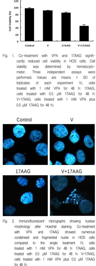
1. Co-treatment with VPA and 17AAG augmented the reduction in viability of HOS cells.
Single treatment of 1 mM VPA or 0.5 μM 17AAG for 48 h reduced slight viability of HOS cells (VPA, 90.03% ; 17AAG, 84.00%). Co-treatment of VPA and 17AAG significantly reduced cell viability compared to the effect of each single treatment (co-treatment, 44.70%) (Fig. 1).
2. Co-treatment with VPA and 17AAG augmented the nuclear condensation and fragmentation in HOS cells.
To explore whether nuclear condensation and fragmentation were induced, Hoechst staining, a hallmark of apoptosis, was conducted. The co-treatment with VPA and 17AAG showed a variety of condensed and fragmented nuclei compared to the single treatment (Fig. 2).
0 20 40 60 80 100 120
Control V 17AAG V+17AAG
Cell Viability (%)