J Korean Ophthalmol Soc 2018;59(8):711-717 ISSN 0378-6471 (Print)⋅ISSN 2092-9374 (Online)
https://doi.org/10.3341/jkos.2018.59.8.711
Original Article
반복각막진무름 환자에서 다이아몬드 연마기의 치료 효과
Effects of Diamond Burr in Patients with Recurrent Corneal Erosion
김현주⋅고재웅
Hyun Ju Kim, MD, Jae Wong Koh, MD, PhD
조선대학교 의과대학 안과학교실
Department of Ophthalmology, Chosun University College of Medicine, Gwangju, Korea
Purpose: The purpose of this study is to evaluate the recurrence rate and treatment effect of conservative treatment and surgical treatment with diamond burr in reccurent corneal erosion.
Methods: Between January 2010 and October 2017, 67 patients who were diagnosed with repeated corneal erosion, 55 patients underwent conservative treatment and 12 patients underwent surgical treatment with a diamond burr were evaluated for the du- ration of previous treatment, recurrence frequency, symptom severity, visual acuity, and the effect of treatment depending on recurrence. Surgical treatment was performed when corneal epithelial loosening occurred in more than 10% of the cases and the patient required surgical treatment. Conservative treatment was continued in patients who did not want surgical treatment.
Results: There were no differences in the mean age, sex, recurrence frequency, symptom severity, and duration of treatment. All 67 patients were unilateral, and 55 patients underwent conservative treatment. Of these, 23 patients (41.8%) showed improve- ment, and 32 patients relapsed within 6 months. Ten eyes (83.3%) of 12 eyes treated with a diamond burr showed improvement and two eyes recurred within 6 months. The recurrence rate was lower in the diamond burr group than in the conservative group (p = 0.011). In comparison with diamond burr and conservative treatment, the visual acuity of the diamond burr group improved statistically significantly compared to the group with conservative treatment (p = 0.002).
Conclusions: In patients with recurrent corneal erosion, surgical treatment with diamond bur was effective in improving visual acuity and preventing recurrence of recurrent corneal erosion than conservative treatment.
J Korean Ophthalmol Soc 2018;59(8):711-717 Keywords: Diamond burr, Recurrent corneal erosion
■Received: 2018. 3. 15. ■ Revised: 2018. 5. 21.
■Accepted: 2018. 7. 20.
■Address reprint requests to Jae Wong Koh, MD, PhD Department of Ophthalmology, Chosun University Hospital,
#365 Pilmun-daero, Dong-gu, Gwangju 61453, Korea Tel: 82-62-220-3190, Fax: 82-62-225-9839
E-mail: [email protected]
* This study was supported by research fund from Chosun University, 2015.
* Conflicts of Interest: The authors have no conflicts to disclose.
ⓒ2018 The Korean Ophthalmological Society
This is an Open Access article distributed under the terms of the Creative Commons Attribution Non-Commercial License (http://creativecommons.org/licenses/by-nc/3.0/) which permits unrestricted non-commercial use, distribution, and reproduction in any medium, provided the original work is properly cited.
반복각막진무름(recurrent corneal erosion)은 각막상피와 각막상피바닥막 사이, 바닥막과 실질층 사이의 바닥막복합
체(basement membrane complex)의 약화로 반복적으로 각 막의 층이 분리되는 질환이다.1 각막상피가 반복적으로 벗 겨짐으로 인해 안통, 충혈, 눈물흘림, 눈부심 등의 증상이 나타나며, 이는 자고 일어났을 때 각막상피가 약하게 부착 된 곳에서 각막상피가 파괴되는 현상과 연관된다. 이러한 각막상피부착의 이상은 외상, 각막이식, 굴정교정수술과 같 은 각막에 관련된 수술 후에 발생할 수 있고 안구건조증, 각막염, 신경인성 각막염, 당뇨병 수포각막병증 등에 의해 서도 발생될 수 있다.2-6 반복각막진무름은 초기의 적절한 치료에도 불구하고 재발이 반복되는 난치성 질환으로, 반 복각막진무름의 보존적 치료로는 무보존제 인공눈물, 고삼 투압제제 등의 점안,7 치료콘택트렌즈 및 압박안대,8 자가
혈청 점안9,10 등이 알려져 있다. 또한 외상 후 발생한 반복 각막진무름에 대해 substance P-derived peptide와 in- sulin-like growth factor 1을 사용하여 호전시킨 보고가 있 으며, metalloproteinase 억제제인 독시사이클린제제 복용과 스테로이드 안약을 병용하여 치료한 논문도 발표되었 다.11-13
그러나 이런 보존적인 치료법에 반응을 보이지 않는 경우 수술적인 치료를 고려해야 한다. 수술 치료에는 흔히 죽은 조직제거술(debridement), 표층각막절제술(superficial kera- tectomy), 일회용 주사바늘이나 미세전기소작기를 이용한 앞기질천자술(anterior stromal puncture)이나 neodymium yttrium aluminum garnet (Nd:YAG) 레이저를 이용한 앞기 질천자술, 알코올 또는 다이아몬드 연마기를 이용한 표층 각막절제술, 엑시머레이저를 이용한 치료레이저각막절제술 (phototherapeutic keratectomy, PTK), 양막이식술 등이 시 행되고 있다.2,14-19
주사바늘을 이용한 앞기질천자술(anterior stromal punc- ture)은 효과적이고 재발률이 낮지만, 각막천공의 위험성이 있고 각막에 영구적인 반흔이 남게 되므로 반흔이 시축에 있는 경우 시력감소를 일으킬 수 있다.20,21 죽은조직제거술 은 효과가 적고, 각막상피바닥막제거술은 바닥막에 상흔을 남길 수 있는 단점이 있다.22-24 엑시머레이저를 이용한 치 료레이저각막절제술은 반흔을 최소화하며 광학적으로 부 드러운 표면이 가능하여 병변이 시축을 침범하고 기존에 근시를 가진 환자에서 좋은 치료법이나, 고가의 레이저 장 비를 갖추어야 하는 단점이 있다.6,25-27
다이아몬드 연마기를 사용하여 표층각막절제술을 시행 한 방법은 저렴하고 시술이 간단하며, 영구적인 각막 흉터 가 발생하지 않고, 각막천공의 가능성과 재발률 또한 매우 낮은 장점이 있어 해외에서는 발표된 적이 있으나 국내에 서는 보고된 바 없어, 이에 저자들은 재발각막진무름 환자 를 대상으로 다이아몬드 연마기를 이용한 수술 치료 방법 에 따른 재발률과 치료 효과에 대해 알아보고자 하였다.28
대상과 방법
2007년 1월부터 2017년 10월 사이에 조선대학교병원 안 과에서 임상적으로 반복각막진무름을 진단받은 환자 67명 을 대상으로 하였다. 보존 치료를 시행한 55명과 다이아몬 드 연마기를 사용한 수술 처치를 시행한 12명을 성별, 연 령, 관찰 기간, 증상 정도, 수술 전후 최대교정시력, 동반된 안과적 질환, 외상력, 재발의 유무 등을 환자의 의무기록을 이용하여 후향적 연구 조사를 시행하였다. 전체 환자들의 반복각막진무름의 원인을 외상에 의한 경우, 각막변성에
의한 경우, 당뇨가 동반된 경우, 안과 시술을 시행한 경우, 원일을 알 수 없는 경우로 나누어 그 분포를 비교하였다. 본 연구는 조선대학교병원 연구윤리 심의위원회(instituitional review board, IRB)로부터 승인을 받았다.
눈을 뜨면서 발생하는 통증 또는 눈을 비비거나 하는 등 의 작은 충격 후에 발생하는 안통, 이물감, 눈물흘림, 눈부 심, 시력감소 등의 자각적인 증상과 세극등현미경검사에서 각막상피 결손, 짓무름, 울퉁불퉁하고 특징적인 회색 상피 병변 또는 바닥막 변성이 관찰되고, 문진상 외상 등에 의한 각막진무름의 과거력이 있는 경우 반복각막진무름으로 정 의하였다. 수술 전 최대교정시력은 logMAR 시력을 사용하 여 초진 시 또는 각막진무름이 발생하여 내원할 당시의 시 력을 측정하였고, 수술 후 최대교정시력은 다이아몬드 연 마기 수술 후 6개월째 시력을 측정하였다.
치료를 시행하기 전 증상의 정도는 병변의 크기를 stage 1에서 stage 4로 나누었다. 각막의 전체 표면에서 1/4에 해 당하는 범위에 병변이 있을 때 stage 1, 2/4 범위에 병변이 있을 때 stage 2, 3/4 범위는 stage 3, 각막전체에 병변이 있 을 때는 stage 4로 구분하였다. 크기 반복각막진무름이 치 료 후 6개월 이내에 재발되는 경우 치료되지 않음으로 하 였고, 6개월 이상 반복각막진무름이 발생하지 않고 유지될 때 치료됨으로 하였다. 수술 처치는 각막상피의 헐거워짐 이 전체의 10% 이상을 차지하고 환자가 수술적 처치를 원 한 경우에 시행하였다. 수술 처치를 원하지 않았던 환자의 경우 보존 치료를 지속하였다. 보존 치료로는 항생제 안약, 인공누액 제제, 고삼투압제, 혈청제제 점안약과 치료용 콘 택트렌즈를 사용하였다. 기본적으로 모든 환자에서 항생제 점안약으로 Cravit® (levofloxacin 0.5%, Santen, Osaka, Japan) 또는 Vigamox® (moxicloxacin 0.5%, Alcon, Fort Worth, TX, USA)를 하루 4회, 인공눈물로는 hyaluronic acid 0.3%
(Hyaluni®, Taejoon, Seoul, Korea)를 하루 8회 사용하였다.
이 외에도 일부 환자에서 solcorin 연고(Solcorin ophthalmic gel®, Hanlim Pharm. Co., Seoul, Korea; 38안)를 하루 4회, 5% Sodium chloride (Muro128®, Bausch & Lomb, Rochester, NY, USA; 29안)를 하루 4회 사용하였다. 치료용 콘택트렌 즈는 모든 환자에서 사용하였다.
다이아몬드 연마기를 이용한 시술은 시술 전 모든 환자 에게 동의를 얻었고, 시술 방법과 시술 후 처치는 다음과 같다. 먼저 0.5% proparacaine (Alcaine®, Alcon, Fort Worth, TX, USA)으로 점안마취를 하고 수술용 면도날(sterile stai- ness steel surgical blade no.15, Swann-Morton, Sheffield, England)을 이용해 짓무름이 발생된 상피를 제거했다. 다이 아몬드 연마기(Katena Ophthalmic burr, Denvile, NJ, USA) (Fig. 1)로 짓무른 부위가 충분히 포함될 수 있도록 전체 각
Figure 2. Combined ocular disease in RCE. Combined ocular
disease at the time of diagnosis of recurrent corneal erosion.RCE = recurrent corneal erosion; DES = dry eye syndrome;
MGD = meibomian gland disease.
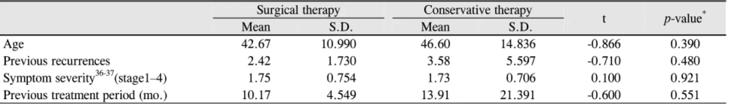
Surgical therapy Conservative therapy
t p-value*
Mean S.D. Mean S.D.
Age 42.67 10.990 46.60 14.836 -0.866 0.390
Previous recurrences 2.42 1.730 3.58 5.597 -0.710 0.480
Symptom severity36-37(stage1–4) 1.75 0.754 1.73 0.706 0.100 0.921
Previous treatment period (mo.) 10.17 4.549 13.91 21.391 -0.600 0.551
S.D. = standard deviation; mo. = months.
*t-test, p < 0.05, p < 0.01.
Table 1. Demographic data of recurrent corneal erosion patients about age, previous recurrences, symptom severity, and duration of
previous treatmentFigure 1. Gross image of diamond burr. The diamond burr tip
used in superficial keratectomy.막표면을 부드럽게 연마했고, 한 지역에서 지나치게 세게 누르거나 오래 머물지 않고 둥글게 원형운동으로 시행하였 다.
시술 후에는 치료콘택트렌즈를 착용하고, Vigamox® (Alcon)를 하루 4회, Muro128® (Bausch & Lomb) 4회, sol- corin 연고 4회, hyaluronic acid 0.3% 무보존제 인공누액을 하루 8회 사용하도록 하였다. 수술 후 각막상피가 정상화되 면 치료콘택트렌즈를 제거하였다. 안약은 2주 이상 유지하 였으며, 짓무름 호전 후 항생제 사용은 중지하였으나, 5%
NaCl 점안액 4회, Solcorin 점안액 4회, 무보존제 인공누액 은 수술 후 3개월간 사용 후 중지하였다. 각막상피의 재생 및 반복각막진무름의 재발에 대해 정기적으로 외래 경과를 확인하였고 최소 6개월 이상 관찰하였다. 치료군별 재발률에 따른 치료 효과에 대한 통계학적 검정은 chi-square test를 통 하여 시행하였다. 치료 방법에 따른 환자군의 시술 전 시력 과, 시술 후의 시력 비교는 T-검정을 사용하였다. 자료의 통계처리는 SPSS 통계프로그램(version 18.0; IBM Corp., Armonk, NY, USA)을 이용하였으며, p값이 0.05 미만인 경 우를 통계적인 의의가 있는 것으로 간주하였다.
결 과
총 67명 중 남자 40명, 여자 27명이 모두 단안에 발생한 반복각막진무름으로 진단되어 치료받았다. 동반된 안 질환 으로 건성안 25안, 마이봄샘기능 장애 25안, 각막변성 3안,
포도막염 1명이 있었으며, 27안에서는 동반된 안 질환이 없었다(Fig. 2). 다이아몬드 연마기를 사용한 환자군의 평균 나이는 42.67 ± 10.9세이며, 보존 치료를 시행한 환자군의 평균 나이는 46.60 ± 14.8세였으며, 두 군의 유의한 차이는 없었다. 본원에서 초진 시 문진한 재발 횟수를 살펴보면, 다이아몬드 연마기를 사용한 군은 평균 2.42 ± 1.7회이고, 보존 치료를 시행한 군의 평균 재발 횟수는 3.58 ± 5.59회 로 역시 통계적으로 유의한 차이를 보이지 않았다. 치료 전 증상 정도는 다이아몬드 연마기를 이용한 군이 1.75 ± 0.75 stage였으며, 보존 치료를 시행한 군은 1.73 ± 0.7 stage였 고, 본원에서 치료를 시행하기 전 시행한 기존 치료 기간은 다이아몬드 연마기를 이용한 군 10.1 ± 4.5개월, 보존 치료 군 13.9 ± 21.3개월로 증상 정도와 기존 치료 기간에서도 두 군 간의 통계적으로 유의한 차이는 없었다(Table 1).
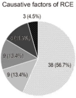
반복각막진무름의 발생 원인으로는 손톱, 나뭇가지 등에 의한 외상이 원인인 경우가 38안(56.7%)으로 가장 많았고, 8안(11.9%)에서는 과거 다래끼 제거술 등의 간단한 안과 시술을 받은 과거력이 있었으며, 각막상피이상증이 있는 경우가 3안(4.5%), 당뇨가 있었던 환자는 9안(13.4%)이었 으며, 원인을 알 수 없는 경우가 9안(13.4%)이었다(Fig. 3).
다이아몬드 연마기를 사용한 표층각막절제술을 시행한
Figure 4. Comparison of surgical and conservative treatment.
A comparison of the recurrences between the surgical treat- ment with diamond burr group and conservative treatment group.
Figure 3. Causative factors. Causative factors in 67 eyes of 67
patients with recurrent corneal erosion. RCE = recurrent cor- neal erosion.Surgical therapy Conservative therapy t p-value†
Mman S.D. Mean S.D.
At the time of diagnosis (V/A) 0.71 0.204 0.55 0.346 1.542 0.128
After 6 months (V/A) 0.95 0.090 0.81 0.259 3.250 0.002*
There was no statistically significant difference in visual acuity between the two groups at the first visit (p = 0.128). After 6 months, statisti- cally significant improvement in visual acuity in the surgical treatment group (p = 0.002).
S.D. = standard deviation; V/A = visual acuity.
*p < 0.05; †t-test.
Table 2. The best corrected visual acuity before treatment and after 6 months of treatment with diamond burr and conservative treat-
ment group환자 12명은 시술 후 1일, 1주, 3주, 1개월, 3개월, 6개월 간 격으로 경과관찰하였고, 보존 치료를 시행한 55명은 1주, 3 주, 1개월, 3개월, 6개월 간격으로 경과관찰하였다. 다이아 몬드 연마기를 사용한 군과 보존 치료를 시행한 군의 초진 시와 치료 후 6개월 후의 최대교정시력을 비교하였다. 치료 전 두 군의 최대교정시력의 차이는 통계학적으로 유의하지 않았지만(p=0.128), 6개월 후 최대교정시력은 보존 치료를 시행한 군에 비해 다이아몬드 연마기를 사용한 군에서 통계 적으로 유의하게 시력이 호전됨을 보였다(p=0.02) (Table 2).
각 군별로 치료 전과 치료 후 6개월째를 비교하였을 때, 다 이아몬드 연마기를 사용한 군은 치료 전 평균시력 logMAR 0.71 ± 0.20에서 logMAR 0.81 ± 0.25 (p<0.001), 보존 치료 를 시행한 군은 logMAR 0.55 ± 0.34에서 치료 후 logMAR 0.81 ± 0.25 (p<0.001)로 두 군에서 모두 통계적으로 유의 하게 시력호전을 보였다.
치료 후 6개월 동안 재발하지 않았을 경우 치료 성공, 6 개월 내에 재발했을 경우 치료 실패로 하여 치료 방법에 따 른 치료 효과 여부를 확인한 결과 다이아몬드 연마기를 사 용한 12안 중 2안에서 6개월 내에 재발하였고, 보존적 치료 를 시행한 55안 중 32안에서 6개월 내에 재발하여 다이아 몬드 연마기를 사용하여 치료한 경우 보존적 치료에 비해 재발률이 통계적으로 유의하게 낮음(p=0.011)을 알 수 있
었다(Fig. 4). 시술 후 세극등현미경검사에서 각막 혼탁은 관찰되지 않았으며 시력저하, 눈부심, 빛번짐 등의 증상도 유발하지 않았으며, 기타 유의한 합병증 발생은 관찰되지 않았다.
고 찰
정상적으로 각막상피는 반결합체(hemidesmosome), 바닥 판(basal lamina), 투명판(lamina lucida), 고정섬유소(anchoring fibril)들로 구성된 바다막복합체(basement membrane com- plex)에 의해 전측 각막실질에 속하는 보우만막에 부착되어
있다.29,30 반복각막진무름은 각막상피세포와 바닥막을 연결
하는 반결합체(hemidesmosome)와 바닥막과 각막실질 사이 를 부착시키는 고정미세섬유(anchoring fibril)로 구성된 바 닥막 복합체(basement membrane complex)의 부착력 약화 로 인해 표층각막의 외상에 의해 바닥막이 손상되거나 각 막이영양증 등의 유전적 이상으로 인해 반복적으로 각막상 피의 탈락이 일어나는 질환이다.1,27 반복각막진무름은 125 년 전 Hansen에 의해 덴마크어로 처음 알려졌으며, 치료 방 법으로는 인공누액, 항생제 안약, 고삼투압제, 연고, 윤활제,
안대 등의 보존적 치료부터 이런 보존 치료에 반응하지 않는 환자들에 대한 적극적 치료로 표층각막절제술, 앞기질천자 술, alcohol을 이용한 화학적 박리법(alcohol delamination), Nd:YAG laser 치료, 엑시머 레이저를 이용한 치료레이저각 막절제술(PTK), 다이아몬드 연마기를 이용한 표층각막절 제술 등이 있다.27 현재 시행되고 있는 수술 방법 중 죽은조 직제거술은 간단하게 시행할 수 있으나 다른 수술에 비하 여 재발률이 높아 단독으로 시행하기에 바람직하지 않다.
Nd:YAG 레이저나 주사바늘을 이용한 전부기질천자술은 각막상피와 전부기질 사이에 부착을 강화시키는 방법으로 보존 치료에 비하여 효과적이나 각막혼탁이 발생하는 단점 이 있고, Nd:YAG 레이저의 경우 레이저 장비가 필요하다 는 점으로 인해 접근이 어렵다는 단점이 있다.31
수술 치료는 재발 횟수, 증상의 정도, 동반된 각막변성 또는 질환의 유무, 병변의 크기 및 위치, 환자의 요구 등을 고려해서 시행하는데10,32 본 연구에서는 각막상피의 헐거워 짐이 전체의 10% 이상을 차지하고 환자가 수술 처치를 원 한 경우에 시행하였다. 1987년 Buxton and Constad24가 각 막상피변성 환자에서 재발성각막진무름 환자에서 다이아 몬드 연마기를 사용하여 재발률을 3%로 낮췄다는 보고를 시작으로 다이아몬드 연마기를 사용하여 재발성각막진무 름을 효과적으로 치료한 여러 연구 결과들이 발표되었다.
Soong et al28은 재발성각막진무름 환자 47명 54안을 대상 으로 다이아몬드 연마기를 사용하여 표층 각막절제술을 시 행하였다. 치료 전 최대교정시력 20/26에서 치료 후 20/22 시력으로(p=0.002) 상승하였고, 치료 전후의 구대면렌즈대 응치 변화는 거의 없었으며, 경면현미경검사에서도 내피세 포의 손상이 보이지 않아, 다이아몬드 연마기를 사용한 표 층 각막절제술이 치료 효과뿐만 아니라 안전하며, 경제적 으로 효율적인 치료 방법으로, 전측각막실질 미세천자술이 나 Nd:YAG 레이저 또는 엑시머 레이저를 사용한 방법을 대체할 수 있는 좋은 방법이라고 보고하였다. Sridhar et al33은 앞바닥막이상증이 있는 재발성각막진무름 환자 39명 42안을 대상으로 엑시머레이저를 이용한 광학적 각막절제 술과 다이아몬드 연마기를 사용하여 치료한 경과를 비교하 였다. 이 연구에서 두 가지 방법 모두 재발성각막진무름을 치료하는 효과적인 방법이지만, 다이아몬드 연마기를 사용 하는 방법이 간단하고, 저비용 고효율적이며, 재발률 및 술 후 각막 혼탁 발생률이 낮아, 광학적 각막절제술보다 이점 이 더 많은 수술적 방법이라고 하였다.33
앞선 연구 결과들처럼, 본 연구에서도 67안 중 보존 치료 만을 지속 시행한 환자군 55명에서 완치된 환자는 23명뿐 이었다. 재발률을 계산하면 12안 중 2안, 즉 16.7%에서 재 발하여, Soong et al28의 연구에서 재발률 11.1% 및 Sridhar
et al33의 연구에서 재발률 11.1%로 타 연구들에 비해 재발 률은 높으나 대상안(12안)이 적어서 나타나는 결과의 차이 로 보인다.
다이아몬드 연마기를 사용하여 재발성각막진무름을 치 료하는 데 여러 장점이 있다. 첫째, 경제적이며, 쉽게 다룰 수 있고 특별한 기술을 필요로 하지 않는다. 둘째, 레이저 처럼 비싼 장비를 필요로 하지 않는다. 셋째, 블레이드를 이용한 각막절제술 및 엑시머 레이저를 이용한 각막절제술 등 재발성각막진무름의 다른 수술 처치에 비해 재발률이 낮으며, 수술 후 굴절이상 및 각막 혼탁 등 합병증 발생이 적다. 넷째, 재수술이 쉽고 각막천공의 가능성이 없다.28 물 론 다이아몬드 연마기를 사용한 수술 후 3-4일간의 통증 발 생 및 당뇨나 신경인성 각막 등에서 각막상피 결손이 지속 적으로 발생할 수 있는 단점이 있지만, 이는 다른 수술 처 치에서도 발생할 수 있는 부작용이므로 다이아몬드 연마기 는 재발성각막진무름 치료에 매우 유용한 방법이 될 수 있 겠다.
REFERENCES
1) Ramamurthi S, Rahman MQ, Dutton GN, Ramaesh K. Pathogenesis, clinical features and management of recurrent corneal erosions.
Eye (Lond) 2006;20:635-44.
2) Ko BY, Lee GW. Clinical results of phototherapeutic keratectomy for refractory recurrent corneal erosion. J Korean Ophthalmol Soc 2011;52:392-400.
3) Reidy JJ, Paulus MP, Gona S. Recurrent erosions of the cornea: ep- idemiology and treatment. Cornea 2000;19:767-71.
4) Lee SH, Kim TI, Chung SH, et al. A case of combined bacterial ker- atitis with recurrent corneal erosion. J Korean Ophthalmol Soc 2007;48:449-54.
5) Han MS, Lee JH, Lee SJ. Therapeutic effect of topical autologous serum in recurrent punctate corneal erosion. J Korean Ophthalmol Soc 2004;45:1639-44.
6) Suh Y, Kim MS. The longterm evaluation of recurrent corneal erosion. J Korean Ophthalmol Soc 2002;43:1570-6.
7) Shin DY, Chung SH. Efficacy of anterior stromal puncture using 5% NaCl eye drops for prolonged time in recurrent corneal erosion syndrome. J Korean Ophthalmol Soc 2017;58:503-8.
8) Watson S, Lee H. Interventions for recurrent corneal erosion: a Cochrane Systematic review. Eye (Lond) 2013;27:1330-1.
9) del Castillo JM, de la Casa JM, Sardiña RC, et al. Treatment of re- current corneal erosions using autologous serum. Cornea 2002;21:
781-3.
10) Ziakas NG, Boboridis KG, Terzidou C, et al. Long-term follow up of autologous serum treatment for recurrent corneal erosions. Clin Experiment Ophthalmol 2010;38:683-7.
11) Benitez-Del-Castillo JM, Rodríguez-Bayo S, Fontan-Rivas E, et al. Treatment of recurrent corneal erosion with substance P-derived peptide and insulin-like growth factor I. Arch Ophthalmol 2005;123:
1445-7.
12) Dursun D, Kim MC, Solomon A, Pflugfelder SC. Treatment of re- calcitrant recurrent corneal erosions with inhibitors of matrix met- alloproteinase-9, doxycycline and corticosteroids. Am J Ophthalmol 2001;132:8-13.
13) Wang L, Tsang H, Coroneo M. Treatment of recurrent corneal ero- sion syndrome using the combination of oral doxycycline and top- ical corticosteroid. Clin Experiment Ophthalmol 2008;36:8-12.
14) Singh RP, Raj D, Pherwani A, et al. Alcohol delamination of the corneal epithelium for recalcitrant recurrent corneal erosion syn- drome: a prospective study of efficacy and safety. Br J Ophthalmol 2007;91:908-11.
15) Ryan G, Lee GA, Maccheron L. Epithelial debridement with dia- mond burr superficial keratectomy for the treatment of recurrent corneal erosion. Clin Exp Ophthalmol 2013;41:621-2.
16) Avni Zauberman N, Artornsombudh P, Elbaz U, et al. Anterior stromal puncture for the treatment of recurrent corneal erosion syn- drome: patient clinical features and outcomes. Am J Ophthalmol 2014;157:273-9.e1.
17) Tsai TY, Tsai TH, Hu FR, Hou YC. Recurrent corneal erosions treated with anterior stromal puncture by neodymium: yttrium-alu- minum-garnet laser. Ophthalmology 2009;116:1296-300.
18) Kim SY, Ko BY. Evaluation of anterior stromal puncture using Nd:
YAG laser for refractory recurrent corneal erosion. J Korean Ophthalmol Soc 2015;56:331-8.
19) Choi M, Jung JW, Seo KY, et al. Comparison of Nd:YAG laser ver- sus conservative management in the treatment of recurrent corneal erosion. J Korean Ophthalmol Soc 2015;56:687-93.
20) Hsu JK, Rubinfeld RS, Barry P, Jester JV. Anterior stromal puncture.
Immunohistochemical studies in human corneas. Arch Ophthalmol 1993;111:1057-63.
21) Lee SW, Choi TH. Anterior stromal puncture with 26-gauge needle for recurrent corneal erosion: a report of five cases. J Korean
Ophthalmol Soc 2003;44:511-6.
22) Galbavy EJ, Mobilia EF, Kenyon KR. Recurrent corneal erosions.
Int Ophthalmol Clin 1984;24:107-31.
23) Trobe JD, Laibson PR. Dystrophic changes in the anterior cornea.
Arch Ophthalmol 1972;87:378-82.
24) Buxton JN, Constad WH. Superficial epithelial keratectomy in the treat- ment of epithelial basement membrane dystrophy. Ann Ophthalmol 1987;19:92-6.
25) Trokel SL, Srinivasan R, Braren B. Excimer laser surgery of the cornea. Am J Ophthalmol 1983;96:710-5.
26) Park JW, Kim JH. Phototherapeutic keratectomy for granular cor- neal dystrophy. J Korean Ophthalmol Soc 2003;44:2465-72.
27) Das S, Seitz B. Recurrent corneal erosion syndrome. Surv Ophthalmol 2008;53:3-15.
28) Soong HK, Farjo Q, Meyer RF, Sugar A. Diamond burr superficial keratectomy for recurrent corneal erosions. Br J Ophthalmol 2002;86:
296-8.
29) Gipson IK, Spurr-Michaud SJ, Tisdale AS. Anchoring fibrils forma complex network in human and rabbit cornea. Invest OphthalmolVis Sci 1987;28:212-20.
30) Judge D, Payant J, Frase S, Wood TO. Anterior stromal micro punc- ture electron microscopic changes in the rabbit cornea. Cornea 1990;9:152-60.
31) Bae KH, Ahn M, Cho NC, You IC. Clinical presentation and treat- ment outcomes of recurrent corneal erosion. J Korean Ophthalmol Soc 2016;57:555-61.
32) Thomas OW. Recurrent erosion. Tr Am Ophth Soc 1984;82:850-98.
33) Sridhar MS, Rapuano CJ, Cosar CB, et al. Phototherapeutic kera- tectomy versus diamond burr polishing of bowman’s membrane in the treatment of recurrent corneal erosions associated with anterior basement membrane dystrophy. Ophthalmology 2002;109:674-9.
= 국문초록 =
반복각막진무름 환자에서 다이아몬드 연마기의 치료 효과
목적: 반복각막진무름 환자를 보존 치료와 다이아몬드 연마기를 사용한 수술 처치로 치료한 임상 효과를 문헌고찰과 더불어 알아보고 자 한다.
대상과 방법: 2010년 1월부터 2017년 10월까지 본원 안과에서 반복각막진무름을 진단받은 환자 67명 67안을 대상으로 보존 치료를 시행한 55명과 다이아몬드 연마기를 이용한 수술 치료를 시행한 12명의 기존 치료 기간, 재발 횟수, 증상 정도, 초진 시와 6개월 후 시력, 재발 여부에 따른 치료 효과 등을 후향적으로 분석하였다. 수술 처치는 각막상피의 헐거워짐이 전체의 10% 이상을 차지하고 환자가 수술 처치를 원한 경우에 시행하였다. 수술 처치를 원하지 않았던 환자의 경우 보존 치료를 지속하였다.
결과: 환자들의 평균 나이, 성별 및 재발 횟수, 증상 정도, 기존 치료 기간에 따른 두 군의 차이는 없었다. 단안의 총 67안 중, 보존 치료를 시행한 환자는 55안으로 이 중 23안(41.8%)에서 호전을 보였고, 32안에서 6개월 내에 다시 재발하였다. 다이아몬드 연마기를 이용한 12안 중 10안(83.3%)에서 호전을 보였고 2안에서 6개월 내에 재발하였다. 다이아몬드 연마기를 사용한 군에서 재발률이 보존 치료를 시행한 군에 비해 통계적으로 유의하게 더 낮았다(p=0.011) 다이아몬드 연마기를 사용한 군과 보존 치료를 시행한 군과의 비 교에서는 다이아몬드 연마기를 사용한 군의 시력이 보존 치료를 시행한 군에 비해 통계적으로 유의하게 호전되었다(p=0.002).
결론: 다이아몬드 연마기를 이용한 수술 치료는 보존 치료에 비해 반복각막진무름 환자의 재발률이 낮고, 시력호전이 좋은 효과적인 치료법임을 알 수 있었다.
<대한안과학회지 2018;59(8):711-717>