ISSN 0378-6471 (Print)⋅ISSN 2092-9374 (Online)
https://doi.org/10.3341/jkos.2019.60.10.929
Original Article
서로 다른 두 가지 스펙트랄 도메인 빛간섭단층촬영기를 이용한 눈물띠의 반복성과 재현성 비교
Repeatability and Reproducibility of Tear Meniscus Evaluations Using Two Different Spectral Domain-optical Coherence Tomography
김진하⋅최규룡⋅전루민⋅한경은
Jin Ha Kim, MD, Kyu Ryong Choi, MD, PhD, Roo Min Jun, MD, PhD, Kyung Eun Han, MD, PhD
이화여자대학교 의과대학 안과학교실
Department of Ophthalmology, Ewha Womans University College of Medicine, Seoul, Korea
Purpose: To evaluate the repeatability and reproducibility of inferior tear meniscus measurements using two different spectral domain-optical coherence tomography (OCT), and to compare the inter-device agreements between these devices.
Methods: Two examiners evaluated the tear meniscus depth (TMD) and tear meniscus height (TMH) of 20 eyes in 20 normal subjects using Cirrus OCT and Spectralis OCT with the examiners calculating the TMD and TMH. We analyzed intra-examiner repeatability, inter-examiner reproducibility, and inter-device agreement.
Results: The average TMD measurements using the Cirrus OCT and Spectralis OCT devices were 151.25 ± 41.53 μm and 139.10 ± 40.56 μm by examiner 1, respectively, and 152.03 ± 42.77 μm and 138.35 ± 39.70 μm by examiner 2, respectively. The TMHs were 291.90 ± 100.19 μm and 245.43 ± 66.44 μm by examiner 1, respectively, and 288.25 ± 98.72 μm and 244.23 ± 60.69 μm by examiner 2, respectively. The TMDs and TMHs measured using these OCT devices were not statistically significant for in- tra-examiner and inter-examiner measurements (all, p > 0.05). These devices showed high repeatability (intraclass correlation coefficient ≥ 0.991) for intra-examiner TMD and TMH measurements and the inter-examiner coefficient of variation ranged from 2.04% to 4.32%. The 95% limits of agreement between the two devices were -66.13 to 91.95 μm for TMD and -127.18 to 217.68 μm for TMH.
Conclusions: Both OCT devices are useful for conducting inferior tear meniscus measurements. The inter-device agreement was poor, and the devices were not interchangeable.
J Korean Ophthalmol Soc 2019;60(10):929-934
Keywords: Cirrus optical coherence tomography, Spectralis optical coherence tomography, Tear meniscus depth, Tear me- niscus height
■Received: 2019. 4. 18. ■ Revised: 2019. 5. 10.
■Accepted: 2019. 9. 24.
■Address reprint requests to Kyung Eun Han, MD, PhD Department of Ophthalmology, Ewha Womans University Medical Center, #1071 Anyangcheon-ro, Yangcheon-gu, Seoul 07985, Korea
Tel: 82-2-2650-5153, Fax: 82-2-2654-4334 E-mail: [email protected]
* This work was supported by the National Research Foundation of Korea (NRF) grant funded by the Korea government (MSIT) (No. NRF-2017R1C1B1011577).
*Conflicts of Interest: The authors have no conflicts to disclose.
ⓒ2019 The Korean Ophthalmological Society
This is an Open Access article distributed under the terms of the Creative Commons Attribution Non-Commercial License (http://creativecommons.org/licenses/by-nc/3.0/) which permits unrestricted non-commercial use, distribution, and reproduction in any medium, provided the original work is properly cited.
건성안은 눈의 불편감, 시력장애, 안구 표면에 잠재적으 로 손상을 줄 수 있는 눈물층의 불안정성을 유발하는 눈물 층과 안구 표면의 복합적인 질환으로 이는 눈물층의 삼투 압과 안구 표면의 염증 반응의 증가와 관계가 있다.1 건성 안은 임상적으로 가장 흔하게 접할 수 있는 안질환 중 하나 로 유병율은 연구마다 차이는 있으나 약 7.4-33.7%로 보고 되고 있다.2,3 이를 진단할 수 있는 방법은 쉬르머검사, 눈물 막파괴시간, 눈물삼투압검사 등이 있는데 이 중 가장 흔히
사용되고 있는 눈물막파괴시간은 눈물층의 안정성을 간편 하고 쉽게 측정할 수 있으나 특이도(specificity)가 낮다는 단점이 있으며, 쉬르머검사는 눈물의 분비량을 측정할 수 있으나 민감도(sensitivity)와 반복성(repeatability)가 낮은 한계점을 지니고 있다.4,5
눈물의 양은 안구 표면과 눈꺼풀의 사이에 생기는 공간 에 고이는 눈물띠를 이용하여 가늠할 수 있다. 눈물띠 내의 눈물의 양은 안구 표면에 생기는 전체 눈물 양의 약 75-90% 정도이며,6,7 눈을 깜빡일 때 눈물띠 내에 고여있던 눈물이 안구 표면을 덮게 된다. 눈물띠의 높이(tear me- niscus height, TMH)는 전체 눈물의 양과 직접적인 관련성 이 있어,8 건성안을 진단하는 데 유용한 도구가 될 수 있 다.9 눈물띠를 측정하는 방법에는 strip meniscometry, grati- cule millimeter scale 등이 있으나 strip meniscometry의 경 우 검사 과정에서 검사지에 의한 자극으로 인해 반복성이 낮을 수 있고, 세극등현미경과 플루오레신 염색을 이용한 graticule millimeter scale는 정확도가 낮다는 단점이 있다.10 그렇기에 비침습적인 빛간섭단층촬영기를 이용하여 정확 하고 객관적으로 눈물띠를 측정하려는 시도가 계속되고 있 다. 기존의 연구에서 타임 도메인 빛간섭단층촬영 기기와 스펙트랄 도메인 빛간섭단층촬영 기기를 이용한 눈물띠의 측정치는 반복성과 재현성(reproducibility)이 높아 눈물띠 측정에 유용한 진단적 도구로 사용될 수 있다고 보고되었
으나11-13 스펙트랄 도메인 빛간섭단층촬영 기기에 속하는
Cirrus OCT (Carl zeiss Meditec Inc, Dublin, CA)와 Spectralis HRA+OCT (Vista, Heidelberg, Germany)로 측정한 눈물띠 수치를 임상적으로 사용하였을 때 각각의 기기로 측정한 눈물띠의 반복성과 재현성, 그리고 일치도에 대한 연구는 아직 이루어진 것이 없다. 이에 본 연구에서는 서로 다른 스펙트랄 도메인 빛간섭단층촬영기를 이용하여 눈물띠의 검사자 내, 검사자 간 반복성과 재현성, 검사 장치 간의 일 치도를 알아보았다.
대상과 방법
2016년 5월부터 8월 사이에 이화여자대학교 목동병원에 내원한 정상인 20명 20안을 대상으로 시행하였으며 건성안 증상이 있거나 안외상이나 수술의 과거력, 콘텍트렌즈 착 용자인 경우는 대상에서 제외하였다. 모든 참여자는 자발 적으로 연구에 참여하였으며, 연구 전 이에 대한 자세한 설 명을 듣고 동의서를 작성하였다. 본 연구는 본원의 임상시 험윤리위원회(Institutional Review Board, IRB)의 승인하에 전향적 연구로 진행되었으며 헬싱키선언을 준수하였다(승 인 번호: EUMC 2016-04-002).
모든 대상자는 빛간섭단층촬영기로 촬영할 때 필요시 눈을 자연스럽게 깜빡일 수 있도록 하였고 눈을 뜬 상태에서 3초 이내에 눈물띠를 촬영하였다. 검사실의 온도는 24.7-26.1°C, 습도는 51-54%로 유지되었다. 눈물띠 촬영은 불을 끈 후 정면 주시 상태에서 검사를 시행하였으며, 우안의 아래 눈 물띠를 각막의 6-12시 방향을 수직으로 지나도록 촬영하였 다. Cirrus HD-OCT의 anterior segment 5 line raster mode 를 이용하면 1회 촬영 시 세로 길이 3 mm를 0.25 mm 간격 으로 5개의 눈물띠 이미지를 획득할 수 있다. 본 연구에서 는 이 5개의 눈물띠 이미지 중 각막의 12-6시 방향을 수직 으로 지나는 선에 가장 가까운 이미지를 선택하여 분석에 이용하였다. Spectralis SD-OCT는 sclera mode를 이용하여 ART frame 20, 길이 15° (384 A-scans)로 각막의 12-6시 방향을 수직으로 지나는 1개의 이미지를 획득하였다.
눈물띠 깊이(tear meniscus depth, TMD)는 아래눈꺼풀과 각막이 만나는 지점에서부터 공기와 눈물띠 면에서 가장 낮은 부위의 가장 높은 층까지의 수직 거리로 정의하였으 며, 눈물띠 높이(TMH)는 공기와 눈물띠 면이 아래눈꺼풀 과 만나는 지점과 각막과 만나는 지점의 수직 거리로 정의 하였다(Fig. 1). Cirrus OCT는 프로그램에 내장된 소프트웨 어를 이용하여 TMD를 측정한 후 이를 기반으로 Image J software version 1.6.0_24 (National Institutes of Health, Bethesda, MD, USA)를 사용하여 TMH를 계산하였으며, Spectralis OCT는 프로그램에 내장된 소프트웨어를 이용하 여 TMD와 TMH를 측정하였다. 각각의 이미지는 2명의 검 사자가 TMD와 TMH를 2회씩 측정하여 검사자 내 반복성 (intra-rater repeatability), 검사자 간 재현성(inter-rater re- producibility)을 분석하였다. 또한 서로 다른 두 가지 장치 에서 얻은 측정치의 평균, 신뢰도, 일치도를 분석하였다.
통계 분석은 IBM SPSS statistics ver.23 (IBM SPSS, Armonk, NY, USA) 프로그램을 이용하였으며, p-value가 0.05 미만일 때 통계적으로 유의한 것으로 정의하였다.
반복성과 재현성은 반복성 계수(repeatability coefficient [Rco]), 개체 내 표준편차(within-subject standard deviation [Sw]), 변동계수(coefficient of variation [CoV]), 급내상관 계수(intraclass correlation coefficient [ICC])를 사용하여 분 석하였다.14,15 CoV란 표준 편차를 산술 평균으로 나눈 값으 로 평균값이 서로 다른 자료를 비교할 때 쓰이는 계수이며, ICC란 두 가지 이상의 검사 방법이나 평가자에 의한 측정 값의 신뢰도를 분석하는 방법이다. ICC는 0.4 미만은 좋지 않음, 0.4-0.6은 보통, 0.6-0.75는 좋음, 0.75-1.00은 매우 좋 음으로 분류된다. 두 기기 간의 일치도는 Bland-Altman analysis를 이용하였으며, 각 측정치의 평균 비교는 Wilcoxon signed rank test를 시행하였다.
A B
Figure 1. Tear meniscus depth (TMD) and tear meniscus height (TMH). Measurement of TMD and TMH in the Cirrus optical co-
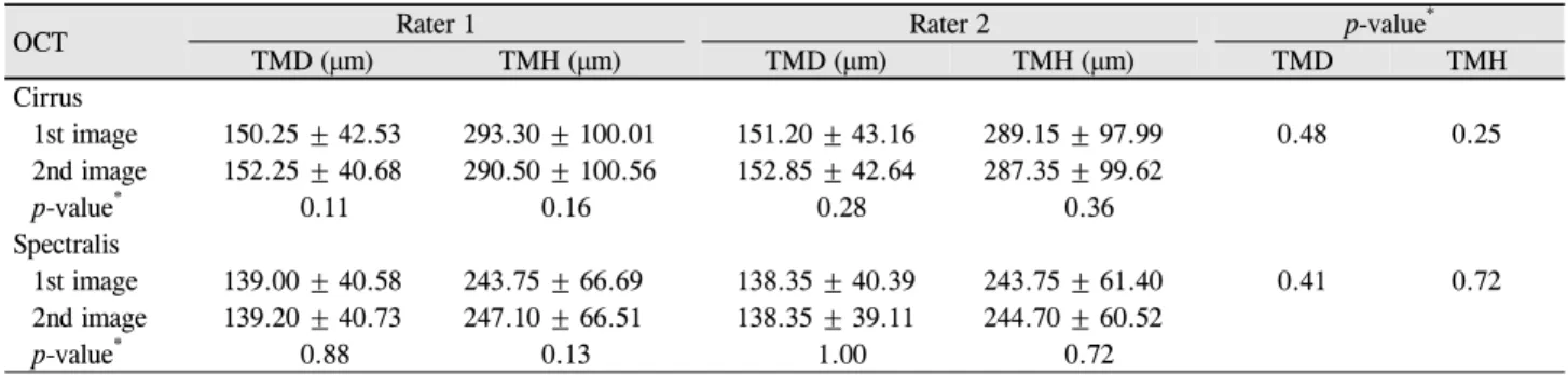
herence tomography (OCT) (A) and Spectralis OCT (B).OCT Rater 1 Rater 2 p-value*
TMD (μm) TMH (μm) TMD (μm) TMH (μm) TMD TMH
Cirrus
1st image 150.25 ± 42.53 293.30 ± 100.01 151.20 ± 43.16 289.15 ± 97.99 0.48 0.25
2nd image 152.25 ± 40.68 290.50 ± 100.56 152.85 ± 42.64 287.35 ± 99.62
p-value* 0.11 0.16 0.28 0.36
Spectralis
1st image 139.00 ± 40.58 243.75 ± 66.69 138.35 ± 40.39 243.75 ± 61.40 0.41 0.72
2nd image 139.20 ± 40.73 247.10 ± 66.51 138.35 ± 39.11 244.70 ± 60.52
p-value* 0.88 0.13 1.00 0.72
Values are presented as mean ± standard deviation.
OCT = optical coherence tomography; TMD = tear meniscus depth; TMH = tear meniscus height.
*Wilcoxon signed rank test.
Table 1. Measurement of tear meniscus depth and tear meniscus height using Cirrus OCT and Spectralis OCT
결 과
총 20명의 피험자(남자 7명, 여자 13명)의 평균 나이는 29.6세였으며 Cirrus OCT와 Spectralis OCT로 측정하여 평 가자 2명에 의해 분석된 눈물띠의 깊이 및 높이는 Table 1 에 기재하였다. Cirrus OCT로 측정한 평균 TMD는 검사자 1에서 151.25 ± 41.53 μm (90.00-219.50 μm), TMH는 291.90 ± 100.19 μm (167.00-529.50 μm)였으며, 검사자 2에서 TMD는 152.03 ± 42.77 μm (91.50-222.50 μm), TMH 는 288.25 ± 98.72 μm (150.00-498.00 μm)였다. Spectralis OCT로 측정한 평균 TMD는 검사자 1에서 139.10 ± 40.56 μm (82.50-223.50 μm), TMH는 245.43 ± 66.44 μm (136.00- 385.50 μm)였으며, 검사자 2에서 TMD는 138.35 ± 39.70 μm (80.00-217.00 μm), TMH는 244.23 ± 60.69 μm (131.50- 383.50 μm)였다. 검사자 내 평균은 모든 값에서 유의한 차 이가 없었으며, 검사자 간 평균 또한 유의한 차이가 없었다 (모두 p>0.05).
검사자 내 반복성은 TMD에서 Cirrus OCT의 CoV (%)는 2.48, 3.08, Spectralis OCT의 CoV (%)는 2.91, 2.16이었으며 TMH에서 Cirrus OCT의 CoV (%)는 2.06, 1.97, Spectralis OCT의 CoV (%)는 2.69, 3.38이었다(Table 2). ICC는 모두 0.991 이상의 결과를 보였다.
검사자 간 재현성을 분석하였을 때 TMD의 CoV (%)는 Cirrus OCT에서 2.24, Spectralis OCT에서 2.04로 측정되었 고, TMH의 CoV (%)는 Cirrus OCT에서 3.34로 Spectralis OCT에서 4.32으로 측정되었다(Table 3). ICC는 모두 0.986 이상의 결과를 보였다.
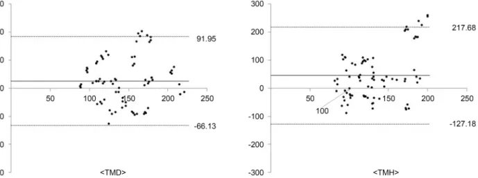
TMD에서 두 기기 간의 평균은 Cirrus OCT에서 Spectralis OCT보다 12.91 μm 더 크게 측정되었고(p=0.01), TMH는 Cirrus OCT에서 45.25 μm 더 크게 측정되었다(p<0.01).
Bland-altman 그림은 두 기기 간의 일치도가 좋지 못함을 보여주고 있으며, TMD의 95% limit of agreement (LoA)는 -66.13 μm에서 91.95 μm, TMH는 -127.18 μm에서 217.68 μm 로 측정되었다(Fig. 2).
Figure 2. Bland-Altman plots. Bland-Altman plots of the tear meniscus depth tear meniscus depth and tear meniscus height tear me-
niscus height difference between Cirrus optical coherence tomography (OCT) and Spectralis OCT. TMD = tear meniscus depth;TMH = tear meniscus height.
Characteristic Sw Rco CoV (%) ICC ICC 95% CI TMD
Cirrus
Rater 1 3.76 10.40 2.48 0.996 0.990-0.998 Rater 2 4.69 12.98 3.08 0.994 0.985-0.998 Spectralis
Rater 1 4.04 11.20 2.91 0.995 0.987-0.998 Rater 2 2.99 8.29 2.16 0.997 0.993-0.999 TMH
Cirrus
Rater 1 6.01 16.65 2.06 0.998 0.995-0.999 Rater 2 5.69 15.77 1.97 0.998 0.995-0.999 Spectralis
Rater 1 6.59 18.27 2.69 0.995 0.988-0.998 Rater 2 8.26 22.87 3.38 0.991 0.977-0.996 TMD = tear meniscus depth; Sw = within-subject standard devia- tion; Rco = repeatability coefficient; CoV = coefficient of varia- tion; ICC = intraclass correlation coefficient; CI = confidence in- terval; TMH = tear meniscus height.
Table 2. Intra-rater repeatability of tear meniscus depth and
tear meniscus height measurementsCharacteristic Sw Rco CoV(%) ICC (95% CI) TMD
Cirrus 3.40 9.43 2.24 0.997 (0.992-0.999) Spectralis 2.83 7.84 2.04 0.998 (0.994-0.999) TMH
Cirrus 9.69 26.85 3.34 0.995 (0.988-0.998) Spectralis 10.58 29.31 4.32 0.986 (0.965-0.994) TMD = tear meniscus depth; TMH = tear meniscus height; Sw = within-subject standard deviation; Rco = repeatability coefficient;
CoV = coefficient of variation; ICC = intraclass correlation co- efficient; CI = confidence interval.
Table 3. Inter-rater reproducibility of tear meniscus depth and
tear meniscus height measurements고 찰
건성안을 진단하는 방법에는 쉬르머검사, 눈물 오스몰 농도 측정, 눈물막파괴시간 등이 있으며 비침습적인 눈물 띠 측정을 이용하여 건성안을 진단하려는 시도 또한 계속 되고 있다. TMH는 눈물의 양을 직접적으로 측정할 수 있 는 방법으로, 2002년 Jones et al16이 제안한 빛간섭단층촬 영기를 이용하여 이를 직접 촬영한 이미지로 분석하는 방 법이 널리 이용되고 있다.17 아래 눈물띠는 눈물의 양을 비 교적 잘 반영한다고 알려져 있어 본 연구에서는 빛간섭단
층촬영기를 이용하여 아래 눈물띠의 높이와 너비를 측정하 였다. 또한 눈물띠를 정확하게 측정하고 질 좋은 이미지 획 득을 위해 빛간섭단층촬영기 촬영 전 눈을 깜빡이도록 하 였고, 여러 장의 이미지를 획득하여 분석하였다. 본 연구에 서는 2개의 이미지를 분석하거나 여러 이미지 분석값의 평 균을 사용하지 않고 1개의 이미지를 2회 분석하는 방법을 선택하였는데, 눈물막이 눈물의 생성 및 증발, 눈물막파괴 시간, 눈 깜빡임을 비롯하여 온도 및 습도 등 여러 요인에 영향을 받기 때문에 이에 의한 오차를 줄이고자 하였다.
Arriola-Villalobos et al13은 30안의 정상안을 대상으 로 아래 눈물띠 측정 시 Keratograph (OculusOptikgerate GmbH, Wetzlar, Germany)와 Spectralis OCT를 비교하였는 데, Keratograph의 아래 눈물띠 측정값은 중간 정도의 반복 성(CoV≥15.6%)이 관찰되었으나 Spectralis OCT는 매우 높 은 반복성(CoV≤13.3%)으로 관찰되었고, Spectralis OCT의 재현성(CoV≤0.12%)은 Keratograph (CoV≥0.19%)보다 나
은 것으로 나타났다. 또한 29안의 정상안을 대상으로 Swept source OCT인 Triton OCT (Topcon, Tokyo, Japan)와 Spectralis OCT를 비교하였을 때,18 Triton OCT (Topcon)의 아래 눈물띠 측정값 역시 중간 정도의 반복성(CoV≥16.9%) 로 측정되었고 Triton OCT의 재현성(CoV≤7.2%)은 Spectralis OCT의 재현성(CoV=10.8%)보다 나은 것으로 나타났다. 두 연구 모두에서 Spectralis와 다른 OCT 간의 일치도는 좋지 못한 것으로 나타났다.
Zhou et al19은 20안의 정상안을 대상으로 다른 종류의 스펙트랄 도메인 빛간섭단층촬영기인 RTVue OCT (Optovue Inc., Fremont, CA, USA)를 이용하여 아래 눈물띠를 측정 하였고 RTVue의 측정값은 높은 반복성(CoV≤14.4)을 보였 다. Chan et al12은 20안의 정상안을 대상으로 Cirrus OCT 와 타임 도메인 빛간섭단층촬영기인 Visante OCT (CarlZeiss Meditech, Dublin, CA, USA)를 비교하였는데 Cirrus OCT (The pooled standard deviation [Sp] = 15 μm)가 visante OCT (Sp = 29 μm)에 비해 반복성이 더 높다고 보고하였으며 두 기기의 일치도는 좋지 못한 것(LoA = -158 to 150 μm) 으로 나타났다.
본 연구에서 빛간섭단층촬영기로 아래 눈물띠를 측정하 였을 때 Cirrus OCT와 Spectralis OCT로 측정한 아래 눈물 띠의 깊이 및 높이는 검사자 내 및 검사자 간 유의한 차이 를 보이지 않았다. 검사자 내 반복성은 CoV≤3.37%로 매우 높게 나타났으며, 검사자 간 재현성 또한 CoV≤4.32%로 나 타났다. 검사자 내 신뢰도 및 검사자 간 신뢰도는 ICC≥0.991 이상으로 높게 나타났다. Cirurs OCT는 Spectralis OCT에 비해 TMD는 12.91 μm, TMH는 45.25 μm 더 크게 측정되는 결과를 보였고 두 기기 간 일치도는 TMD (LoA = -66.13 to 91.95 μm), TMH (LoA = -127.18 to 217.68 μm)로 모두 좋 지 못한 것으로 나타나 두 기계는 서로 대치하여 사용할 수 없을 것으로 생각된다. 이는 기존의 빛간섭단층촬영기를 이용한 연구 결과들과 비슷한 결과이다. 본 연구에 사용된 두 가지 스펙트랄 도메인 빛간섭단층촬영기의 축 해상도 (axial resolution)는 Cirrus OCT가 5 μm, Spectralis OCT가 7 μm로 달라 Spectralis OCT의 이미지 해상도가 Cirrus OCT에 비해 다소 낮아 이미지 분석 시 어려움이 있었으나 해상도에 따른 차이점은 없었다.
눈물의 양을 객관적으로 측정할 수 있는 빛간섭단층촬영 기를 이용한 아래 눈물띠 촬영은 현재 건성안 진단에 널리 쓰이는 쉬르머검사, 눈물막파괴시간검사, 안구표면질환지 수 등과 함께 추후 건성안 진단에 유용하게 사용될 수 있을 것으로 생각된다. 눈물띠 측정은 Cirrus OCT 및 Spectralis OCT 모두 반복성, 재현성이 높아 두 기기 모두 진단 도구 로 사용할 수 있으나 두 기기 간의 일치도는 좋지 못한 것
으로 나타나 서로 대치하여 사용할 수는 없을 것으로 생각 한다.
REFERENCES
1) Lemp MA, Baudouin C, Baum J, et al. The definition and classi- fication of dry eye disease: report of the Definition and Classification Subcommittee of the International Dry Eye WorkShop (2007).
Ocul Surf 2007;5:75-92.
2) Lin PY, Tsai SY, Cheng CY, et al. Prevalence of dry eye among an elderly Chinese population in Taiwan: the Shihpai Eye Study.
Ophthalmology 2003;110:1096-101.
3) McCarty CA, Bansal AK, Livingston PM, et al. The epidemiology of dry eye in Melbourne, Australia. Ophthalmology 1998;105:1114-9.
4) Kallarackal GU, Ansari EA, Amos N, et al. A comparative study to assess the clinical use of fluorescein meniscus time (FMT) with tear break up time (TBUT) and Schirmer’s tests (ST) in the diag- nosis of dry eyes. Eye 2002;16:594-600.
5) Senchyna M, Wax MB. Quantitative assessment of tear pro- duction: a review of methods and utility in dry eye drug discovery.
J Ocul Biol Dis Infor 2008;1:1-6.
6) Mishima S, Gasset A, Klyce SD Jr, Baum JL. Determination of tear volume and tear flow. Invest Ophthalmol 1966;5:264-76.
7) Yokoi N, Bron AJ, Tiffany JM, et al. Relationship between tear vol- ume and tear meniscus curvature. Arch Ophthalmol 2004;122:1265-9.
8) Mainstone JC, Bruce AS, Golding TR. Tear meniscus measure- ment in the diagnosis of dry eye. Curr Eye Res 1996;15:653-61.
9) Lim KJ, Lee JH. Measurement of the tear meniscus height using 0.25% fluorescein sodium. Korean J Ophthalmol 1991;5:34-6.
10) Ibrahim OM, Dogru M, Ward SK, et al. The efficacy, sensitivity, and specificity of strip meniscometry in conjunction with tear func- tion tests in the assessment of tear meniscus. Invest Ophthalmol Vis Sci 2011;52:2194-8.
11) Raj A, Dhasmana R, Nagpal RC. Anterior segment optical coher- ence tomography for tear meniscus evaluation and its correlation with other tear variables in healthy individuals. J Clin Diagn Res 2016;10:NC01-4.
12) Chan HH, Zhao Y, Tun TA, Tong L. Repeatability of tear meniscus evaluation using spectral-domain Cirrus(R) HD-OCT and time-do- main Visante(R) OCT. Cont Lens Anterior Eye 2015;38:368-72.
13) Arriola-Villalobos P, Fernández-Vigo JI, Díaz-Valle D, et al.
Assessment of lower tear meniscus measurements obtained with keratograph and agreement with fourier-domain optical-coherence tomography. Br J Ophthalmol 2015;99:1120-5.
14) Vaz S, Falkmer T, Passmore AE, et al. The case for using the re- peatability coefficient when calculating test-retest reliability. PLoS One 2013;8:e73990.
15) Kong KA. Statistical methods: reliability assessment and method comparison. Ewha Med J 2017;40:9-16.
16) Jones LW, Rahman S, Leech R, et al. Determination of inferior tear meniscus height and inferior tear meniscus volume using optical coherence tomography. Invest Ophthalmol Vis Sci 2004;45:144.
17) Bitton E, Keech A, Simpson T, Jones L. Variability of the analysis of the tear meniscus height by optical coherence tomography.
Optom Vis Sci 2007;84:903-8.
18) Arriola-Villalobos P, Fernández-Vigo JI, Díaz-Valle D, et al.
= 국문초록 =
서로 다른 두 가지 스펙트랄 도메인 빛간섭단층촬영기를 이용한 눈물띠의 반복성과 재현성 비교
목적: 서로 다른 스펙트랄 도메인 빛간섭단층촬영기(optical coherence tomography, OCT)로 측정한 아래 눈물띠 측정치의 반복성, 재현성, 일치도를 알아보고자 하였다.
대상과 방법: 총 20명 20안을 Cirrus OCT와 Spectralis OCT를 이용하여 아래 눈물띠를 촬영하고, 2명의 검사자가 눈물띠의 너비와 높이를 각각 측정하였다. 이를 토대로 검사자 내 반복성, 검사자 간 재현성, 장치 간 일치도를 분석하였다.
결과: Cirrus OCT와 Spectralis OCT를 이용하여 측정한 눈물띠의 너비는 각각 검사자 1에서 151.25 ± 41.53 μm, 139.10 ± 40.56 μm, 검사자 2에서 152.03 ± 42.77 μm, 138.35 ± 39.70 μm였다. 높이는 검사자 1에서 291.90 ± 100.19 μm, 245.43 ± 66.44 μm, 검사자 2에서 288.25 ± 98.72 μm, 244.23 ± 60.69 μm로 측정되었다. 눈물띠 너비와 높이의 측정값은 검사자 내 및 검사자 간 유의한 차이 가 관찰되지 않았다(모두 p>0.05). 검사자 내 반복성은 두 기기 모두 높은 신뢰도를 보였고(급내상관계수 ≥0.991) 검사자 간 변이계 수는 2.04%에서 4.32%였다. 두 장치 간 95% 일치도 범위는 눈물띠의 너비는 -66.13 to 91.95 μm, 높이는 -127.18 to 217.68 μm였다.
결론: Cirrus OCT와 Spectralis OCT는 아래 눈물띠 측정에 유용하게 사용될 수 있다. 다만 두 기기 간 일치도는 좋지 못하므로 서로 대치하여 사용할 수는 없다.
<대한안과학회지 2019;60(10):929-934>
김진하 / Jin Ha Kim
이화여자대학교 의과대학 안과학교실 Department of Ophthalmology, Ewha Womans University College of Medicine Lower tear meniscus measurements using a new anterior segment
swept-source optical coherence tomography and agreement with fourier-domain optical coherence tomography. Cornea 2016;36:
183-8.
19) Zhou S, Li Y, Lu AT, et al. Reproducibility of tear meniscus meas- urement by Fourier-domain optical coherence tomography: a pilot study. Ophthalmic Surg Lasers Imaging 2009;40:442-7.