A Simple Method for Reconstruction of the Temporalis Muscle Using Contourable Strut Plate after Pterional
Craniotomy: Introduction of the Surgical Techniques and Analysis of Its Efficacy
Jin-Hack Park, Yoon-Soo Lee, Sang-Jun Suh, Jeong-Ho Lee, Kee-Young Ryu, Dong-Gee Kang Department of Neurosurgery, Daegu Fatima Hospital, Daegu, Korea
Objective : Pterional craniotomy (PC) using myocutaneous (MC) flap is a simple and efficient technique; however, due to subsequent inferior displace- ment (ID) of the temporalis muscle, it can cause postoperative deformities of the muscle such as depression along the inferior margin of the tem- poral line of the frontal bone (DTL) and muscular protrusion at the in- ferior portion of the temporal fossa (PITF). Herein, we introduce a simple method for reconstruction of the temporalis muscle using a contourable strut plate (CSP) and evaluate its efficacy.
Materials and Methods : Patients at follow-ups between January 2014 and October 2014 after PCs were enrolled in this study. Their postoperative deformities of the temporalis muscle including ID, DTL, and PITF were evaluated. These PC cases using MC flap were classified according to two groups; one with conventional technique without CSP (MC Only) and an- other with reconstruction of the temporalis muscle using CSP (MC + CSP).
Statistical analyses were performed for comparison between the two groups.
Results : Lower incidences of ID of the muscle (p < 0.001), DTL (p <
0.001), and PITF (p = 0.001) were observed in the MC + CSP than in the MC Only group. The incidence of acceptable outcome was markedly higher in the MC + CSP group (p < 0.001). ID was regarded as a causa- tive factor for DTL and PITF (p < 0.001 in both).
Conclusion : Reconstruction of the temporalis muscle using CSP after MC flap is a simple and efficient technique, which provides an outstanding outcome in terms of anatomical restoration of the temporalis muscle.
J Cerebrovasc Endovasc Neurosurg.
2015 June;17(2):93-100 Received : 24 February 2015 Revised : 24 February 2015 Accepted : 3 June 2015 Correspondence to Yoon-Soo Lee
Department of Neurosurgery, Daegu Fatima Hospital, 99 Ayang-ro, Dong-gu, Daegu 701-724, Korea
Tel : 82-53-940-7330, 7339 Fax : 82-53-954-7417
E-mail : [email protected] ORCID : http://orcid.org/0000-0003-2506-0168
This is an Open Access article distributed under the terms of the Creative Commons Attribution Non- Commercial License (http://creativecommons.org/li- censes/by-nc/3.0) which permits unrestricted non- commercial use, distribution, and reproduction in any medium, provided the original work is properly cited.
Keywords Contourable strut plate, Deformities, Myocutaneous flap, Pterional, Temporalis, Temporal line
INTRODUCTION
Frontotemporal craniotomy, also known as "pterional craniotomy" (PC), provides an optimal microscopic exposure and a wide open working space for manipu- lation of intracranial structures, and it has been wide-
ly used in the field of neurosurgery for treatment of lesions in the anterior and posterior circulations.9) When Yasargil17) first described standard techniques and procedures for PC in his publication in 1984, sub- galeal dissection was used for separation and mobi- lization of the temporalis muscle. Because subgaleal
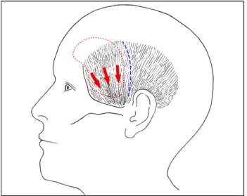
Fig. 1. The illustration depicts the course of fibers of the tempo- ralis muscle and the direction of displacement, or sliding, (red arrows) of unattached temporalis muscle after pterional craniot- omy (PC). The size and shape of PC (red dotted line) and the incision line on the temporalis muscle (blue dotted line) are also shown.
dissection of the temporalis muscle bears significant risk of injury to the frontal branches of the facial nerve, various surgical techniques have been adopted such as interfascial and subfascial dissection. However, interfascial dissection is somewhat complex and time-consuming, and, because the facial nerve some- times courses into the interfascial space, it still cannot eliminate the risk of facial nerve injury. Subfascial dis- section is also time-consuming, and may result in in- jury to muscle fibers and intramuscular bleeding.
These two techniques require transection of the tem- poralis muscle to leave a cuff for closure, which caus- es functional and cosmetic problems by muscle fib- rosis and atrophy.1)5)6)11)15)18) To minimize the risk of facial nerve injury and temporalis muscle atrophy, an- other technique of dissecting the temporalis muscle and skin as one flap, known as myocutaneous (MC) flap, was introduced, and this technique is now com- monly used.6) Although it is a simple and quick meth- od, inadequate anatomical restoration of the tempora- lis muscle can cause cosmetic problems such as de- pression along the inferior margin of the temporal line of the frontal bone and muscular protrusion at the inferior portion of the temporal fossa due to sub-
sequent inferior displacement, or sliding, of the tem- poralis muscle, and is eventually exacerbated by un- expected atrophy and fibrosis (Fig. 1, 2).
According to previous reports, the majority of pa- tients with even minor postoperative deformities ex- perience cosmetic complexes and functional handicaps.
Therefore, neurosurgeons should be aware of not only the clinical outcomes but also the cosmetic and func- tional outcomes after intracranial procedures.7)
This study was conducted in order to introduce a simple and quick surgical method for reconstruction of the temporalis muscle using a contourable strut plate (CSP) and to evaluate the efficacy of this techni- que by analyzing the clinical results in patients who underwent PC using MC flap in comparison with those treated with previous techniques without CSP.
MATERIALS AND METHODS
Data collection and patient enrollment
Patients who visited the outpatient department (OPD) at our institute between January 2014 and October 2014 for follow-ups after PCs were candi- dates for this study. All of them underwent PCs per- formed by a single neurosurgeon using MC flap with or without CSP for treatment of cerebral aneurysms.
Patients who underwent additional cosmetic proce- dures such as injections or fat grafts were excluded.
Outcomes after reconstruction of the temporalis mus- cle were evaluated at OPD by gross inspection and al- so on the axial images of three-dimensional computed tomography (CT) scan at least more than three months after surgery, and the results were assigned to one or more of the following categories: (1) inferior displacement (ID), or sliding, of the temporalis mus- cle, (2) depression along the inferior margin of the temporal line of the frontal bone (DTL), (3) muscular protrusion at the inferior portion of the temporal fos- sa (PITF), or (4) none. The patient's profiles, sides of lesions, diagnoses, presence of deformities after re- construction of the temporalis muscle, and complica- tions such as instrument failures or postoperative in-
A B C
D E F
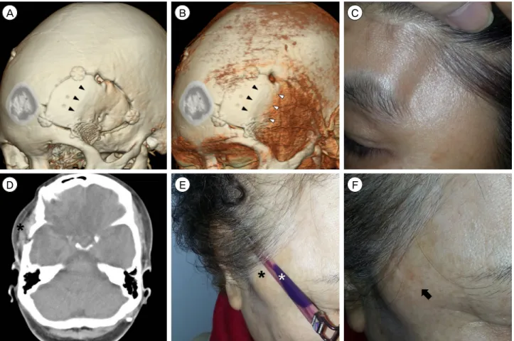
Fig. 2. Three-dimensional computed tomography (CT) scans (A, B) of a patient demonstrate the temporal line of the frontal bone (black arrowheads) and the displaced attachment site of the temporalis muscle (white arrowheads). A photograph (C) of the same pa- tient shows a marked depression along the inferior margin of the temporal line of the frontal bone. The axial CT image (D) of a pa- tient demonstrates a muscular protrusion at the inferior portion of the temporal fossa (black asterisk). Photographs (E, F) of the same patient show that this temporal protrusion (black asterisk) may cause discomfort when wearing glasses (white asterisk). Note the imprint on the skin (black arrow) after taking the glasses off.
Fig. 3. The photograph shows the structure of a contourable strut plate (Synthes GmbH, Oberdorf, Switzerland). It is malleable and easily bent into various shapes which fit on the underlying bony contour.
fections were recorded in detail. These prospectively maintained databases including medical records and radiological images were reviewed retrospectively.
These PC cases using MC flap were classified ac- cording to two groups; one with conventional techni- que without CSP (MC Only) and another with re- construction of the temporalis muscle using CSP (MC + CSP). In both groups, keyhole site defect was re- constructed using "temporal mesh floating techniques"
as described by Lee et al.10) For the MC + CSP group, one titanium CSP (Synthes GmbH, Oberdorf, Switzerland) of 35 × 12.5 mm in size and 0.4 mm in thickness with eight screw holes was used. It is malle- able and easily bent into various shapes which fit on the underlying bony contour (Fig. 3).
Technique for reconstruction of the temporalis muscle using CSP
After fixation of the craniotomy bone using instru- ments such as mini-plates, titanium clamps, or burr
A B C
D E
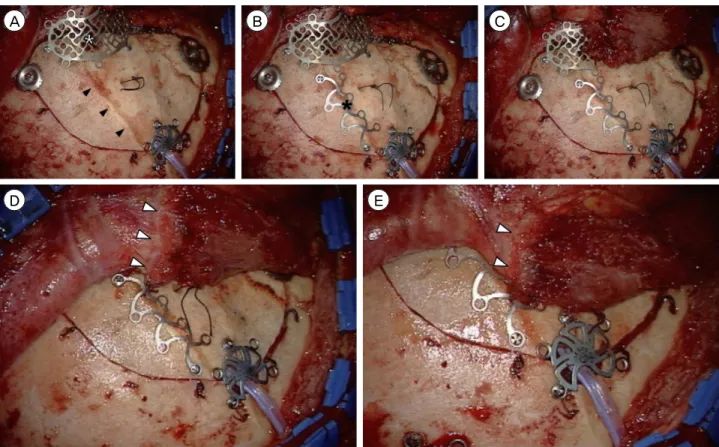
Fig. 4. Intraoperative photographs demonstrate the technique for reconstruction of the temporalis muscle using a contourable strut plate (CSP). (A) After fixation of the craniotomy bone using instruments such as mini-plates, titanium clamps, or burr hole covers, key- hole defect is repaired by temporal mesh floating technique (white asterisk). The temporal line of the frontal bone is indicated by black arrowheads. (B) A CSP (black asterisk) is slightly bent into the contour of the temporal line of the frontal bone and fixed to the tem- poral line using two low profile self-tapping screws. (C) The antero-inferior portion of the temporalis muscle is sutured over the tempo- ral mesh. (D, E) The edge of the temporalis fascia and muscle (white arrowheads) is sutured and fixed to the CSP, which is the site of original attachment.
hole covers, keyhole defect was repaired by temporal mesh floating technique.10) CSP was slightly bent into the contour of the temporal line of the frontal bone, and fixed to the temporal line using two low profile self-tapping screws. The antero-inferior portion of the temporalis muscle was sutured over the temporal mesh. The edge of the temporalis fascia and muscle was sutured again and fixed to the CSP, which is the site of original attachment (Fig. 4).
Statistical analysis
Statistical analyses were performed using Statistical Package for the Social Sciences (SPSS) version 20.0 (SPSS Inc., Chicago, IL, USA). For comparison of MC Only and MC + CSP groups, Pearson's chi-square test and Fisher's exact test were performed for the catego-
rical variables, and Student's t-test for the continuous variables. Simple regression analyses were performed to determine the cause-and-effect relationships be- tween the variables. The results were considered sig- nificant for probability value (p value) less than 0.05.
RESULTS
Overall 106 PC cases in 95 patients with 134 aneur- ysms were finally enrolled in this study. Eleven pa- tients underwent PCs on both sides in the different sessions for treatment of multiple bilateral aneurysms, and each side was separately evaluated. Eighty cases (18 males and 62 females, mean age of 55.0 ± 9.2 years) were enrolled in the MC Only group, and 26 (10 males and 16 females, mean age of 57.7 ± 9.6
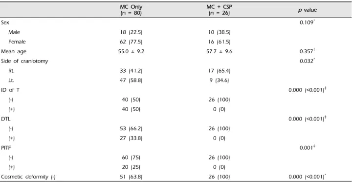
MC Only
(n = 80) MC + CSP
(n = 26) p value
Sex 0.109*
Male 18 (22.5) 10 (38.5)
Female 62 (77.5) 16 (61.5)
Mean age 55.0 ± 9.2 57.7 ± 9.6 0.357†
Side of craniotomy 0.032*
Rt. 33 (41.2) 17 (65.4)
Lt. 47 (58.8) 9 (34.6)
ID of T 0.000 (<0.001)‡
(-) 40 (50) 26 (100)
(+) 40 (50) 0 (0)
DTL 0.000 (<0.001)‡
(-) 53 (66.2) 26 (100)
(+) 27 (33.8) 0 (0)
PITF 0.001‡
(-) 60 (75) 26 (100)
(+) 20 (25) 0 (0)
Cosmetic deformity (-) 51 (63.8) 26 (100) 0.000 (<0.001)*
Values are presented as number (%) or mean ± SD.
*Chi-square test, †t-test, ‡Fisher’s exact test.
MC = group with myocutaneous flap only; MC + CSP = group with myocutaneous flap plus reconstruction of the temporalis muscle using a contourable strut plate; ID = inferior displacement; T = the temporalis muscle; DTL = depression along the inferior margin of the temporal line of the frontal bone; PITF = muscular protrusion at the inferior portion of the temporal fossa; Cosmetic Deformity (-) = absence of definite cosmetic deformities of the temporalis muscle
Table 1. Subgroup analysis between MC Only and MC + CSP groups
years) in the MC + CSP group. There were no stat- istical differences in sex and age between the two groups (p = 0.109 by chi-square test and p = 0.357 by t-test respectively). However, there was statistical dif- ference in the side of craniotomy (p = 0.032 by chi-square test), left dominant in the MC Only group and right dominant in the MC + CSP group, although this difference would not have actually affected the surgical outcomes (Table 1).
ID of the temporalis muscle was detected on the three-dimensional CT scans in 40 cases (37.7%); 40 (50%) in the MC Only group and none (0%) in the MC + CSP group, and the incidences were statistically different between the two groups (p < 0.001 by Fisher's exact test) (Fig. 2A, B). DTL was observed in 27 cases (25.5%); 27 (33.8%) in the MC Only group and none (0%) in the MC + CSP group, and the in- cidences were statistically different between the two groups (p < 0.001 by Fisher's exact test) (Fig. 2C).
PITF was observed in 20 cases (18.9%); 20 (25%) in the MC Only group and none (0%) in the MC + CSP group, and the incidences were also statistically dif- ferent (p = 0.001 by Fisher's exact test) (Fig. 2D-F).
Fifty-one cases (63.8%) in the MC Only group and all 26 cases (100%) in the MC + CSP group did not have DTL and PITF regardless of presence of ID of the temporalis muscle on the CT scans, indicating absence of definite cosmetic deformities of the temporalis muscle (Fig. 5). The incidences of absence of the de- formities were also statistically different (p < 0.001 by chi-square test) (Table 1).
Of 40 cases with ID of the temporalis muscle, 27 cases (67.5%) had DTL, 19 (47.5%) had PITF, 18 (45%) had both DTL and PITF, and 12 (30%) had neither DTL nor PITF. Only in one case, PITF was observed even without definite ID of the temporalis muscle.
The results of simple regression analyses between ID and DTL, and ID and PITF showed statistically sig-
Independent variable Dependent variable p value
ID of T DTL 0.000 (<0.001)*
PITF 0.000 (<0.001)*
*Simple regression analysis.
ID = inferior displacement; T = the temporalis muscle; DTL = depression along the inferior margin of the temporal line of the frontal bone;
PITF = muscular protrusion at the inferior portion of the temporal fossa
Table 2. Simple regression analysis for the cause-and-effect relationships between the variables
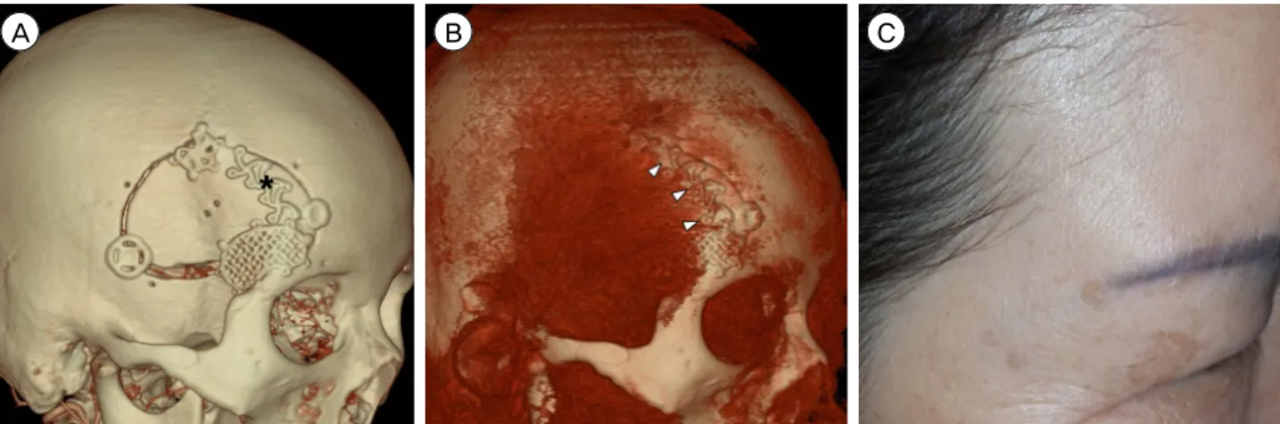
A B C
Fig. 5. Three-dimensional computed tomography (CT) scans (A, B) of a patient after reconstruction of the temporalis muscle using a contourable strut plate (CSP) (black asterisk) show anatomical restoration of the temporalis muscle at the temporal line (white arrow- heads). A photograph (C) of the same patient shows an outstanding outcome without any deformities of the temporalis muscle.
nificant cause-and-effect relationships (p < 0.001 in both), indicating that ID of the temporalis muscle could be a cause of DTL and PITF (Table 2).
There was no occurrence of postoperative wound infection in either group. There were no instrument failures such as screw loosening, displacement of CSP, implant protrusion, or scalp perforation in the MC + CSP group.
DISCUSSION
Reconstruction of the temporalis muscle after PC has been a challenging task for neurosurgeons be- cause there have been no clear solutions completely preventing postoperative deformities of the temporalis muscle. The following causes of temporalis muscle deformities have been suggested: (1) denervation by nerve injury, (2) muscle ischemia by prolonged re- traction or interruption of blood supply, (3) direct in-
jury to temporalis muscle fibers, and (4) inappropriate repair of the temporalis muscle to the site of original attachment.8)12)13)19) Therefore, many surgical mod- ifications of the temporalis muscle reconstruction have been introduced, and the two key points of these modifications to minimize postoperative deformities are (1) preservation of anatomical structures such as the nerves, arteries, and muscle fibers, and (2) restora- tion of the muscle alignment as similar to the pre- operative condition as possible.
The temporalis muscle is innervated by the deep temporal nerves, terminal branches of the mandibular division of the trigeminal nerve, and they course in the deep portion at the medial aspect of the muscle superficial to the periosteum. The blood supply to the temporalis muscle is by the deep temporal arteries and the middle temporal artery. The deep temporal arteries, branches of the maxillary artery, lie super- ficial to the periosteum, and supply blood to the ante-
rior and middle portion of the muscle from the me- dial side. The middle temporal artery, a branch of the superficial temporal artery, passes through the super- ficial temporal fascia, and supplies blood to the mus- cle from the posterolateral side.1)8)15)20) Injury of these complex structures of nerves and arteries along with muscle fibers can occur during muscle dissection, cau- terization for hemostasis, and extensive or prolonged muscle retraction. The muscle damaged by denerva- tion, loss of blood supply, or direct injury gradually necrotizes, undergoes fibrosis and atrophy, and even- tually changes to fat tissues.2)6)8)13)
Several previous articles reported that making an MC flap has an advantage in the preservation of the frontal branches of the facial nerve, although with a disadvantage in limited exposure over interfascial or subfascial dissection.14) In the era of minimally in- vasive microsurgery, with the development of instru- ments and techniques, a slight limitation of exposure is no longer a problem. Moreover, with just some modifications in the direction of muscle retraction, the operation field can be sufficiently exposed.16) The ad- vantages of making an MC flap over interfascial or subfascial dissection exist also in the extent of dissection. To make an MC flap, dissection is required only on the medial side of the temporalis muscle. For interfascial or subfascial dissection, extensive dis- section on the medial and lateral side of the muscle and transection of the fibers to leave a cuff are re- quired, and the dissected pedicle of muscle is con- tinuously retracted and distorted throughout the surgery. These procedures increase the risk of injury to the muscle fibers, nerves, and arteries. For these reasons, temporalis muscle atrophy is more frequent in interfascial dissection than MC flap.6)
However, dissection of an MC flap may cause prob- lems from subsequent inferior displacement, or slid- ing, of the temporalis muscle after detachment of the muscle from the temporal line of the frontal bone (Fig. 2). Unattached temporalis muscle tends to slide following the direction of the muscle fibers during the activity of mastication (Fig. 1). According to the cur-
rent study, 50% of patients who underwent MC flap without adequate reconstruction developed ID of the temporalis muscle, 33.8% showed DTL, and 25%
showed PITF. Patients with these temporalis deform- ities experience cosmetic complexes and functional handicaps. The PITF may cause discomfort when wearing glasses (Fig. 2D-F). Acceptable cosmetic out- comes were achieved in only 63.8% of patients with- out adequate reconstruction, while the outcomes were dramatically improved by the techniques described in this study (Fig. 5). Therefore, restoration of the muscle alignment is an essential step for MC flap. Multiple methods were introduced for anatomical restoration of the temporalis muscle. Spetzler and Lee16) introduced a surgical modification that leaves a muscle cuff along the superior temporal line at dissection, and secures the muscle cuff with the dissected temporalis muscle at reconstruction. However, this method again re- quires transection of the muscle leading to similar problems in interfascial or subfascial dissection. Zager et al.19) suggested a technique to suture the temporalis muscle around multiple partially advanced micro- screws at the temporal line. Other reports also de- scribed techniques to suture the muscle to multiple small holes at the temporal line.3)4)8) However, these surgical modifications were attempted after subfascial dissection. In addition, multiple partially advanced screws may cause protrusions at the forehead. Making multiple superficial holes and passing threads through them seems too complex and time-consuming.
In this study, reconstruction of the temporalis mus- cle using CSP after MC flap led to an outstanding outcome. There were no failures of operation due to limited exposure after MC flap. In all cases, anatomi- cal restoration of the temporalis muscle at the tempo- ral line was achieved. Postoperative muscle atrophy and fibrosis were negligible by minimizing the extent of dissection around the temporalis muscle. No cases of DTL or PITF were observed after this technique by preventing ID of the temporalis muscle which was proven to be the causative factor in this study. In ad- dition, this technique is simple and fast. Fixation of
CSP using only two low profile self-tapping screws and suturing over the CSP take less than 5 minutes and are feasible even for beginners.
CONCLUSION
Restoration of the alignment of the temporalis mus- cle is an essential step after PC. Reconstruction using CSP after MC flap provides an outstanding outcome.
This technique is simple and fast, and it provides ana- tomical restoration of the temporalis muscle at the temporal line and prevents postoperative deformities of the temporalis muscle. Neurosurgeons should be aware of such undesirable cosmetic and functional outcomes after PC, and these deformities must be pre- vented by adequate reconstruction.
Disclosure
This work is not financially supported. The authors have no personal financial or institutional interest in any of the drugs, materials, or devices described in this article.
REFERENCES
1. Ammirati M, Spallone A, Ma J, Cheatham M, Becker D.
An anatomicosurgical study of the temporal branch of the facial nerve. Neurosurgery. 1993 Dec;33(6):1038-43;
discussion 1044.
2. Blaisdell FW, Steele M, Allen RE. Management of acute lower extremity arterial ischemia due to embolism and thrombosis. Surgery. 1978 Dec;84(6):822-34.
3. Bowles AP Jr. Reconstruction of the temporalis muscle for pterional and cranio-orbital craniotomies. Surg Neurol.
1999 Nov;52(5):524-9.
4. Brunori A, DiBenedetto A, Chiappetta F. Transosseous reconstruction of temporalis muscle for pterional craniot- omy: technical note. Minim Invasive Neurosurg. 1997 Mar;40(1):22-3.
5. Coscarella E, Vishteh AG, Spetzler RF, Seoane E, Zabramski JM. Subfascial and submuscular methods of temporal muscle dissection and their relationship to the frontal branch of the facial nerve. Technical note. J Neurosurg. 2000 May;92(5):877-80.
6. de Andrade Júnior FC, de Andrade FC, de Araujo Filho
CM, Carcagnolo Filho J. Dysfunction of the temporalis muscle after pterional craniotomy for intracranial aneurysms.
Comparative, prospective and randomized study of one flap versus two flaps dieresis. Arq Neuropsiquiatr. 1998 Jan;56(2):200-5.
7. Im TS, Lee YS, Suh SJ, Lee JH, Ryu KY, Kang DG. The efficacy of titanium burr hole cover for reconstruction of skull defect after burr hole trephination of chronic subdural hematoma. Korean J Neurotrauma. 2014 Oct;10(2):76-81.
8. Kadri PA, Al-Mefty O. The anatomical basis for surgical preservation of temporal muscle. J Neurosurg. 2004 Mar;100(3):517-22.
9. Kang HJ, Lee YS, Suh SJ, Lee JH, Ryu KY, Kang DG.
Comparative analysis of the mini-pterional and supraorbital keyhole craniotomies for unruptured aneurysms with numeric measurements of their geometric configurations.
J Cerebrovasc Endovasc Neurosurg. 2013 Mar;15(1):5-12.
10. Lee MS, Lee YS, Lee JH, Ryu KY, Kang DG. The effi- cacy of temporal mesh plate floating techinique for key- hole site depression after frontotemporal craniotomy. J Korean Neurotraumatol Soc. 2011 Oct;7(2):78-82.
11. Matsumoto K, Akagi K, Abekura M, Ohkawa M, Tasaki O, Tomishima T. Cosmetic and functional reconstruction achieved using a split myofascial bone flap for pterional craniotomy. Technical note. J Neurosurg. 2001 Apr;94(4):
667-70.
12. Miyazawa T. Less invasive reconstruction of the temporalis muscle for pterional craniotomy: modified procedures.
Surg Neurol. 1998 Oct;50(4):347-51.
13. Oikawa S, Mizuno M, Muraoka S, Kobayashi S.
Retrograde dissection of the temporalis muscle prevent- ing muscle atrophy for pterional craniotomy. Technical note. J Neurosurg. 1996 Feb;84(2):297-9.
14. Rhoton AL Jr. Anatomy of saccular aneurysms. Surg Neurol. 1980 Jul;14(1):59-66.
15. Salas E, Ziyal IM, Bejjani GK, Sekhar LN. Anatomy of the frontotemporal branch of the facial nerve and in- dications for interfascial dissection. Neurosurgery. 1998 Sep;43(3):563-8.
16. Spetzler RF, Lee KS. Reconstruction of the temporalis muscle for the pterional craniotomy. Technical note. J Neurosurg. 1990 Oct;73(4):636-7.
17. Yaşargil MG. Microneurosurgery. Vol.Ⅰ. New York, NY:
Thieme, 1984, p. 217-20.
18. Yaşargil MG, Reichman MV, Kubik S. Preservation of the frontotemporal branch of the facial nerve using the interfascial temporalis flap for pterional craniotomy.
Technical article. J Neurosurg. 1987 Sep;67(3):463-6.
19. Zager EL, DelVecchio DA, Bartlett SP. Temporal muscle microfixation in pterional craniotomies. Technical note. J Neurosurg. 1993 Dec;79(6):946-7.
20. Ziccardi VB, Mu L, Schneider RE, Sanders I. Innervation pattern of the temporalis muscle. J Craniofac Surg. 1998 Mar;9(2):185-9.