Introduction
Environmental exposure to heavy metals is a public health hazard that has been linked to cognitive and neurological deficits [1]. Exposure to heavy metals can occur through contaminated food, water, air, or in industrial work settings
[2]. To date, the majority of research investigating the delete- rious effects of environmental toxins on neurocognition has focused on heavy metals such as lead and mercury [2]. Of increasing interest in recent years are the health risks and/or benefits of exposure to the transition metal known as vana- dium.
Vanadium (NaVO3) as a potentially toxic environmental pollutant induces oxidative damage in the central nervous system [3, 4]. Vanadium deposition in the brain might be associated with the pathogenesis of certain neurological dis- orders, and this, after long-term exposure can result in more severe pathology. Most research on vanadium neurotoxic-
Corresponding author:
Adedamola Adediran Fafure
Department of Anatomy, College of Medicine and Health Sciences, Afe Babalola University, Ado Ekiti 360201, Nigeria
E-mail: adedamolaf@abuad.edu.ng
Ficus exasperata Vahl leaves extract
attenuates motor deficit in vanadium-induced parkinsonism mice
Adeshina O Adekeye
1,2, Gold J Irawo
1, Adedamola Adediran Fafure
11Department of Anatomy, College of Medicine and Health Sciences, Afe Babalola University, Ado Ekiti, Nigeria, 2Department of Anatomy, Faculty of Basic Medical Sciences, College of Medicine, Olabisi Onabanjo University, Ago Iwoye, Nigeria
Abstract: Medicinal herbs have played significant roles in the treatment of various diseases in humans and animals.
Sodium metavanadate is a potentially toxic environmental pollutant that induces oxidative damage, neurological disorder, Parkinsonism and Parkinson-like disease upon excessive exposure. This study is designed to investigate the impact of saponin fraction of Ficus exasperata Vahl leaf extract (at 50 and 100 mg/kg body weight for 14 days at different animal groupings) on vanadium treated mice. Animals were randomly grouped into five groups. Control (normal saline), NaVO3 (10 mg/kg for 7 days), withdrawal group, NaVO3+Vahl (low dose) and NaVO3+Vahl (high dose). The animals were screened for motor coordination using rotarod and PBTs and a post mortem study was conducted by quantitatively assessing the markers of oxidative stress such as lipid peroxidation, catalase, glutathione activities, and also through immunohistochemistry via glia fibrillary acidic protein, tyrosine hydroxylase and dopamine transporter to study the integrity of astrocytes and dopaminergic neurons of the substantia nigra (SNc). Vanadium-exposed group showed a decreased motor activity on the neurobehavioural tests as well as an increase in markers of oxidative stress. Saponin fraction of F. exasperata Vahl leaves extract produced a statistically significant motor improvement which may be due to high antioxidant activities of saponin, thereby providing an ameliorative effect on the histoarchitecture of the SNc. It can be inferred that the saponin fraction of F. exasperata Vahl leaves extract to possesses ameliorative, motor-enhancing and neurorestorative benefit on motor deficit in vanadium-induced parkinsonism mice.
Key words: Saponins, Immunohistochemistry, Parkinsonian disorders, Vanadium, Neurodegerative disease
Received September 26, 2019; 1st Revised December 15, 2019; 2nd Revised December 21, 2019; Accepted December 24, 2019
ity has been conducted after acute exposure; however, some populations are exposed to this Vanadium for a lifetime [5].
Exposure to neurotoxic metals such as vanadium occurs through various sources including heavy metals mining [6], combustion products of vanadium bearing fuel oils [7], forest fires and volcanic emissions. Large quantities of vanadium compounds have also been reported to be released into the environment mainly through the burning of fossil fuels hav- ing vanadium contaminated crude as seen in oil-producing communities such as Venezuela, the Arabian Gulf, the Gulf of Mexico and the Nigerian Niger Delta [8, 9].
Medicinal herbs have been known to be effective follow- ing their roles in the treatment of various diseases in humans and animals [10]. Sandpaper leaf, also known as Ficus ex- asperate Vahl leaf (FEVL) is a type of medicinal plant with various medicinal properties [11]. It is widespread in tropical Africa from Mozambique, Zambia, and northern Angola to Senegal, Ethiopia and also in the southern part of the Ara- bian Peninsula and India [12].
The toxicology analysis and phytochemical constituents of Ficus exasperata revealed the presence of flavonoids, tan- nins, saponin, alkaloids, and glycosides [4, 13]. Fafure et al.
[4] demonstrated that F. exasperata Vahl improved motor activities in mice exposed to manganese chloride. Available reports in western Nigeria indicate that leaves of F. exas- perata Vahl exhibit antiulcer, hypotensive, hypoglycemic, hypolipidemic, anti-inflammatory, anxiolytic, oxytocin in- hibiting, anticonvulsant, antinociceptive, antimicrobial, an- ticandidal, insecticidal and pesticidal activities [14, 15], and the decoctions and infusions of F. exasperata leaf have been used traditionally in the management and treatment of dif- ferent human diseases including diabetes mellitus, hyperten- sion, and certain cardiovascular dysfunctions [16]. Although sandpaper leaf has been shown to possess many properties, there is a dearth of information on the effect of saponin fraction on the anti-inflammatory properties of sandpaper leaf. Hence, this study sought to investigate in mice whether saponin fraction of F.exasparata Vahl leaves could counter- act the noxious effects induced by a subchronic treatment of vanadium which can serve as a readily accessible and inex- pensive alternative for treating parkinsonism/Parkinson-like diseases. The animals were tested for motor coordination using the rotarod test (RT) and parallel bar test (PBT), and the integrity of dopaminergic (DA) neurons of the substantia nigra (SNc) was evaluated with histological/immunohisto- chemical approaches.
Materials and Methods
Animal procurement and careForty male adult Balb/c mice weighing between 25-35 g were used for this study. The mice were collected from the animal handling facility of the department of anatomy from the University of Delta State, Nigeria. The mice were allowed to acclimatized for two weeks and had free access to rodent chow (purchased from the ABUAD feed mill, Ado Ekiti, Nigeria) and water; they were also exposed to 12 hours by 12 hours dark and light period.
Collection and identification of plant
F. exasperata Vahl leaves were collected during its blos- soming stage in February from farmland in Ikole Ekiti South-Western Nigeria. The plant was identified at the Uni- versity of Lagos Herbarium as F. exasperata Vahl leaves with herbarium number 7786.
Preparation of plant materials
The F. exasperata Vahl leaves were dried for five days and then pulverized into a fine powder using an electric blender.
Saponin was extracted from the fine powder (250 g) in the Abuad chemistry Laboratory. A 250 g of the fine powder was weighed into a big beaker while 100 ml of 20% ethanol was added and mixed properly; it was placed in a water bath at 55ºC for 4 hours. It was continuously stirred for 15 minutes each for the four hours until it became concentrated. Di ethyl ether was added to it after the 4 hours (not in the water bath) and stirred vigorously to get pure saponin, after which N-butanol was also added and stirred. A total of 5% sodium chloride was also put and allowed to decant; it was then fil- tered to get the saponin extract, which was later left in the water bath at 60ºC until it was properly dried.
Research ethical approval
Ethical clearance for this study was obtained from the Health Research Ethics Committee (HREC) of the Afe Babalola University, Ado Ekiti, Ekiti State, Nigeria (AB/
EC/19/02/001).
Experimental design and administration
The mice were randomly divided into four groups (n=10);
Group A (control group) received normal saline for 21 days, Group B with subgroup B1 (Vanadium) and B2 (Withdrawal).
Group B1 received normal saline for 14 days and NaVO3 for
seven days. Group B2 received normal saline for seven days, NaVO3 intraperitoneally and withdrew for seven days. Group C received 10 mg/kg of NaVO3 intraperitoneally for 7 days followed by 50 mg/kg of Saponin fraction of F. exasperata Vahl leave extract orally for 14 days; Group D received 10 mg/kg of NaVO3 intraperitoneally for seven days followed by 100 mg/kg of Saponin fraction of F. exasperata Vahl leave extract orally for 14 days.
After administration, five mice from each group were sac- rificed by cervical dislocation. Their brains were harvested and transferred into 30% sucrose in a frozen environment to preserve the chemicals for biochemical analysis (malondi- aldehyde [MDA], catalase [CAT], and glutathione [GSH]).
Also, another five mice from each group were sacrificed by cervical dislocation and their brains were perfused with 10%
formal saline and quickly transferred to a specimen bottle containing 10% formal saline.
Neurobehavioral study
Motor activity and coordination were measured in the animals using rotarod (IITC Life Science, Woodland Hills, CA, USA), and PBT. All tests were done on mice using a digi- tal video recorder and were analyzed later for motor activity and coordination.
Rotarod test
Motor coordination was assessed on the mice using ro- tarod following treatment with Vanadium. Each animal was placed on the rotarod, and the time spent on the rotating bar was recorded as the latency of fall. This is a test of motor learning and coordination. Mice are placed on a rod made of PVC doweling (~5-cm in diameter) that has an etched surface. The rod is then activated so that it accelerates at a defined rate. The mice will begin to walk/run until they can- not stay on any longer, at which point they fall from the ro- tating rod (approximately 30-cm). With repeated exposure, mice show increased latencies, which is a measure of motor coordination and cerebellar learning. The latency of fall was calculated, which is the time the animals will fall off of the rotating bar and the number of times the animals cling to the rotating bar and rotate with it is termed passive rotation.
Parallel bar test
Two metal bars, each 1meter long and 2 mm thick were placed on an elevated wooden platform, 50 cm high above the floor. Subsequently, the animals were placed perpen-
dicular to the axis of the metal bars at the midpoint of their length (i.e. 0.5 m mark). With the use of a timer, the duration for each animal to complete a 90-degree turn on the double bar was recorded (as the latency of turn [LOT]) in seconds for 3 minutes trial. Also, the time taken for an animal to cover the 0.5 m to one end of the bars was determined.
Immunohistochemistry
The experimental animals were sacrificed 48 hours after the last injection, and neurobehavioural studies were done, based on previous studies that revealed significant activation of astroglia. Mice were sacrificed under ketamine anesthesia and transcardially perfused with paraformaldehyde (4% in 0.1 M phosphate buffer, pH 7.4). For immunohistochemistry studies, sections from the SNc (10 µm thick) were coronally cut on a microtome and immunoreacted with primary antibodies directed against glial fibrillary acidic protein (GFAP)- to mark the astrocytes, tyrosine hydroxylase (TH 1:400, Bio-Techne, Oxford, UK) to mark the DA neurons, and dopamine transporter (DAT) to mark the dopaminergic transporter. For diaminobenzidine visualization of GFAP, the proper biotinylated secondary antibody (goat anti-mouse IgG for GFAP from Vector, United Kingdom) was used, and the avidin-biotin-peroxidase protocol (ABC; Vector Labo- ratories, Inc., Peterborough, UK) was applied. The 5 µm- sections were mounted on gelatin-coated slides, dehydrated and cover-slipped.
Microscopy analysis
All sections were studied under a Leica DM 750 micro- scope (Leica Microsystems Heidelberg, Mannheim, Germa- ny) connected to a digital camera (Leica ICC 50) and a com- puter. Photomicrographs of stained sections were obtained, reported and imported into Image J image analysis software (National Institute of Health, Bethesda, MD, USA) for analy- sis.
Statistical analysis
Relative brain weight and motor coordination perfor- mance of the treated group were compared with those of control using the One-way ANOVA followed by Newman- Keuls for post hoc in case of significance. All results pre- sented here are mean±SEM and are considered statistically significant at P<0.05.
Results
Phytochemical analysis of gutenbergia nigritana leaves
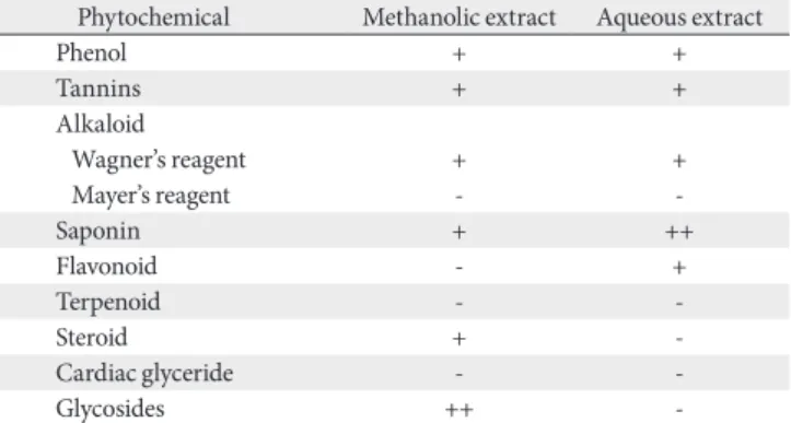
Methanol and aqueous F. exasperata Vahl leaves extracts contained: phenol, tannins, steroids, flavonoids, saponins, alkaloids and glycosides which have been identified by other researchers in various plants and in different parts of plants [17, 18] but the activities of phenol, tannin alkaloids, saponin and glycosides were more pronounced with the methanolic extract as seen in Table 1. Saponins are group of steroid or triterpenoid glycosides, also found to be effective antimicro- bial substances in vitro against a wide array of microorgan- isms by inhibiting the membrane bound enzymes [19]. They have been reported to possess substantial anti-carcinogenic activities due to their anti-oxidant and inflammatory prop-
erties [20].
Assessment of motor coordination performance
Animals with good motor coordination are expected to spend more time on the rotating bar [4, 21]. Vanadium group shows significantly decrease motor activity when compared to the control group, while the F. exasperata Vahl group and post-treated groups (NaVO3+Vahl) show a significant increase in the retention time as they spent more time on the rotating bar compared to vanadium exposed group (**P<0.01). However, the withdrawal group showed a signifi- cant increase in the retention time on the rotating bar com- pared to the vanadium exposed group (Fig. 1).
Significant increases in LOT scores were considered as ab- normal motor coordination when the treatment groups were compared against control, as shown in Fig. 2 below. Vana- dium exposed mice exhibited increased LOT (time to turn) and total time spent during the PBT when compared to the control group. There was a significant decrease in the LOT of post-treated groups and the withdrawal group when com- pared with the vanadium exposed group. However, control (saline) group exhibited a decrease in time of turn compared to the vanadium exposed group (*P<0.05, **P<0.01).
Analysis of biochemical assay Lipid peroxidation
Fig. 3 showed no significant difference of MDA level when
Table 1. Phytochemical analysis of Ficus exasperata vahl leaves
Phytochemical Methanolic extract Aqueous extract
Phenol + +
Tannins + +
Alkaloid
Wagner’s reagent + +
Mayer’s reagent - -
Saponin + ++
Flavonoid - +
Terpenoid - -
Steroid + -
Cardiac glyceride - -
Glycosides ++ -
-, not detected; +, present in low concentration; ++, present in high concentration.
Control 15
10
5
Latencyoffall(sec)
0
Vanadium Withdrawal NS
NaVO +Vahl
(LD)
3
NaVO +Vahl
(HD)
3
Fig. 1. Graph showing the latency of fall of the experimental animals (P-value=0.894). HD, high dose; LD, low dose; NS, not significant.
Control 150
100
50
Latencyofturn(sec)
0
Vanadium Withdrawal
*
**
*
**
NaVO +Vahl
(LD)
3
NaVO +Vahl
(HD)
3
Fig. 2. Graph showing the latency of turn of the experimental animals (P-value=0.029: F-value=9.216). HD, high dose; LD, low dose.
*P<0.05, **P<0.01.
comparing control group with the vanadium, withdrawal, NaVO3+Vahl (low dose) and NaVO3+Vahl (high dose) group.
However, the group exposed to vanadium showed increased level of MDA, while a reduction in the level of MDA was seen
in NaVO3+Vahl (high dose) group when they were compared with the control group.
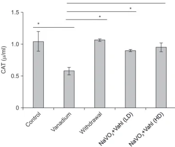
Catalase
Fig. 4 showed significance difference in the CAT level when normal saline group (control) was compared with vanadium (*P<0.05). When the Vanadium group (NaVO3) was also compared with the withdrawal group, it showed a significant difference of *P<0.05, this also goes for the NaVO3+Vahl (low dose) and NaVO3+Vahl (high dose) group (*P<0.05).
Glutathione
Vanadium group in Fig. 5 expressed significantly lower level of GPX when compared with the normal saline group (control), and when NaVO3+Vahl (low dose) group was com- pared with control group (*P<0.05).
Immunohistochemistry
Immunohistochemical studies evaluating the neuroin- flammatory level and DA activities in a section of the SNc, and ameliorative roles of F. exasperata Vahl leaves extract after exposure to Vanadium was observed to be significant on the glia following the uses of primary antibodies directed against GFAP, DAT, and TH (Figs. 6–9).
Control 15
10
(MDA/ml) 5
0
Vanadium Withdrawal NS
NaVO +Vahl
(LD)
3
NaVO +Vahl
(HD)
3
Fig. 3. Graph representing the concentration of MDA (units) per mg in brain tissue. There is no statistical significant different in the MDA between the control and vanadium treated groups. Sodium metavanadate and low dose of Ficus exasperata Vahl (NaVO3+Vahl LD), Sodium metavanadate and low dose of F. exasperata Vahl (NaVO3+Vahl HD). HD, high dose; LD, low dose; MDA, malondialdehyde.
Control 1.5
1.0
TCA(/ml) 0.5
0
Vanadium Withdrawal
*
*
*
*
NaVO +Vahl
(LD)
3
NaVO +Vahl
(HD)
3
NaVO +Vahl
(LD)
3
NaVO +Vahl
(HD)
3
Fig. 4. Graph representing the concentration of CAT (units) per mg in brain tissue homogenate. There is statistical significant different in the CAT between the control and vanadium and Ficus exasperata treated groups. Sodium metavanadate and low dose of F. exasperata Vahl (NaVO3+Vahl LD), Sodium metavanadate and low dose of F.
exasperata Vahl (NaVO3+Vahl HD). CAT, catalase; HD, high dose;
LD, low dose. *P<0.05.
Control 15
10
5
GSH(g/mgprotein)
0
Vanadium Withdrawal
*
*
NaVO +Vahl
(LD)
3
NaVO +Vahl
(HD)
3
Fig. 5. Graph representing the concentration of GSH (units) per mg in brain tissue homogenate. There is statistical significant different in the catalase between the control and vanadium and Ficus exasperata treated groups. Sodium metavanadate and low dose of F. exasperata Vahl (NaVO3+Vahl LD), Sodium metavanadate and low dose of F.
exasperata Vahl (NaVO3+Vahl HD). GSH, glutathione; HD, high dose, LD, low dose. *P<0.05.
Tyrosine hydroxylase
Control group (A) and NaVO3+Vahl group (D) low dose, showed normal DA neurons which are evenly distributed, while the vanadium exposed group (B) displayed disruption of DA neurons, withdrawal group (C) shows the presence of scanty distribution of DA neurons, while NaVO3+Vahl group (E) high dose, revealed increase level of DA neurons as com- pared to the vanadium exposed group.
Dopamine transporter
Vanadium group (B) shows increased expression of DAT activity as compared to the control group, while withdrawal group (C) revealed little expression of DAT. However, NaVO3+Vahl group (D) low dose, revealed little expression of DAT, while NaVO3+Vahl group (E) high dose, revealed increased DAT activity. Black arrow indicates the DA trans- porter.
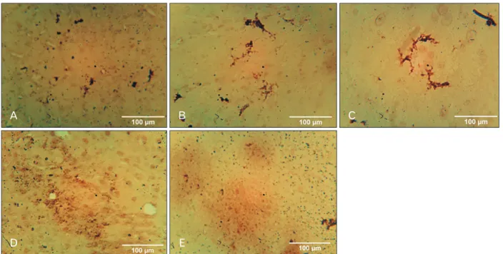
Glial fibrillary acidic protein
Vanadium (B) and withdrawal group (C) showed the pres- ence of well-marked activated astrocytes, while control group (A) revealed no trace of astrocyte activation. NaVO3+Vahl group (D) low dose and NaVO3+Vahl group (E) high dose,
A B C
D E
Fig. 7. Photomicrograph of the SNc immunostained for TH (×100). Control group (A) and NaVO3+Vahl group (D) low dose, showed normal DA neurons which are evenly distributed, while the vanadium exposed group (B) displayed degeneration and disruption of DA neurons, withdrawal group (C) shows the presence of scanty distribution of normal DA neurons, while NaVO3+Vahl group (E) high dose, revealed slight recovery of degenerated DA neurons. Black arrows indicate the TH cells (DA neurons). DA, dopaminergic; SNc, substantia nigra; TH, tyrosine hydroxylase.
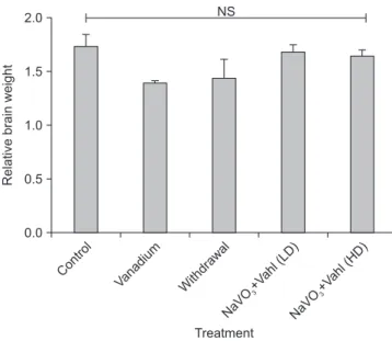
Control 2.0
1.5
1.0
Relativebrainweight 0.5
0.0
Vanadium Withdrawal NS
Treatment NaVO
+Vahl (LD)
3
NaVO +Vahl
(HD)
3
Fig. 6. Bar chart showing the relative brain weight of the experimental animals (P-value=0.458: F-value=1.918). There is no statistical significant different in the relative brain weight between the groups. Sodium metavanadate and low dose of Ficus exasperata Vahl (NaVO3+Vahl LD), Sodium metavanadate and low dose of F. exasperata Vahl (NaVO3+Vahl HD). HD, high dose; LD, low dose; NS, not significant.
A B C
D E
Fig. 8. Photomicrograph of the SNc immunostained for DAT (×100). Control (A) group revealed little expression of the DAT. Vanadium group (B) shows increased expression of DAT activity, while withdrawal group revealed little expression of DAT. However, NaVO3+Vahl group (D) low dose, revealed little expression of DAT, while NaVO3+Vahl group (E) high dose, revealed increased DAT activity. Black arrows indicat the dopaminergic transporter. DAT, dopamine transporter; SNc, substantia nigra.
A B C
D E
Fig. 9. Photomicrograph of the SNc immunostained for GFAP. From the vanadium Group (B) and withdrawal group (C), it can be observed that Vanadium-induced astroglia activation as shown by the presence of well-marked activated astrocytes, while control group (A) revealed no trace of astrocyte activation. NaVO3+Vahl group (D) low dose and NaVO3+Vahl group (E) high dose, revealed no traces of activated astrocytes. GFAP, glial fibrillary acidic protein; SNc, substantia nigra.
revealed no traces of activated astrocytes.
Phytochemical analysis
The activities of phenol, tannin alkaloids, saponin and glycosides were more pronounced with the methanolic extract. However, saponin is seen to be more pronounced following the phytochemical of this extract (F. exasperata Vahl).
Discussion
In the present investigation, we provide evidence for the motor benefit of the plant extract in a non-classical mouse model of Parkinson’s disease (PD). The benefit is related to the protective effect of the extract on DA neurons via an evidenced anti-oxidant activity. Our data suggest that the extract could have a beneficial effect in the treatment of PD.
As previously reported [22, 23], vanadium reduced loco- motor activity and motor coordination using the RT [24].
The FEVL extract and withdrawal group demonstrate an increase in the retention time by spending a remarkable time on the rotating bar, which indicates an improvement in mo- tor coordination. Also, another behavioral test used to assess motor coordination is a PBT which measures the LOT and the results obtained suggest a significant decrease in loco- motor activities in the withdrawal group as well as when the extract is used after vanadium, and this suggests an ame- liorative role of extract. Excessive exposure of the brain to amounts of trace elements like vanadium increase oxidative stress which later causes neurodegeneration and increased incidence of Parkinsonism and PD [5, 25].
Previous research done in our laboratory reviewed that FEVL extract alone at different dosages improves motor defi- cits in heavy metal consumption [4]. However, deleterious health effects induced by this transition element found in sodium metavanadate (NaVO3) compounds are seen in our environment especially due to heavy metal mining and in oil-producing communities where the burning of fossil fuel exist. This can be linked to dopaminergic neurons insult as a result of oxidative stress that leads to a decrease in dopamine levels in the brain region such as Striatum, SNc and globus pallidus which are implicated in vanadium toxicity [5].
The induction of lipid peroxidation in the brain following vanadium consumption is a testament to the role oxidative stress plays in the neurotoxicity of the metal [26, 27]. To as- certain this neurological effects being observed in humans [5,
28], MDA (an intermediate in the lipid peroxidation process) results showed an increased level in the vanadium-exposed group, and contribute to neuronal cell death and continued exposure to excess free radicals derived from some endog- enous or exogenous neurotoxic species [29, 30]. However, when looking at the FEVL and those group that was with- drawn after some exposure, this in addition to the results above revealed a statistical reduction in MDA level. Reduction of lipid peroxidation may protect against oxidative neurode- generation caused by vanadium exposure. Also, a significant increase in the activity of CAT level in vanadium-intoxicated mice was recorded, and this was seen to be reduced in the two groups that received FEVL extract. This is by following Shi and Dalal [31], which suggests that vanadium treatment may result in the increased formation of oxygen-free radi- cals, leading to oxidative stress. GSH as one of the markers of oxidative stress plays a major role in many cellular functions, which include detoxification of reactive oxygen species (ROS) in the brain, but abnormal levels of GSH is considered to be a sign of many diseases including neurotoxicity [32]. There was also a significant increase in the GSH level of the vana- dium-exposed mice when compared with the control group, as seen in Fig. 5 (P<0.05).
However, the significant increase of GSH noticed in some treatment groups points to an ameliorative intervention of FeLV extract which indicates that the saponin fraction of FeLV extract has a strong antioxidant and anti-inflammato- ry properties to reduce the activities of these oxidative stress markers following the acute administration of vanadium.
The changes in GSH and CAT, as well as increase in activi- ties of MDA observed in the SNc suggest a correlation be- tween loss of antioxidant response and an increase in oxida- tive stress caused by exposure to vanadium. MDA, GSH, and CAT constitute the main components of the antioxidant de- fense system, and modification in their expressions reflects potential oxidative stress [33]. The mechanism of action of the saponin fraction of FEVL extracts also affects the mono- amine oxidase and DAT pathway to enhance and improve motor deficit in mice.
Dopamine, which is an important neurotransmitter in the brain, plays a modulatory role in the coordination of movement, reward, and cognition [34]. Alterations in DA neurotransmission are key hallmarks in the pathogenesis of several diseases such as Parkinson’s disease and addic- tion [35]. Results from this study can further substantiate that exposure to vanadium can lead to loss of dopaminergic
neurons and a reduction of dopamine in the SNc [34]. While, withdrawal group showed a marked elimination of the dis- tribution of noxious agent (vanadium) from the SNc, indi- cated by several dopaminergic neurons increase. However, the group with FEVL extract after exposure to vanadium revealed the perfect distribution of dopaminergic neurons, and this is by following the findings of Chinyere [36], which stipulate the possibility of the extract to have either inhibited the release or antagonize the action of these inflammatory mediators. On the other hand, the plasma membrane DAT which is considered to be a reliable marker of presynaptic do- paminergic terminal loss [37] that can transport potentially toxic agents into cells or elevate the intracellular concentra- tion of dopamine to detrimental levels. Immunohistochemi- cally, vanadium revealed increased expression of DAT when compared to the control group and withdrawal group, which revealed a little expression of DAT activity. However, the effi- cacy of the extract revealed little expression of DAT, which is dose-dependent. This finding agrees with numerous neuro- chemical studies of dopamine markers and is consistent with the hypothesis that those areas expressing the highest levels of DAT are most susceptible to neurotoxicity [37].
Inflammatory responses in the central nervous system are elicited by non-neuronal glial cells, chiefly astrocyte and microglia, whose activities underlie several neurodegenera- tive disease pathogeneses [38]. Astrocytes play a key role in the regulation of the neuronal activities, synaptic plasticity and also some key elements in the brain parenchyma de- fense against oxidative and toxic insults [39]. The number of visible astrocytes and the complexity of their processes may also be related to the extent of neural injury. In this cur- rent investigation, Vanadium-induced astrogliosis as shown by the presence of well-marked activated astrocytes, when compared to the control group, which revealed no trace of astrocyte activation. However, post-treated groups revealed no traces of activated astrocytes, which is as a result of the ameliorative effect of saponin fraction of FEVL extract. This corroborates with the findings of Garcia et al. [40] which stipulate that vanadium-induced astrogliosis has reported in the cerebellum and hippocampus after exposure to sodium metavanadate for five days, indicating a rapid response of astrocytes to this challenge.
In conclusion, taken together, This work has shown im- proved motor coordination with F. exasperata Vahl leave ex- tract treatment after the vanadium-exposure. The results of the immunohistochemical and biochemical studies also re-
vealed a well-marked expression of astrocytes and increased in the markers of oxidative stress, respectively, which become more pronounced during the process of neurodegeneration with vanadium. Therefore, from the present study, it can be deduced that vanadium accumulation in the brain can lead to astrocyte activation, which might induce neurotoxicity.
However, inflammations actively contribute to neuronal damage and death, but the intervention of F. exasperata Vahl extract showed a remarkable improvement.
In conclusion, this study has shown that the saponin frac- tion of F. exasperata Vahl leaves has a powerful ameliorative effect on the motor deficits caused by vanadium in the SNc of male adult mice hence improve locomotor activity and motor coordination.
ORCID
Adeshina O Adekeye:
https://orcid.org/0000-0003-0691-149X
Gold J Irawo: https://orcid.org/0000-0002-7823-2413 Adedamola Adediran Fafure:
https://orcid.org/0000-0002-6335-8793
Author Contributions
Conceptualization: AAF. Data analysis and interpreta- tion: AAF. Drafting of the manuscript: GJI. Critical revision of the manuscript: AOA. Approval of the final version of the manuscript: all authors.
Conflicts of Interest
No potential conflict of interest relevant to this article was reported.
Acknowledgements
The authors gratefully acknowledge the assistance of Mrs. Peters from the Chemistry Department of Afe Babalola University for invaluable assistance in the preparation of saponin fraction from F. exasperata Vahl leaves and also do appreciate the support of Writing paper workshop commit- tee at SONA 2019 supported by IBRO/ISN.
References
1. Hsu HW, Bondy SC, Kitazawa M. Environmental and dietary exposure to copper and its cellular mechanisms linking to Al- zheimer’s disease. Toxicol Sci 2018;163:338-45.
2. Liu J, Lewis G. Environmental toxicity and poor cognitive out- comes in children and adults. J Environ Health 2014;76:130-8.
3. Racette BA, McGee-Minnich L, Moerlein SM, Mink JW, Vi- deen TO, Perlmutter JS. Welding-related parkinsonism: clinical features, treatment, and pathophysiology. Neurology 2001;56:8- 13.
4. Fafure AA, Adekeye AO, Enye LA, Tijani AA, Ajao MM, Edem EE. Ficus exasperata vahl improves manganese-induced neuro- toxicity and motor dysfunction in mice. Anat J Afr 2018;7:1206- 19.
5. Afeseh Ngwa H, Kanthasamy A, Anantharam V, Song C, Witte T, Houk R, Kanthasamy AG. Vanadium induces dopaminergic neurotoxicity via protein kinase Cdelta dependent oxidative signaling mechanisms: relevance to etiopathogenesis of Parkin- son’s disease. Toxicol Appl Pharmacol 2009;240:273-85.
6. Moskalyk RR, Alfantazi AM. Processing of vanadium: a re- view. Miner Eng 2003;16:793-805.
7. Amorim FA, Welz B, Costa AC, Lepri FG, Vale MG, Ferreira SL. Determination of vanadium in petroleum and petroleum products using atomic spectrometric techniques. Talanta 2007;
72:349-59.
8. Olopade JO, Connor JR. Vanadium and neurotoxicity: a review.
Curr Top Toxicol 2011;7:33-9.
9. Fortoul TI, Rodriguez-Lara V, González-Villalva A, Rojas-Le- mus M, Cano-Gutiérrez G, Ustarroz-Cano M, Colín-Barenque L, Bizarro-Nevares P, García-Pealez I, Montaño LF, Jimenez- Martinez RS, Lopez-Valdez N, Ruiz-Guerrero ML, Meléndez- García NA, García-Ibarra FA, Martínez-Baez V, Zapata Alfaro D, Muñiz-Rivera-Cambas A, López-Zepeda LS, Quezada- Maldonado EM, Cervantes-Yépez S. Inhalation of vanadium pentoxide and its toxic effects in a mouse model. Inorganica Chim Acta 2014;420:8-15.
10. Roy A, Jauhari N, Bharadvaja N. Medicinal plants as a potential source of chemopreventive agents. In: Akhtar MS, Swamy MK, editors. Anticancer plants: natural products and biotechnologi- cal implements. Singapore: Springer; 2018. p. 109.
11. Odiba PA, Yusuf D, Ali E, Yusuf MI, John E. Effect of aqueous extract of Ficus exasperata leaf on the body weight and haema- tological parameters of Wistar rats. J Appl Sci Environ 2012;3:
80-3.
12. Ahmed F, Toume K, Ohtsuki T, Rahman M, Sadhu SK, Ishi- bashi M. Cryptolepine, isolated from Sida acuta, sensitizes human gastric adenocarcinoma cells to TRAIL-induced apop- tosis. Phytother Res 2011;25:147-50.
13. Ijeh II, Ukweni AI. Acute effect of administration of ethanol extracts of Ficus exasperata vahl on kidney function in albino rats. J Med Plant Res 2007;1:27-9.
14. Akah PA, Orisakwe OE, Gamaniel KS, Shittu A. Evaluation of
Nigerian traditional medicines: II. effects of some Nigerian folk remedies on peptic ulcer. J Ethnopharmacol 1998;62:123-7.
15. Ogunleye RF. Effectiveness of the leaf powder of Ficus exas- perata Vahl. (Moraceae) in suppressing thepopulation of two major storage insect pests. Cont J Biol Sci 2011;4:6-11.
16. Ayinde BA, Omogbai EK, Amaechina FC. Pharmacognosy and hypotensive evaluation of Ficus exasperata Vahl (Moraceae) leaf. Acta Pol Pharm 2007;64:543-6.
17. Devbhuti D, Gupta JK, Devbhuti P, Bose A. Phytochemical and acute toxicity study on Tinospora tomentosa Miers. Acta Pol Pharm 2009;66:89-92.
18. Tijjani MB, Bello IA, Aliyu AB, Olurishe T, Maidawa SM, Ha- bila JD, Balogun EO. Phytochemical and antibacterial studies of root extract of Cochlospermum tinctorium A. Rich. (Cochlo- spermaceae). Res J Med Plant 2009;3:16-22.
19. Sparg SG, Light ME, van Staden J. Biological activities and dis- tribution of plant saponins. J Ethnopharmacol 2004;94:219-43.
20. Nandakumar V, Singh T, Katiyar SK. Multi-targeted preven- tion and therapy of cancer by proanthocyanidins. Cancer Lett 2008;269:378-87.
21. Adekeye AO, Adumah C, Fafure AA, Ajao M, Sabiu S, Shallie P, Adefule AK. Evaluation of the effects of gutenbergia nigritana leaves extract on cerebellum of adult mice and its implication on manganese toxicity. World J Pharm Pharm Sci 2018;7:55-68.
22. Mustapha O, Oke B, Offen N, Sirén AL, Olopade J. Neurobe- havioral and cytotoxic effects of vanadium during oligoden- drocyte maturation: a protective role for erythropoietin. Envi- ron Toxicol Pharmacol 2014;38:98-111.
23. Folarin O, Olopade F, Onwuka S, Olopade J. Memory deficit recovery after chronic vanadium exposure in mice. Oxid Med Cell Longev 2016;2016:4860582.
24. Carter RJ, Morton J, Dunnett SB. Motor coordination and bal- ance in rodents. Curr Protoc Neurosci 2001;Chapter 8:Unit 8.
12.
25. Zwolak I. Vanadium carcinogenic, immunotoxic and neuro- toxic effects: a review of in vitro studies. Toxicol Mech Methods 2014;24:1-12.
26. Folarin OR, Snyder AM, Peters DG, Olopade F, Connor JR, Olopade JO. Brain metal distribution and neuro-inflammatory profiles after chronic vanadium administration and withdraw- al in mice. Front Neuroanat 2017;11:58.
27. Igado OO, Olopade JO, Adesida A, Aina OO, Farombi EO.
Morphological and biochemical investigation into the possible neuroprotective effects of kolaviron (Garcinia kola bioflavo- noid) on the brains of rats exposed to vanadium. Drug Chem Toxicol 2012;35:371-80.
28. Fatola OI, Olaolorun FA, Olopade FE, Olopade JO. Trends in vanadium neurotoxicity. Brain Res Bull 2019;145:75-80.
29. Dexter DT, Carter CJ, Wells FR, Javoy-Agid F, Agid Y, Lees A, Jenner P, Marsden CD. Basal lipid peroxidation in substantia nigra is increased in Parkinson’s disease. J Neurochem 1989;52:
381-9.
30. Nissanka N, Moraes CT. Mitochondrial DNA damage and re- active oxygen species in neurodegenerative disease. FEBS Lett
2018;592:728-42.
31. Shi X, Dalal NS. Vanadate-mediated hydroxyl radical genera- tion from superoxide radical in the presence of NADH: Haber- Weiss vs Fenton mechanism. Arch Biochem Biophys 1993;307:
336-41.
32. Lee S, Li J, Zhou X, Yin J, Yoon J. Recent progress on the devel- opment of glutathione (GSH) selective fluorescent and colori- metric probes. Coord Chem Rev 2018;366:29-68.
33. Monleau M, De Méo M, Frelon S, Paquet F, Donnadieu-Claraz M, Duménil G, Chazel V. Distribution and genotoxic effects after successive exposure to different uranium oxide particles inhaled by rats. Inhal Toxicol 2006;18:885-94.
34. Ooms M, Celen S, De Hoogt R, Lenaerts I, Liebregts J, Vanhoof G, Langlois X, Postnov A, Koole M, Verbruggen A, Van Laere K, Bormans G. Striatal phosphodiesterase 10A availability is altered secondary to chronic changes in dopamine neurotrans- mission. EJNMMI Radiopharm Chem 2017;1:3.
35. Schmitt KC, Reith ME. Regulation of the dopamine transport-
er: aspects relevant to psychostimulant drugs of abuse. Ann N Y Acad Sci 2010;1187:316-40.
36. Chinyere NH. Studies on anti-inflammatory properties of the leaf extracts of ficus exasperata Vahl (Moraceae) [Doctoral dis- sertation]. Nsukka: University of Nigeria Nsukka; 2012.
37. Miller GW, Staley JK, Heilman CJ, Perez JT, Mash DC, Rye DB, Levey AI. Immunochemical analysis of dopamine transporter protein in Parkinson's disease. Ann Neurol 1997;41:530-9.
38. Fang SC, Hsu CL, Yen GC. Anti-inflammatory effects of phe- nolic compounds isolated from the fruits of Artocarpus hetero- phyllus. J Agric Food Chem 2008;56:4463-8.
39. Heller JP, Rusakov DA. Morphological plasticity of astroglia:
understanding synaptic microenvironment. Glia 2015;63:2133- 51.
40. Garcia GB, Biancardi ME, Quiroga AD. Vanadium (V)-induced neurotoxicity in the rat central nervous system: a histo-immu- nohistochemical study. Drug Chem Toxicol 2005;28:329-44.