INTRODUCTION
Aspergillosis of the respiratory tract has diverse manifesta- tions that range from hypersensitivity disorders to rapidly inva- sive disseminated disease.1,2 These can be classified into 3 dis- tinct clinical categories, viz. allergic aspergillosis, saprophytic colonization, and invasive aspergillosis (Table 1). Different pre- sentations of the allergic form, usually seen in atopic individu- als, include Aspergillus-induced asthma (AIA), allergic bron- chopulmonary aspergillosis (ABPA), and allergic Aspergillus si- nusitis (AAS). This review focuses on ABPA and highlights some of the other Aspergillus-related respiratory disorders.
ASPERGILLUS-INDUCED ASTHMA AND SEVERE ASTHMA WITH FUNGAL SENSITIZATION
Patients with asthma who have a positive immediate (type I) IgE-mediated hypersensitivity to Aspergillus are grouped as AIA. A wide variation to the tune of 16% to 38% has been ob- served in Aspergillus sensitization among asthmatics across the
world.3-5 One of the initial studies found a then considered “un- expected” finding of more severe airway obstruction in patients with AIA.3 In our study of 105 patients with asthma, positive skin reactivity to Aspergillus antigens was noted in 30 subjects (28.5%).5 The disease was more severe in these patients with AIA as evidenced by a statistically significant higher mean du- ration of illness (P<0.001), mean eosinophil count (P<0.0001), mean total IgE (P<0.05), and more usage of oral corticosteroids per year (P<0.004).
The term ‘severe asthma with fungal sensitization’ (SAFS) was coined for a subset of asthmatics that demonstrated sensitiza- tion to fungal antigens and had frequent exacerbations of asth-
Allergic Bronchopulmonary Aspergillosis: A Perplexing Clinical Entity
Ashok Shah,
1* Chandramani Panjabi
21Department of Pulmonary Medicine, Vallabhbhai Patel Chest Institute, University of Delhi, Delhi, India
2Department of Respiratory Medicine, Mata Chanan Devi Hospital, New Delhi, India
This is an Open Access article distributed under the terms of the Creative Commons Attribution Non-Commercial License (http://creativecommons.org/licenses/by-nc/3.0/) which permits unrestricted non-commercial use, distribution, and reproduction in any medium, provided the original work is properly cited.
In susceptible individuals, inhalation of Aspergillus spores can affect the respiratory tract in many ways. These spores get trapped in the viscid spu- tum of asthmatic subjects which triggers a cascade of inflammatory reactions that can result in Aspergillus-induced asthma, allergic bronchopulmo- nary aspergillosis (ABPA), and allergic Aspergillus sinusitis (AAS). An immunologically mediated disease, ABPA, occurs predominantly in patients with asthma and cystic fibrosis (CF). A set of criteria, which is still evolving, is required for diagnosis. Imaging plays a compelling role in the diagno- sis and monitoring of the disease. Demonstration of central bronchiectasis with normal tapering bronchi is still considered pathognomonic in pa- tients without CF. Elevated serum IgE levels and Aspergillus-specific IgE and/or IgG are also vital for the diagnosis. Mucoid impaction occurring in the paranasal sinuses results in AAS, which also requires a set of diagnostic criteria. Demonstration of fungal elements in sinus material is the hall- mark of AAS. In spite of similar histopathologic features, co-existence of ABPA and AAS is still uncommon. Oral corticosteroids continue to be the mainstay of management of allergic aspergillosis. Antifungal agents play an adjunctive role in ABPA as they help reduce the fungal load. Saprophyt- ic colonization in cavitary ABPA may lead to aspergilloma formation, which could increase the severity of the disease. The presence of ABPA, AAS, and aspergilloma in the same patient has also been documented. All patients with Aspergillus-sensitized asthma must be screened for ABPA, and AAS should always be looked for.
Key Words: Allergic Aspergillus sinusitis; allergic bronchopulmonary aspergillosis; allergic fungal sinusitis; aspergilloma; Aspergillus; asthma
Correspondence to: Ashok Shah, Professor, Department of Pulmonary Medicine, Vallabhbhai Patel Chest Institute, University of Delhi, Delhi 110 007 P.O. Box 2101, India.
Tel: +91-11-2543 3783; Fax: +91-11-2766 6549;
E-mail: [email protected]
Received: April 20, 2015; Accepted: May 15, 2015
•There are no financial or other issues that might lead to conflict of interest.
Allergy Asthma Immunol Res. 2016 July;8(4):282-297.
http://dx.doi.org/10.4168/aair.2016.8.4.282 pISSN 2092-7355 • eISSN 2092-7363
ma that necessitated admission to the hospital.6 Diagnostic cri- teria for SAFS include: (1) severe (poorly controlled) asthma, (2) either a positive skin prick test result for fungi (but not neces- sarily to Aspergillus species) or in vitro demonstration of anti- fungal IgE of at least 0.4 kU/L, (3) total serum IgE concentration
<1,000 kU/L.6 Unlike in ABPA, patients with SAFS do not have mucoid impaction or bronchiectasis. Another important differ- ence between SAFS and ABPA is that while severe asthma is one of the diagnostic criteria for SAFS, ABPA also develops in those with mild or moderate asthma. Greenberger7 has also highlighted the divergence between SAFS and ABPA.
Given the persistent fungal colonization of the bronchi in pa- tients with allergic aspergillosis, a potential role for antifungal therapy has been suggested.8 Significantly better asthma quali- ty of life scores were found when itraconazole was adminis- tered to patients with SAFS.9 However, a recent study using vori- conazole for 3 months in moderate-to-severe asthmatics sensi- tized to Aspergillus fumigatus (Af) did not show any benefit.10 The authors suggested that the beneficial effects of itraconazole in previous studies could possibly be due to its pharmacokinet- ic effects on corticosteroids.
ABPA
ABPA is the most significant manifestation of allergic aspergil- losis that occurs worldwide but has not received the impor- tance that it deserves.11 Most commonly seen in patients with asthma and cystic fibrosis (CF), ABPA is caused by hypersensi-
tivity to Aspergillus antigens. In susceptible hosts, an allergic re- sponse is evoked by repeated inhalation of Aspergillus spores.
The fungal antigens, chiefly of Af, elicit mainly a type I (IgE-me- diated) reaction that is responsible for the disease presentation.
Type-III (IgG-mediated immune complex) and Type-IV (cell mediated) responses have also been implicated, but tissue in- vasion does not occur.12 When fungi other than Aspergillus are responsible for such a condition, it is termed as allergic bron- chopulmonary mycoses (ABPM).13,14 Based on specific patho- physiological mechanisms, it has been proposed that ABPA/
ABPM be classified as a distinct endotype of asthma.15
Although 63 years have passed since this disease was first de- scribed in England,16 we are still unable to fathom the reason why only a few patients with asthma are affected with ABPA. In- dividual host genetic susceptibility appears more significant than environmental factors in the causation of ABPA in these subjects. Moreover, although familial preponderance is very common in asthma, occurrence of ABPA among family mem- bers is rare.17,18 We have detected familial occurrence in 4 pairs (4.9%) of first-degree relatives. One patient each in 3 of these 4 pairs also had concomitant AAS.19
Epidemiology of ABPA
The exact prevalence of this disease is still not known, and this is most likely due to the lack of a uniform diagnostic criterion and standardized tests.20 This potentially destructive lung dis- ease is yet to be included in the ninth revision of the Interna- tional Classification of Diseases published in 1996.21 Prior to 1968, when ABPA was unknown outside Europe, the preva- lence of definite ABPA among asthmatics was around 8%-11%, while that of probable ABPA was approximately 22%.22 After the first report from the United States in 1968,23 awareness regard- ing ABPA grew across all continents. Between 1983 and 1986, Greenberger and Patterson24 from the United States found ABPA in 32 (6%) out of 531 asthmatic patients having immedi- ate cutaneous reactivity to Aspergillus antigens. In other stud- ies, ABPA was detected in as many as 25% to 37% of asthmatics with a positive skin prick test to Af.25 Among 105 patients with bronchial asthma, we noted a significantly longer duration of illness, earlier age of onset of asthma as well as rhinitis, higher mean total leucocyte counts, absolute eosinophil counts, and total serum IgE values in 8 patients diagnosed with ABPA when compared to those with Aspergillus sensitization only without ABPA.5
Western estimates suggest that ABPA complicates up to 6% of all chronic cases of asthma.26 The prevalence of ABPA in pa- tients with underlying CF ranges from 2% to 15%.27 Denning et al.28 in a scoping review based on the published studies on asthma and ABPA, attempted to ascertain the global burden of ABPA. The prevalence of ABPA in adult asthmatics, as analyzed from 5 prospective studies having at least 50 patients with asth- ma, was found to be 2.5% (range 0.72%-3.5%). Based on this, Table 1. Aspergillus-associated respiratory disorders1,2
I. Upper respiratory tract 1. Allergic aspergillosis
- Allergic Aspergillus sinusitis (AAS) 2. Saprophytic colonisation
- Sinus fungal balls 3. Invasive disease
- Acute fulminant invasive sinusitis - Chronic invasive sinusitis - Granulomatous invasive sinusitis II. Lower respiratory tract
1. Allergic aspergillosis
- (IgE mediated) Aspergillus induced asthma (AIA) - Allergic bronchopulmonary aspergillosis (ABPA) - Hypersensitvity pneumonitis
2. Saprophytic colonisation - Aspergilloma simple
complex (chronic cavitary pulmonary aspergillosis) 3. Invasive disease
- Invasive pulmonary aspergillosis acute
subacute (chronic necrotising pulmonary aspergillosis)
the authors deduced that adult patients with ABPA across the globe could “potentially exceed 4.8 million.”28
Since there were no consensus-based guidelines on ABPA so far, the International Society for Human and Animal Mycology (ISHAM), in September 2011, constituted a Working Group on ABPA complicating asthma.29 Data on Aspergillus sensitization and ABPA published since circa 2000 was collected by the ISH- AM Working Group.29 The prevalence of Aspergillus sensitiza- tion among patients with asthma ranged from 5.5%-38.5%, and the prevalence of ABPA in asthma varied between 2.5% and 22.3% with a pooled prevalence of 8.4%.
Immunopathogenesis
Immune mediated mechanisms of lung destruction in ABPA are not fully understood. Af antigens elicits a polyclonal anti- body response which is largely responsible for elevated levels of total IgE as well as Af-IgE and Af-IgG antibodies.30,31 Increased interleukin (IL)-4, IL-5, IL-10, and IL-13 production due to the cellular Th-2 immunologic response suggests an immunocom- petent host.32,33 We have identified antibodies to a cytotoxic ri- bonuclease antigen (18 kD) and an elastinolytic protease anti- gen (45kD) in Indian patients with ABPA.34,35 Genetic risk fac- tors include expression of HLA-DR2 and HLA-DR5 genotypes, while HLA-DQ2 protected against ABPA.36,37 In subjects with CF, increased chances of Aspergillus colonization of the airways and subsequent development of ABPA were found in those with CF transmembrane conductance regulator gene muta- tions.38-40 Surfactant protein-A2 polymorphisms,41 elevated lev- els of mannan-binding lectin due to the 1011A allele,42 and toll- like receptor polymorphisms43 also play an important role in the development of ABPA. An immunoproteomics approach would help identify synthetic peptide antigens of Af for skin testing, serodiagnosis, and potentially immunotherapy.44,45 Diagnosis
As our understanding of the disease is improving, diagnostic criteria continue to evolve. In 1952, Hinson et al.16 reported 3 patients of “a kind not previously recognized”. These patients presented with repeated episodes of fever, productive cough, wheezing dyspnoea, and occasional chest pain. Eosinophilia, pulmonary infiltrates in different areas on the chest roentgeno- gram, and Aspergillus mycelium on microscopic examination of the purulent sputum were found during acute attacks. Sac- cular bronchiectasis was noted in 2 cases. Due to certain pecu- liar features not usually observed in patients with pulmonary eosinophilia, the authors suggested to classify their 3 patients as a separate entity. Unlike the acute presentation of pulmo- nary eosinophilia, these patients had a protracted course of ill- ness. It was also observed that production of sputum “plugs”
correlated with clearing of radiologic infiltrates. Mucosal ede- ma and bronchial spasm without any obstructing masses were found on bronchoscopy, while markedly dilated bronchi filled
with sticky, tenacious mucus were necropsy findings in 1 pa- tient. The authors stated that, “….intense eosinophilic infiltra- tion and excessive production of mucus represented an allergic response….” Since fungal masses were not observed, they did not group these patients under “mycetomata.”
Diagnostic criteria
Once the disease was recognized in the United States in 1968 and thence globally, the key diagnostic features have been stan- dardized.46,47 Based on clinical, radiologic, and laboratory fea- tures, a set of 8 major and 3 minor criteria was proposed in 1977 by Rosenberg and Patterson,46 which remains the most well-ac- knowledged criteria (Table 2). Although a set of criteria is re- quired, there is no single test that establishes the diagnosis oth- er than demonstration of central bronchiectasis (CB) with nor- mal tapering bronchi, a feature still considered pathognomonic of ABPA.48,49 However, CB has also been found to extend to the periphery in some segments.50
All the 8 major criteria may not be found at all times. Some of the features may be present only during the acute (stage 1) or the exacerbation (stage 3) states. Moreover, apart from CB and Aspergillus type-1 hypersensitivity, the other parameters are af- fected by therapy with prednisolone. This makes it difficult for all criteria to always be fulfilled in patients with ABPA. In 2002, Greenberger51 advocated a set of minimally essential criteria, which includes (1) asthma, (2) immediate cutaneous reactivity to Af, (3) total serum IgE >1,000 ng/mL (417 kU/L), (4) elevated specific IgE-Af/IgG-Af, and (5) CB in the absence of distal bron- chiectasis. Greenberger7 in 2013 further proposed “truly mini- mal” diagnostic criteria that included items (1), (2), (3), and (5) of the aforementioned minimally essential criteria.
Central or proximal bronchiectasis with normal peripheral bronchi continues to be considered a sine qua non for the diag- nosis of ABPA.48 However, there exists a subset of patients with a milder form of the disease in whom CB may not be present.
These serologically positive patients satisfy the remaining crite- ria for ABPA and are categorised as ABPA-S.52 Specific treatment for ABPA should immediately be commenced so as to delay or prevent further lung damage. Later on when CB develops, the patient is classified as ABPA-CB.53
Even today, there is no agreement on the minimum number of criteria, either major or minor, required to diagnose ABPA.54 The ISHAM Working Group29 has proposed a set of revised cri- teria wherein the items are broadly divided into ‘obligatory’ and
‘other’ criteria (Table 2). Bronchial asthma and CF are identi- fied as predisposing conditions for ABPA in this newly pro- posed set of criteria. The 2 features of the obligatory criteria are as follows: (1) positive immediate (type I) cutaneous hypersen- sitivity to Aspergillus antigen or elevated IgE levels against Af and (2) elevated total IgE levels >1,000 IU/mL. Both of these findings must be present to establish a diagnosis of ABPA. At least 2 out of 3 other criteria viz. (1) presence of precipitating or
IgG antibodies against Af in serum, (2) radiographic pulmonary opacities consistent with ABPA, and (3) total eosinophil count
>500 cells/µL in steroid naïve patients should be fulfilled. How- ever, the Working Group29 has suggested that this newly pro- posed criteria needs “validation and further refinement.”
ABPA in CF
Based on the Epidemiologic Study of Cystic Fibrosis (ESCF) database,55 a set of criteria for the diagnosis of acute ABPA in patients with CF has been laid down. The ESCF criteria adopt- ed included the presence of 2 of the following 3: (1) immediate skin reactivity to Af antigens, (2) precipitating antibodies to Af antigens, and (3) total serum IgE >1,000 IU/mL; and at least 2 of the following 6: (1) bronchoconstriction, (2) peripheral blood eosinophilia >1,000/µL, (3) history of pulmonary infiltrates, (4) elevated specific IgE-Af/IgG-Af, (5) Af in sputum by smear or culture, and (6) response to steroids.
ABPA without asthma
Although ABPA is predominantly a disease of asthmatics, this entity has also been diagnosed in patients without asthma. Af- ter the first such description in 1981,56 more than a score of pa- tients have been documented. A noteworthy aspect of this sub- set of patients was that more than half were initially worked up for bronchogenic carcinoma. Furthermore, the remarkable ra- diologic similarity to pulmonary tuberculosis has important clinical implications in high tuberculous prevalent areas, as the patient reported by us was referred as ‘multidrug-resistant tu- berculosis’ for evaluation.57 The presence of broncholithiasis in ABPA without asthma has also been described.58
Clinical features
This immunologically mediated lung disease is usually indo- lent in nature and has a protracted course of illness. The pre- sentation can range from mild asthma, with very few symp- Table 2. Evolving diagnostic criteria for ABPA
Rosenberg-
Patterson criteria46,47 Minimal essential criteria51 ‘Truly minimal’ criteria7 ISHAM Working Group29 ABPA in CF55 Major criteria
1. Asthma
2. Presence of transient pulmonary infiltrates (fleeting shadows) 3. Immediate cutaneous reactivity to Af 4. Elevated total serum IgE 5. Precipitating antibodies against Af
6. Peripheral blood eosinophilia
7. Elevated serum IgE and IgG to Af
8. Central/proximal bronchiectasis with normal tapering of distal bronchi Minor criteria
1. Expectoration of golden brownish sputum plugs 2. Positive sputum culture for Aspergillus species 3. Late (Arthus-type) skin reactivity to Af
1. Asthma
2. Immediate cutaneous reactivity to Af 3. Total serum IgE >1,000 ng/mL (417 kU/L) 4. Elevated specific IgE-Af/IgG-Af 5. CB in the absence of distal bronchiectasis
1. Asthma
2. Immediate cutaneous reactivity to Af 3. Total serum IgE >1,000 ng/mL (417 kU/L) 4. CB in the absence of distal bronchiectasis
Predisposing conditions 1. Bronchial asthma 2. Cystic fibrosis Obligatory criteria (both should be present) 1. Type I Aspergillus skin test
positive (immediate cuta- neous hypersensitivity to Aspergillus antigen) or el- evated IgE levels against Af
2. Elevated total IgE levels (>1,000 IU/mL)*
Other criteria (at least two of three)
1. Presence of precipitating or IgG antibodies against Af in serum
2. Radiographic pulmonary opacities consistent with ABPA
3. Total eosinophil count
>500 cells/µL in steroid naïve patients (may be historical)
(*If the patient meets all other criteria, an IgE value
<1,000 IU/mL may be acceptable)
Presence of two of the following three:
(i) Immediate skin reactivity to Af antigens,
(ii) Precipitating antibodies to Af antigens,
(iii) Total serum IgE
>1,000 IU/mL;
and at least two of the following six:
(i) Bronchoconstriction, (ii) Peripheral blood
eosinophilia >1,000/µL, (iii) History of pulmonary
infiltrates,
(iv) Elevated specific IgE-Af/
IgG-Af,
(v) Af in sputum by smear or culture,
(vi) Response to steroids
ABPA, allergic bronchopulmonary asperillosis; Af, Aspergillus fumigatus; CB, central bronchiectasis; CF, cystic fibrosis; IgE, immunoglobulin E; IgG, immunoglobulin G;
ISHAM, International Society for Human and Animal Mycology.
toms, to extensive lung disease that may manifest as respiratory failure. Patients encounter repeated episodes of acute exacer- bations that, after treatment, are followed by periods of remis- sions. If left untreated, it more often than not results in a chronic fibrotic lung disease that mimics post tubercular fibrotic sequel- ae.59 Apart from asthma, ABPA may also be associated with oth- er clinical allergic diseases. Although these atopic conditions may manifest at an early age, ABPA is usually seen in the 20s or 30s, but has also been reported in children60,61 and even in in- fants.62 In a patient with poorly controlled asthma and peripher- al eosinophilia, expectoration of golden-brown plugs in the sputum should raise the possibility of ABPA.63 A third of the pa- tients, in spite of extensive radiological lesions, may have few or no symptoms at all.64 Hence, it appears that the severity or chro- nicity of the disease does not correlate with symptomatology.
We reviewed the clinical profile of 113 patients with ABPA, 70 of whom were males.65 The mean age was 32 years, while the mean age of onset of asthma was 21 years. Respiratory symp- toms included cough (99%), breathlessness (99%), expectora- tion (98%), wheezing (97%), and haemoptysis (41%). Nasal symptoms suggestive of upper airways allergy were present in 45%. Expectoration of sputum plugs was reported by 37% of the patients and nasal plugs by 6%. Approximately half of the pa- tients had a personal/family history of atopy. A study from Ko- rea highlighted that ABPA could possibly occur in patients with destructive lung disease due to tuberculosis.66 Physical exami- nation in ABPA may not be fruitful if the patient is asymptomat-
ic. Rhonchi, crepitations, and bronchial breathing may be heard depending on the degree of the lung disease present.
Persistent crackles, which do not clear after either a tussive ef- fort or corticosteroid therapy, suggest extensive fibrosis. These patients may also exhibit cyanosis, digital clubbing, and fea- tures of cor pulmonale. Associated hypertrophic osteoarthrop- athy has also been reported.67
Roentgenologic manifestations
Ever since its first description, different radiologic modalities have played an integral part not only in diagnosing ABPA but also in monitoring the progress of the disease.50,68 Various imag- ing techniques employed over a period of time include plain chest roentgenography, bronchography, and computed tomog- raphy (CT).
Plain chest roentgenography
The wide spectrum of plain chest radiographic appearances, transient or permanent in nature (Table 3), is responsible for the ‘picturesque’ nature of ABPA.68-70 The transient opacities were first recognized by Hinson et al.16 in their seminal descrip- tion of ABPA in 1952 wherein they stated, “…serial radiographs are essential to show the sequence of incidents of lobar or seg- mental collapse and consolidation, first in one part, then in an- other and in either lung.” Such ‘fleeting shadows’ were subse- quently encountered in most patients with ABPA and were in- corporated as 1 of the 8 major criteria enunciated by Rosenberg
Table 3. Radiological changes in ABPA1,25,50
Plain chest radiology Computed tomography findings
Transient changes
• Perihilar infiltrates simulating adenopathy
• Air-fluid levels from dilated central bronchi filled with fluid and debris
• Massive consolidation-unilateral or bilateral
• Radiologic infiltrates
• ‘Toothpaste’ shadows due to mucoid impaction in damaged bronchi
• ‘Gloved finger’ shadows from distally occluded bronchi filled with secretions
• ‘Tramline’ shadows representing oedema of the bronchial walls
• Collapse-lobar or segmental Permanent changes
• Central bronchiectasis with normal peripheral bronchi
• Parallel-line shadows representing bronchial widening
• Ring-shadows 1-2 cm in diameter representing dilated bronchi en face
• Pulmonary fibrosis
• Late changes-cavitation, contracted upper lobes and localised emphysema
Bronchial abnormalities
• Bronchiectasis, usually central, as characterised by the ‘signet ring’
and ‘string of pearls’ appearances
• Dilated bronchi with or without air-fluid levels
• Totally occluded bronchi
• Bronchial wall thickening
• Parallel-line opacities extending to the periphery
• High attenuation mucous plugs Parenchymal changes
• Consolidation
• Non-homogeneous patchy opacities
• Parenchymal scarring of varying extent
• Segmental or lobar collapse
• Cavitation
• Emphysematous bullae Pleural involvement
• Pleural effusions
• Spontaneous pneumothorax
• Bronchopleural fistula
• Pleural fibrosis
• Pleural thickening
and Patterson.46 While evaluating 1,340 chest roentgenograms in 113 patients with ABPA, fleeting shadows were documented in 89%.65 These transient pulmonary infiltrates (Figs. 1 and 2) reflect disease activity and are usually observed in either the acute or the exacerbation stage. Mucoid impaction due to se- cretions in the damaged bronchi, which may clear with or with- out therapy, is responsible for the transient nature of these pul- monary infiltrates. No area of the lung remains unaffected, but the upper lobes are predominantly involved.50 While these ra- diological opacities may be easily mistaken for pulmonary tu- berculosis in countries with high tuberculosis prevalence, fleet- ing shadows can be observed on evaluation of serial radio- graphs of the patient.71 ‘Recurrent fixed shadows’ may ensue when pulmonary infiltrates reappear at the same sites.72
The most commonly observed transient pattern is consolida- tion or non-homogeneous opacities, seen in up to 91% of pa- tients with ABPA.65,69,70 These are produced as a result of paren- chymal infiltration by inflammatory cells, especially eosino- phils. The consolidation often clears after therapy, or some- times even spontaneously, and is not specific to ABPA. Perihilar or ‘pseudohilar’ infiltrates, found in 40%-77% of patients,65,70 are seen surrounding the dilated, central bronchi that are filled with secretions. Such opacities may simulate hilar lymphade- nopathy. However, true hilar adenopathy that resolved after therapy has also been reported in ABPA in adults73 as well as in a child.61
Other transient radiologic features, viz. ‘tramline’ sign, ‘tooth- paste’ shadows, ‘gloved fingers’ opacities, and ‘V-Y’ shaped or
‘wine glass’ shadows are also highly suggestive of ABPA. Bron- chial wall edema, which is due to thickening but without any increase in diameter, gives rise to the ‘tramline’ sign. This fea-
ture, observed in 45%-92% of patients,69,70 is not specific for ABPA as it is also visualized in patients with asthma, CF, and acute left heart failure. ‘Toothpaste’ or bandlike shadows, re- ported in 24%-65% of patients,65,69,70 are formed due to mucoid impaction and retention of respiratory secretions in the distort- ed bronchi. ‘Gloved fingers’ opacities are cast by the expanded and rounded ends of the occluded distal bronchi. Such shad- ows, seen in 11%-23% of patients with ABPA,65,69,70 may disap- pear after coughing or with treatment. Mucoid impaction in the bronchi of the upper lobes may lead to ‘V-shaped’/‘wine glass’
shadows. This radiologic feature was observed in 27% of our patients.65 Air-fluid levels due to secretions and debris in the di- lated central bronchi, found in up to 20% of patients,65,70 are also indicative of ABPA. Collapse, both lobar as well as segmental, are not uncommon in ABPA. Proximal occlusion of the bronchi has caused lobar collapse in 14%-39% of patients.65,69,70 We have earlier documented a patient with concomitant ABPA and AAS presenting as a case of middle lobe syndrome.74 Recently, we reported another patient who presented with middle lobe syn- drome.75 Although there was significant symptomatic improve- ment after 2 weeks, the radiological opacity persisted for 4 months. Subsequently, at 6 months while on therapy with pred- nisolone, the middle lobe reinflated spontaneously.
The irreversible, fibrotic changes in the bronchial walls and parenchyma lead to various permanent opacities, which tend to persist throughout life even when the patient is in remission.
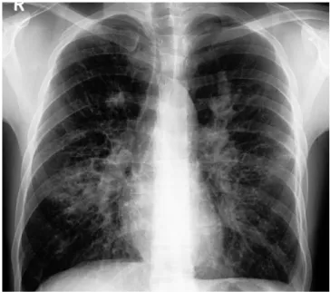
These can be depicted on the chest roentgenogram as paren- chymal fibrosis in the form of reticulolinear markings or honey- Fig. 1. Plain chest roentgenogram showing a left-sided perihilar opacity along
with non-homogeneous infiltrates in all zones of both lung fields.
Fig. 2. Plain chest roentgenogram of the same patient taken 4 months later showing spontaneous resolution of the left-sided perihilar opacity. The bilateral non-homogeneous infiltrates have increased considerably (Fig s. 1 and 2 reveal
‘transient pulmonary infiltrates’ or ‘fleeting shadows’, which are characteristic of ABPA).
combing, contracted upper lobes, cavitation and localized em- physema. The most characteristic permanent change, however, is the occurrence of CB with normal peripheral bronchi.76 This continues to be recognized as a hallmark of the disease.
Demonstration of CB
It is believed that bronchiectasis occurs in areas with previous consolidation. On plain chest roentgenograms, this is visual- ized either as parallel-line opacities, representing widening of the bronchi, or as ring opacities, 1-2 cm in diameter, represent- ing dilated bronchi en face. Parallel-line shadows were ob- served in 65%-70% of patients with ABPA and ring shadows in 45%-68%.65,69,70 Bronchography, once regarded as the gold stan- dard for the demonstration of bronchiectasis but now consid- ered obsolete, gave a one time complete picture of the whole tracheobronchial tree.77
Currently, CT of the thorax, high resolution in particular, is the modality of choice for the demonstration of bronchiectasis.
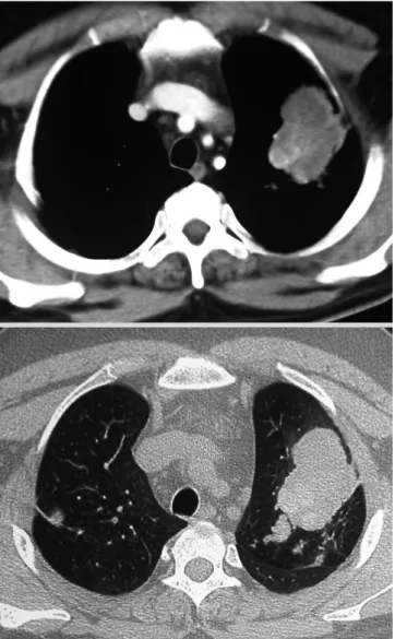
When compared to bronchography, CT had a sensitivity of 83%
and a specificity of 92% in detecting CB in patients with ABPA.77 In children with severe asthma, CT scans helped rapidly and safely establish the diagnosis of ABPA.78 Bronchiectasis on CT is characterised by the ‘string of pearl’ and ‘signet ring’ appear- ances (Fig. 3). Although demonstration of CB with normal pe- ripheral bronchi should continue to be regarded as a sine qua non for the diagnosis of ABPA in patients without CF, extension of CB to the periphery was found in 30% of the lobes and 21% of the segments.50
Other CT appearances (Table 3)
Besides bronchiectasis, the other bronchial abnormalities ob- served on CT include dilated and totally occluded bronchi, air-
fluid levels within dilated bronchi, and bronchial wall thicken- ing. Common parenchymal abnormalities are nonhomoge- neous patchy consolidation and parenchymal scarring of vary- ing extent, segmental or lobar collapse, cavities and emphyse- matous bullae.68 Cavitary lung disease associated with fibrosis in ABPA can often be difficult to differentiate from fibrocavitary lung disease of tuberculous origin.60,67,79
On high-resolution CT, high-attenuation mucous (HAM) plugs (Fig. 4) were reported in 28% of patients with ABPA.80 The ISHAM Working Group29 has highlighted this finding and con- siders HAM a pathognomonic feature of ABPA. In an analysis of 155 patients with ABPA, the presence of HAM was associated with significantly higher levels of eosinophils, total IgE, and IgE- Af at the time of diagnosis.81 Pleural abnormalities have also been observed in up to 43% of patients with ABPA.50 Pleural ef- fusion, most likely attributable to the mechanical effect of lung Fig. 3. Computed tomography of the thorax showing ‘signet ring’ (short, thick
arrow) and ‘string of pearls’ (long, thin arrow) appearances, indicative of central bronchiectasis. Mucoid impaction and dilated bronchi are also visualized.
Fig. 4. High resolution computed tomography of the thorax (mediastinal win- dow and corresponding section on the lung window) showing high attenuation mucous (HAM) impaction.
collapse, was first documented in 1981 in 2 patients with ABPA.82 We have also reported an ipsilateral pleural effusion secondary to lung collapse, which subsequently cleared on re- expansion of the lobe after steroid therapy, in a patient with ABPA, AAS and an operated aspergilloma.83 Spontaneous pneumothorax, bronchopleural fistula, pleural thickening, and pleural fibrosis have also been described.68,72
Laboratory findings
Apart from radiologic investigations, the laboratory findings useful for diagnosing and monitoring ABPA include skin testing with Aspergillus antigens, peripheral eosinophil count, serum total IgE, Af-specific IgE and IgG, and precipitating antibodies against Af. Expectoration of golden brownish sputum plugs, one of the 3 minor criteria laid down by Rosenberg and Patter- son,46 often provides the first clue in patients with asthma and CF. A positive sputum culture for Aspergillus species, another of the minor criteria, was noted in about 58% cases.63
Eosinophil count
Peripheral blood eosinophilia, one of the major criteria, can often be demonstrated. During exacerbations, most patients have an absolute eosinophil count between 1,000 and 3,000 per cumm. However, eosinophilia is also found in many other lung diseases; while a normal eosinophil count may be seen in pa- tients on treatment with corticosteroids. Since this test is not very specific, the ISHAM Working Group29 has included this test under ‘other’ criteria. Sputum eosinophilia may be demonstrat- ed in patients with productive cough.
Skin testing with Aspergillus antigens
Both type I (immediate) and type III (delayed) skin sensitivity with different Aspergillus antigens can be found in patients with ABPA. While the type III response is completely suppressed by steroid therapy, there is little or no effect on the type I reaction.
Both intradermal and prick tests have been used by different researchers to diagnose ABPA. Depending on the geographical area and the manufacturing technique employed, the Aspergil- lus antigen extracts available are not uniform.84 Currently, the prick test is used for the initial screening of ABPA. If the prick test is negative, then intradermal testing, which is more sensi- tive than the prick method, can be performed to elicit Aspergil- lus sensitization. However, higher false positive results are ob- served with intradermal tests.
Immediate skin hypersensitivity is not highly specific for ABPA. This is evidenced by the fact that approximately up to 40% of all asthmatics29 and up to 56% of patients with CF85 are sensitized to Af. To overcome this, recombinant Af allergens have now been cloned, purified and standardized for testing. In one of the initial studies in Af sensitized patients with CF, 3 out of 6 patients with ABPA did not demonstrate hypersensitivity with recombinant Af allergen I/a (rAsp f I/a).86 When skin test-
ing with rAsp f 1, rAsp f 3, rAsp f 4 and rAsp f 6 was performed on 50 patients with CF (12 with ABPA, 17 with Aspergillus sensi- tization without ABPA, and 21 not sensitized to Af), no reactivi- ty to 1:100 or higher dilutions of the cloned rAsp f 4 and rAsp f 6 was found in any of the 38 patients without ABPA.87 The authors suggested that these 2 recombinant allergens are reliable mark- ers for ABPA in CF.
Total serum IgE
Elevated total serum IgE is one of the minimal essential crite- ria51 as well as a component of the “truly minimal” diagnostic criteria7 both proposed by Greenberger. In spite of being recog- nized as a key criterion for diagnosing ABPA, there still remains a disagreement among different research groups in the cutoff level for IgE. In the Rosenberg-Patterson criteria,46 the IgE level for diagnosing ABPA was greater than 1,000 IU/mL (-2,500 ng/
mL). However, in the minimal essential criteria, Greenberger51 has given a reduced serum IgE level (>417 IU/mL or 1,000 ng/
mL) in order to establish the diagnosis. According to the ‘ABPA in CF’ consensus criteria, serum IgE >500 IU/mL is considered diagnostic.27 The ISHAM Working Group29 proposed a cutoff level of 1,000 IU/mL as was initially set forth by Rosenberg and Patterson.46 This was so because the Working Group29 “felt that a cutoff of 500 IU/mL may lead to overdiagnosis of ABPA.” The cutoff IgE value needs to be validated across all populations as it could possibly be affected by both ethnicity and risk of expo- sure to Aspergillus antigens. In an electronic survey conducted by Greenberger et al.54 among members of the American Acad- emy of Allergy, Asthma, and Immunology (AAAAI), informa- tion was sought on the diagnostic criteria and management practices adopted for ABPA. The authors observed that 44.9%
of allergists/immunologists used total IgE concentration ≥417 IU/mL when diagnosing ABPA while 42% respondents regard- ed IgE levels ≥1,000 IU/mL as the cutoff.
Specific IgE/IgG to A. fumigatus
Elevated IgE-Af and IgG-Af is also one of the minimal essen- tial criteria51 for the diagnosis of ABPA. Generally, double the serum values of IgE-Af and IgG-Af are found in patients with ABPA as compared to AIA.47 If the controls’ values are not avail- able for comparison, then very high serum levels of IgE-Af or IgG-Af, in an appropriate clinical setting, may be diagnostic of ABPA. The ISHAM Working Group29 has suggested IgE-Af level
>0.35 kUA/L to be diagnostic.
The radioimmunoassay (RIA) method replaced the previous- ly used enzyme-linked immunosorbent assay (ELISA) for esti- mating IgE-Af and IgG-Af as low levels of IgE-Af could not be detected by ELISA. However, the RIA technique was limited by the short shelf life of the radioisotope and exposure to radioac- tivity. Subsequently, the biotin-avidin-linked immunosorbent assay method was employed in 13 patients with ABPA, 9 with AIA, 12 with aspergilloma, and 9 controls without asthma,
which resulted in significantly higher IgE-Af levels in patients with ABPA, even at very high dilutions of 1:1,000.88 The authors attributed this finding to a polyclonal antibody response to As- pergillus antigens in patients with ABPA but not in those with AIA. Similar to demonstration of skin hypersensitivity, ABPA can be distinguished from AIA with high specificity (100%) and sensitivity (90%) by using recombinant Af allergens.89 High lev- els of specific IgE to recombinant Af allergens also helped de- tect ABPA in patients with underlying CF.90
Precipitating antibodies against A. fumigatus
By the double immunodiffusion technique of Outcherlony, precipitating antibodies against Af could be detected in the un- concentrated serum from 70% of patients.63 Using concentrated serum, this detection rate improved to 92% of patients with a radiological infiltrate.63 These precipitating antibodies have also been found in 10% of asthmatics without ABPA,4 aspergilloma and in different forms of chronic pulmonary aspergillosis (CPA). Denning et al.91 have correlated the presence of compli- cating features like fibrosis and cavitation with high titres of se- rum precipitins in patients with ABPA.
Pulmonary function testing
Pulmonary function testing does not help confirm the diagno- sis of ABPA. In addition to airflow obstruction, a restrictive pat- tern with reduction in total lung capacity (TLC), vital capacity (VC), forced expiratory volume in the first second (FEV1) and reduced diffusion capacity for carbon monoxide (DLCO) may be observed when the patient presents in the acute or the exac- erbation stage.92,93 Normalization of some of these parameters may be noticed after treatment with corticosteroids and also during remission. A significant (P<0.05) reduction in FEV1, FEV1/VC ratio and FEF25-75 was observed in patients with ABPA having mean duration of illness greater than 10 years when compared to those with symptoms less than 10 years’ duration.93 Staging
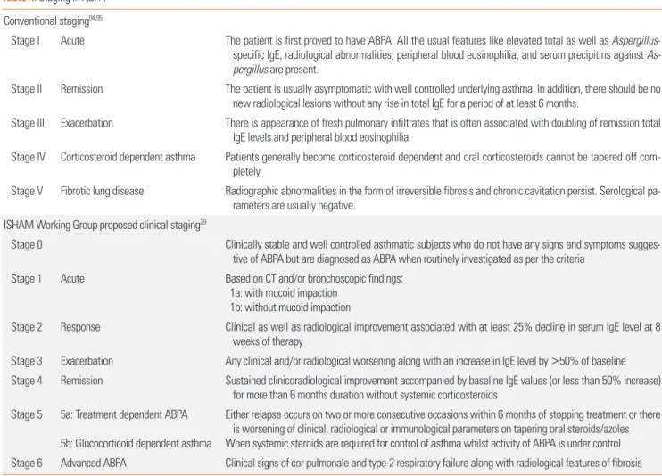
Five stages of ABPA94,95 have been identified, viz. (i) acute, (ii) remission, (iii) exacerbation, (iv) corticosteroid dependent asthma, and (v) fibrotic lung disease (Table 4). Staging of the disease must be done when establishing the diagnosis and should be reassessed during follow-up visits whenever the pa- tient improves or deteriorates. Improvement in symptoms, res-
Table 4. Staging in ABPA Conventional staging94,95
Stage I Acute The patient is first proved to have ABPA. All the usual features like elevated total as well as Aspergillus- specific IgE, radiological abnormalities, peripheral blood eosinophilia, and serum precipitins against As- pergillus are present.
Stage II Remission The patient is usually asymptomatic with well controlled underlying asthma. In addition, there should be no new radiological lesions without any rise in total IgE for a period of at least 6 months.
Stage III Exacerbation There is appearance of fresh pulmonary infiltrates that is often associated with doubling of remission total IgE levels and peripheral blood eosinophilia.
Stage IV Corticosteroid dependent asthma Patients generally become corticosteroid dependent and oral corticosteroids cannot be tapered off com- pletely.
Stage V Fibrotic lung disease Radiographic abnormalities in the form of irreversible fibrosis and chronic cavitation persist. Serological pa- rameters are usually negative.
ISHAM Working Group proposed clinical staging29
Stage 0 Clinically stable and well controlled asthmatic subjects who do not have any signs and symptoms sugges- tive of ABPA but are diagnosed as ABPA when routinely investigated as per the criteria
Stage 1 Acute Based on CT and/or bronchoscopic findings:
1a: with mucoid impaction 1b: without mucoid impaction
Stage 2 Response Clinical as well as radiological improvement associated with at least 25% decline in serum IgE level at 8 weeks of therapy
Stage 3 Exacerbation Any clinical and/or radiological worsening along with an increase in IgE level by >50% of baseline Stage 4 Remission Sustained clinicoradiological improvement accompanied by baseline IgE values (or less than 50% increase)
for more than 6 months duration without systemic corticosteroids Stage 5 5a: Treatment dependent ABPA
5b: Glucocorticoid dependent asthma
Either relapse occurs on two or more consecutive occasions within 6 months of stopping treatment or there is worsening of clinical, radiological or immunological parameters on tapering oral steroids/azoles When systemic steroids are required for control of asthma whilst activity of ABPA is under control Stage 6 Advanced ABPA Clinical signs of cor pulmonale and type-2 respiratory failure along with radiological features of fibrosis
olution of radiologic lesions, as well as a decline in total IgE and blood eosinophilia usually occurs with prednisolone therapy, or at times spontaneously. Exacerbations are entirely asymp- tomatic in approximately one-third of the cases, and may be detected either by the doubling of remission IgE values or dem- onstration of extensive radiologic opacities. Although remission for prolonged periods is not common, we reported an exacer- bation after prolonged remission in a patient with ABPA and an associated aspergilloma.96 Since therapy with prednisolone may mask the characteristic features of the disease, stage IV ABPA is clinically indistinguishable from corticosteroid depen- dent asthma without ABPA.97
In order to refine these stages, the ISHAM Working Group29 has proposed a new clinical staging of ABPA in asthma (Table 4).
Asymptomatic patients diagnosed with ABPA when routinely investigated as per the criteria were categorized as stage 0. This was done to recognize the disease as early as possible so that commencement of appropriate treatment even before the acute presentation (stage 1) could possibly prevent progression to end stage fibrosis. Stage 2 (response) sets in when there is clinical, radiological and serological improvement. However, this newly proposed staging would require prospective validation.
Radiological staging
A newly proposed radiological classification of ABPA that is based on thoracic CT findings has also been tabled by the ISH- AM Working Group.29 This new classification has 4 categories that correlate the immunological severity of ABPA with various CT features. As the disease progresses from mild to moderate to severe, the radiological classification is as follows: (1) serologi- cal ABPA (ABPA-S), (2) ABPA with bronchiectasis (ABPA-B), (3) ABPA with HAM (ABPA-HAM), and (4) ABPA with chronic pleuropulmonary fibrosis (ABPA-CPF). For inclusion in the AB- PA-CPF group, there should be at least 2 other radiologic fea- tures, apart from bronchiectasis and HAM, viz. pulmonary fi- brosis, parenchymal scarring, fibrocavitary lesions, aspergillo- ma, and pleural thickening.
Treatment
As described in the recent AAAAI Committee Report54 on ABPA, the goals of treatment of ABPA are to: (i) control symp- toms of asthma or CF, (ii) prevent or treat pulmonary exacerba- tions of ABPA, (iii) reduce or remit pulmonary inflammation, and (iv) mitigate progression to end-stage fibrotic or cavitary disease. Exclusion of ABPA in family members and identifica- tion of any potential environmental source of the incriminated fungus should also be stressed upon when managing a patient with ABPA. No definite prognostic indicators for progression or regression of the disease have been identified. In order to suc- cessfully achieve these goals, it is important to treat the disease aggressively during the early stages. Oral corticosteroids contin- ue to remain the cornerstone for the management of ABPA. Ap-
propriately designed clinical trials for the treatment of ABPA are lacking. The role of antifungal drugs so far is at best adjunctive.
Corticosteroids
Oral corticosteroids, till date, remain the most effective drugs for treating ABPA.98 The dosing schedule and duration of thera- py for oral steroids remain poorly defined. For Stages I (acute) and III (exacerbation), the most widely accepted protocol is prednisolone 0.5 mg/kg/day given as a single morning dose for the initial 2 weeks and then switched to an alternate-day dose for the next 6-8 weeks.51 Once the total serum IgE declines by at least 35% and resolution of radiologic infiltrates are noted, prednisolone is further tapered by 2.5 to 5 mg every 2 weeks.99 After discontinuation of prednisolone, if achieved, the patient should be monitored every 6 to 8 weeks to ensure that remis- sion is maintained. If there are any features suggestive of re- lapse, treatment should be recommenced as early as possible.
Patients with stage IV ABPA (steroid-dependent asthma) usual- ly require alternate day therapy with prednisolone 10-40 mg for many years to sustain symptom control. Daily prednisolone along with other interventions for the management of cor pul- monale and arterial hypoxemia are needed for patients with end stage lung disease (stage V).26
To minimize the well-known adverse effects of long term ste- roid therapy, we assessed the feasibility of a biweekly dosing schedule in 26 patients with ABPA with or without AAS.100 Fol- lowing an initial dosage of prednisolone 0.5 mg/kg/day for two weeks, patients were alternately prescribed either the conven- tional alternate-day regimen or a twice weekly dosing protocol was adopted. Patients receiving the biweekly regimen also showed a significant improvement in FEV1, total IgE levels, and eosinophil counts. Pulse therapy with intravenous methylpred- nisolone using 10 to 20 mg/kg/day for 3 consecutive days has been shown to be useful in managing severe and sometimes life-threatening exacerbations among children with ABPA and CF.101 In conjunction with antifungal agents, this could be con- sidered in patients not improving with oral steroid therapy. In- haled corticosteroids alone help achieve asthma control, but neither do they prevent symptomatic exacerbations of ABPA nor delay progression of lung damage.
Antifungal agents
The exact role of antifungal agents in the treatment of ABPA is still debated. By reducing the fungal load, antifungal agents help control the antigenic stimulus and thus decrease the in- flammatory response.102 Earlier studies with older antifungal molecules, viz. natamycin, hamycin, amphotericin B, micon- azole, clotrimazole and ketoconazole did not show much promise. Subsequently, studies with itraconazole demonstrat- ed a reduction in daily corticosteroid doses without clinical de- terioration. The Cochrane Database review103 on azoles for ABPA concluded that itraconazole improved clinical outcomes.
The dosage of itraconazole recommended is 200 mg twice daily for 4 to 6 months, which is then tapered over the next 4 to 6 months. By inhibiting steroid metabolism and thereby exacer- bating adrenal suppression, itraconazole might lead to cushin- goid features when used for very long durations.
To avoid drug resistance and possible clinical failure due to suboptimal therapeutic levels, regular monitoring of itracon- azole blood levels is advocated.104 In ABPA, the newer azoles, voriconazole and posaconazole, have improved asthma severi- ty in 70% and 78% of patients respectively.105 However, skin can- cer has been associated with long-term voriconazole therapy.106 It is still not known whether itraconazole and other newer azoles would successfully replace oral steroids as first-line ther- apy for ABPA. The results of a randomized trial on monothera- py of itraconazole vs prednisolone in ABPA (MIPA study) are awaited (clinical trials.gov; NCT01321827).
Omalizumab
Omalizumab, a monoclonal antibody against IgE, has also been tried in the management of ABPA. Initial studies in pa- tients with underlying CF have demonstrated a significant clin- ical improvement with reduction in hospitalisation and exacer- bations.107,108 Usage of oral corticosteroids in these patients also declined. Similar results have also been documented with omalizumab in patients with ABPA due to underlying asth- ma.109,110 This potential of this drug in decreasing or avoiding oral steroids in patients with stage IV (steroid-dependent) dis- ease should be investigated. Randomized trials with omalizum- ab and possibly other antibodies to IL-4Ra (dupilumab), IL 5 (mepolizumab), and IL 13 (lebrikizumab) are needed to assess their routine usage in ABPA.
ABPA and other pulmonary disorders
Rarely, ABPA has also recently been recognized in patients with chronic obstructive pulmonary disease.111,112 The associa- tion of ABPA or an overlap condition that resembles ABPA was described in patients with hyper-IgE syndrome, chronic granu- lomatous disease, and Kartagener’s syndrome.25 In these con- genital immunodeficiency neutrophilic conditions, it is essen- tial to distinguish ABPA from invasive aspergillosis as mistreat- ment with systemic steroids may hasten the invasive process, resulting in increased morbidity.
ABPA AND OTHER ASPERGILLUS–RELATED DISORDERS Although the clinical categories of Aspergillus-associated re- spiratory disorders usually remain mutually exclusive, similar immunopathologic responses may lead to the coexistence of different forms of respiratory aspergillosis. Concomitant occur- rence of ABPA and AAS may not be all that uncommon.18,74,83,113
Aspergilloma formation has also been documented in patients with ABPA.67,79,114 We have twice so far reported concurrent
ABPA, AAS, and aspergilloma in a single patient.83,115 Allergic Aspergillus sinusitis
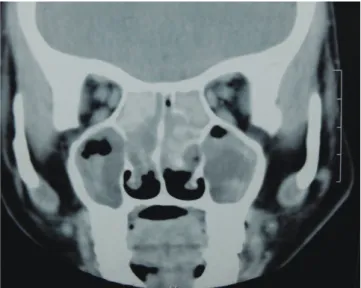
Just as in ABPA, Aspergillus antigens trigger immunologic re- actions in the paranasal sinuses, thereby leading to AAS.116,117 Those patients with rhinitis who were sensitized to Aspergillus are possibly at higher risk of developing AAS.118,119 Here too, a set of criteria is required to establish the diagnosis of AAS. The key components of the diagnostic criteria for AAS are (1) radio- logical evidence of sinusitis of 1 or more paranasal sinuses; (2) necrosed amorphous tissue along with edematous polyps infil- trated by eosinophils on histopathological evaluation of mate- rial from the sinus; (3) demonstration of fungal elements in na- sal discharge or in material obtained at the time of surgery by stain or culture; (4) absence of diabetes, previous or subsequent immunodeficiency disease, and treatment with immunosup- pressive drugs; and (5) absence of invasive fungal disease at the time of diagnosis or subsequently. The other features consid- ered include: (1) peripheral blood eosinophilia; (2) type I and type III cutaneous hypersensitivity to Aspergillus; (3) precipitat- ing antibodies to Aspergillus antigens; (4) elevated total and As- pergillus specific IgE levels; and (5) characteristic CT appear- ances. Features, such as passage of nasal plugs, recurrent nasal polyps, and radiographic evidence of pansinusitis, in patients having an atopic background point toward an allergic fungal phenomenon in the upper airways.113 ‘Allergic mucin,’ which is the characteristic nasal pathologic material comprising eosino- phils, Charcot-Leyden crystals, cellular debris, and scattered fungal hyphae, is the hallmark of this disease.116 The character- istic CT finding is the presence of heterogeneous densities with serpiginous areas of increased attenuation (Fig. 5) on noncon- trast scans.72 Histopathologic confirmation from the inspissated
Fig. 5. Computed tomography of the paranasal sinuses showing hyperdense le- sions in the ethmoid and maxillary sinuses bilaterally, suggestive of inspissated secretions.
mucus is a sine qua non for the diagnosis of AAS. The current approach to therapy includes an initial surgical debridement, followed by postoperative oral corticosteroids and supportive therapy.113,120
Aspergilloma
Chronic lung damage in ABPA, especially the presence of cav- itating lesions, may provide a favourable milieu for aspergillo- ma formation.67,79 Furthermore, steroid therapy could possibly accelerate the development of fungal balls in patients with cavi- tary lung disease.114 Vice versa, it has also been seen that a pre- existing aspergilloma, by functioning as a nidus for antigen stimulation in susceptible individuals, may lead to ABPA subse- quently.121 We have postulated that the coexistence of an asper- gilloma would likely lead to an increase in the severity of un- derlying ABPA.122
CPA
The entity CPA was first highlighted by Denning et al.91 in 2003 when they identified a subset of patients with pre-existing structural lung disease who were chronically affected by Asper- gillus but did not have any vascular or tissue invasion by the fungal hyphae. These patients were either immunocompetent or had only pulmonary (localised) immune suppression. On the basis of the radiological patterns, they were categorized as (1) chronic cavitary pulmonary aspergillosis (CCPA), (2) chron- ic fibrosing pulmonary aspergillosis (CFPA), and (3) chronic necrotizing pulmonary aspergillosis (CNPA). In CCPA, progres- sive cavitation or extension of a pre-existing cavity was noted;
and if left untreated, development of chronic scarring and marked pulmonary fibrosis over time led to CFPA. In the pres- ence of underlying confounding factors like alcoholism, smok- ing, AIDS, diabetes and corticosteroid treatment that caused mild to moderate immune dysfunction, a necrotizing condition developed, usually after enlargement of a thin-walled cavity.
This condition occurred rapidly within weeks or gradually over months, and was labeled as CNPA. The classification of CPA has evolved over the last decade.123,124 As these 3 forms are not easy to distinguish clinically, a new simplified classification for CPA has been proposed: (1) simple aspergilloma, (2) CCPA (complex aspergilloma) or slowly progressive CNPA (more than 3 months’ duration) and (3) subacute invasive pulmonary as- pergillosis or rapidly progressive CNPA of less than 3 months’
duration.123
Diagnostic criteria
Patients with CPA usually have chronic respiratory and con- stitutional symptoms for at least 3 months, progressive enlarge- ment and formation of new pulmonary cavities, positive serum precipitins against Aspergillus or isolation of Aspergillus spp.
from the cavitary lesion, and elevated inflammatory markers (C-reactive protein, plasma viscosity or erythrocyte sedimenta-
tion rate). Diagnostic criteria for CPA also include exclusion of other pulmonary conditions like active tuberculosis and malig- nancy that mimic the symptoms as well as no obvious condi- tions suggesting an immunocompromised state.91
ABPA, SAFS, and CPA
When cavitation, fibrosis, pleural thickening and aspergilloma develop in patients with ABPA, CPA ensues. In a study of 126 patients with CPA, ABPA was the primary underlying condition in 15 subjects (11.9%) while SAFS was incriminated in 2 sub- jects (1.6%).125 A scoping review28 also attempted to estimate the global burden of CPA in patients with ABPA. Applying an annu- al 15% attrition rate while calculating the period prevalence of CPA over 5 years, the global case burden of CPA complicating ABPA was approximately 10% (range, 7%-20%).28
CONCLUSIONS
Sensitization to molds in patients with asthma is known to in- crease the severity of the disease.126 Patients with asthma, eo- sinophilia, and history of repeated ‘pneumonitis’ should be evaluated aggressively for ABPA. This would help avoid diag- nostic delay and prevent steady lung damage leading to end- stage fibrosis. In high tuberculosis-prevalent regions, the strik- ing radiological resemblance often results in erroneous treat- ment with antituberculous drugs. Furthermore, poor access to costly and advanced diagnostic modalities like CT scans and mycoserological tests in low-income countries may hamper es- tablishment of the diagnosis. The occurrence of other Aspergil- lus-related hypersensitivity respiratory disorders must be sought for in all patients with ABPA.127,128
REFERENCES
1. Shah A. Allergic bronchopulmonary aspergillosis. Indian J Chest Dis Allied Sci 1998;40:41-54.
2. deShazo RD, Chapin K, Swain RE. Fungal sinusitis. N Engl J Med 1997;337:254-9.
3. Hendrick DJ, Davies RJ, D’Souza MF, Pepys J. An analysis of skin prick test reactions in 656 asthmatic patients. Thorax 1975;30:2-8.
4. Schwartz HJ, Citron KM, Chester EH, Kaimal J, Barlow PB, Baum GL, et al. A comparison of the prevalence of sensitization to As- pergillus antigens among asthmatics in Cleveland and London. J Allergy Clin Immunol 1978;62:9-14.
5. Maurya V, Gugnani HC, Sarma PU, Madan T, Shah A. Sensitiza- tion to Aspergillus antigens and occurrence of allergic broncho- pulmonary aspergillosis in patients with asthma. Chest 2005;127:
1252-9.
6. Denning DW, O’Driscoll BR, Hogaboam CM, Bowyer P, Niven RM. The link between fungi and severe asthma: a summary of the evidence. Eur Respir J 2006;27:615-26.
7. Greenberger PA. When to suspect and work up allergic broncho- pulmonary aspergillosis. Ann Allergy Asthma Immunol 2013;111:
1-4.
8. Pasqualotto AC, Powell G, Niven R, Denning DW. The effects of antifungal therapy on severe asthma with fungal sensitization and allergic bronchopulmonary aspergillosis. Respirology 2009;14:
1121-7.
9. Denning DW, O’Driscoll BR, Powell G, Chew F, Atherton GT, Vyas A, et al. Randomized controlled trial of oral antifungal treatment for severe asthma with fungal sensitization: The Fungal Asthma Sensitization Trial (FAST) study. Am J Respir Crit Care Med 2009;
179:11-8.
10. Agbetile J, Bourne M, Fairs A, Hargadon B, Desai D, Broad C, et al.
Effectiveness of voriconazole in the treatment of Aspergillus fu- migatus-associated asthma (EVITA3 study). J Allergy Clin Immu- nol 2014;134:33-9.
11. Shah A, Panjabi C. Allergic bronchopulmonary aspergillosis: a re- view of a disease with a worldwide distribution. J Asthma 2002;39:
273-89.
12. Patterson R. Allergic bronchopulmonary aspergillosis and hyper- sensitivity reactions to fungi. In: Fishman AP, Elias JA, Fishman JA, Grippi MA, Kaiser LR, Senior RM, editors. Fishman’s pulmonary diseases and disorders. 3rd ed. New York (NY): McGraw-Hill;
1998. 777-82.
13. Patterson R. Allergic bronchopulmonary aspergillosis: a historical perspective. Immunol Allergy Clin North Am 1998;18:471-8.
14. Chowdhary A, Agarwal K, Kathuria S, Gaur SN, Randhawa HS, Meis JF. Allergic bronchopulmonary mycosis due to fungi other than Aspergillus: a global overview. Crit Rev Microbiol 2014;40:30- 48.
15. Lötvall J, Akdis CA, Bacharier LB, Bjermer L, Casale TB, Custovic A, et al. Asthma endotypes: a new approach to classification of dis- ease entities within the asthma syndrome. J Allergy Clin Immunol 2011;127:355-60.
16. Hinson KF, Moon AJ, Plummer NS. Broncho-pulmonary aspergil- losis; a review and a report of eight new cases. Thorax 1952;7:317- 33.
17. Graves TS, Fink JN, Patterson R, Kurup VP, Scanlon GT. A familial occurrence of allergic bronchopulmonary aspergillosis. Ann In- tern Med 1979;91:378-82.
18. Shah A, Khan ZU, Chaturvedi S, Malik GB, Randhawa HS. Con- comitant allergic Aspergillus sinusitis and allergic bronchopul- monary aspergillosis associated with familial occurrence of aller- gic bronchopulmonary aspergillosis. Ann Allergy 1990;64:507-12.
19. Shah A, Kala J, Sahay S, Panjabi C. Frequency of familial occur- rence in 164 patients with allergic bronchopulmonary aspergillo- sis. Ann Allergy Asthma Immunol 2008;101:363-9.
20. Shah A, Alamoudi O, Al-Mobeireek AF. Allergic bronchopulmona- ry aspergillosis: a view from India. Saudi Med J 2002;23:1559-60.
21. Novey HS. Epidemiology of allergic bronchopulmonary aspergil- losis. Immunol Allergy Clin North Am 1998;18:641-53.
22. Henderson AH, English MP, Vecht RJ. Pulmonary aspergillosis. A survey of its occurrence in patients with chronic lung disease and a discussion of the significance of diagnostic tests. Thorax 1968;23:
513-8.
23. Patterson R, Golbert TM. Hypersensitivity disease of the lung.
Univ Mich Med Cent J 1968;34:8-11.
24. Greenberger PA, Patterson R. Allergic bronchopulmonary asper- gillosis and the evaluation of the patient with asthma. J Allergy Clin Immunol 1988;81:646-50.
25. Shah A, Panjabi C. Allergic aspergillosis of the respiratory tract.
Eur Respir Rev 2014;23:8-29.
26. Greenberger PA. Allergic bronchopulmonary aspergillosis. In:
Middleton E Jr, Reed CE, Ellis EF, Franklin AN Jr, Yunginger JW, Busse WW, edtors. Allergy: principles and practice. 5th ed. St. Lou- is (MO): Mosby; 1998. 981-93.
27. Stevens DA, Moss RB, Kurup VP, Knutsen AP, Greenberger P, Jud- son MA, et al. Allergic bronchopulmonary aspergillosis in cystic fibrosis--state of the art: Cystic Fibrosis Foundation Consensus Conference. Clin Infect Dis 2003;37 Suppl 3:S225-64.
28. Denning DW, Pleuvry A, Cole DC. Global burden of allergic bron- chopulmonary aspergillosis with asthma and its complication chronic pulmonary aspergillosis in adults. Med Mycol 2013;51:
361-70.
29. Agarwal R, Chakrabarti A, Shah A, Gupta D, Meis JF, Guleria R, et al. Allergic bronchopulmonary aspergillosis: review of literature and proposal of new diagnostic and classification criteria. Clin Exp Allergy 2013;43:850-73.
30. Bhatnagar PK, Banerjee B, Shah A, Sarma PU. Probable role of IgG subclasses in patients with allergic bronchopulmonary aspergillo- sis. Serodiagn Immunother Infect Dis 1993;5:123-4.
31. Kurup VP. Immunology of allergic bronchopulmonary aspergillo- sis. Indian J Chest Dis Allied Sci 2000;42:225-37.
32. Wark PA, Gibson PG. Allergic bronchopulmonary aspergillosis:
new concepts of pathogenesis and treatment. Respirology 2001;6:
1-7.
33. Sambatakou H, Pravica V, Hutchinson IV, Denning DW. Cytokine profiling of pulmonary aspergillosis. Int J Immunogenet 2006;33:
297-302.
34. Madan T, Banerjee B, Bhatnagar PK, Shah A, Sarma PU. Identifi- cation of 45 kD antigen in immune complexes of patients of aller- gic bronchopulmonary aspergillosis. Mol Cell Biochem 1997;166:
111-6.
35. Purkayastha S, Madan T, Shah A, Krishnamurthy HG, Sarma PU.
Multifunctional antigens of A. fumigatus and specific antibodies.
Appl Biochem Biotechnol 2000;83:271-83.
36. Chauhan B, Santiago L, Kirschmann DA, Hauptfeld V, Knutsen AP, Hutcheson PS, et al. The association of HLA-DR alleles and T cell activation with allergic bronchopulmonary aspergillosis. J Im- munol 1997;159:4072-6.
37. Chauhan B, Santiago L, Hutcheson PS, Schwartz HJ, Spitznagel E, Castro M, et al. Evidence for the involvement of two different MHC class II regions in susceptibility or protection in allergic bronchopulmonary aspergillosis. J Allergy Clin Immunol 2000;
106:723-9.
38. Miller PW, Hamosh A, Macek M Jr, Greenberger PA, MacLean J, Walden SM, et al. Cystic fibrosis transmembrane conductance regulator (CFTR) gene mutations in allergic bronchopulmonary aspergillosis. Am J Hum Genet 1996;59:45-51.
39. Eaton TE, Weiner Miller P, Garrett JE, Cutting GR. Cystic fibrosis transmembrane conductance regulator gene mutations: do they play a role in the aetiology of allergic bronchopulmonary aspergil- losis? Clin Exp Allergy 2002;32:756-61.
40. de Almeida MB, Bussamra MH, Rodrigues JC. Allergic broncho- pulmonary aspergillosis in paediatric cystic fibrosis patients. Pae- diatr Respir Rev 2006;7:67-72.
41. Saxena S, Madan T, Shah A, Muralidhar K, Sarma PU. Association of polymorphisms in the collagen region of SP-A2 with increased levels of total IgE antibodies and eosinophilia in patients with al- lergic bronchopulmonary aspergillosis. J Allergy Clin Immunol 2003;111:1001-7.