Received February 3, 2016, Revised May 31, 2016, Accepted for publication June 28, 2016
Corresponding author: You Chan Kim, Department of Dermatology, Ajou University School of Medicine, 164 WorldCup-ro, Yeongtong-gu, Suwon 16499, Korea. Tel: 82-31-219-5190, Fax: 82-31-219-5189, E-mail: maychan@
ajou.ac.kr
This is an Open Access article distributed under the terms of the Creative Commons Attribution Non-Commercial License (http://creativecommons.
org/licenses/by-nc/4.0) which permits unrestricted non-commercial use, distribution, and reproduction in any medium, provided the original work is properly cited.
Copyright © The Korean Dermatological Association and The Korean Society for Investigative Dermatology
pISSN 1013-9087ㆍeISSN 2005-3894
Ann Dermatol Vol. 29, No. 2, 2017 https://doi.org/10.5021/ad.2017.29.2.149
ORIGINAL ARTICLE
Low-Level Light Therapy with 410 nm Light Emitting Diode Suppresses Collagen Synthesis in Human Keloid Fibroblasts: An In Vitro Study
Hyun Soo Lee, Soo-Eun Jung, Sue Kyung Kim, You-Sun Kim1, Seonghyang Sohn2, You Chan Kim
Departments of Dermatology and 1Biochemistry, 2Laboratory of Cell Biology, Ajou University School of Medicine, Suwon, Korea
Background: Keloids are characterized by excessive colla- gen deposition in the dermis, in which transforming growth factor β (TGF-β)/Smad signaling plays an important role.
Low-level light therapy (LLLT) is reported as effective in pre- venting keloids in clinical reports, recently. To date, studies investigating the effect of LLLT on keloid fibroblasts are ex- tremely rare. Objective: We investigated the effect of LLLT with blue (410 nm), red (630 nm), and infrared (830 nm) light on the collagen synthesis in keloid fibroblasts. Methods:
Keloid fibroblasts were isolated from keloid-revision surgery samples and irradiated using 410-, 630-, 830-nm light emit- ting diode twice, with a 24-hour interval at 10 J/cm2. After ir- radiation, cells were incubated for 24 and 48 hours and re- al-time quantitative reverse transcription polymerase chain reaction was performed. Western blot analysis was also per- formed in 48 hours after last irradiation. The genes and pro- teins of collagen type I, TGF-β1, Smad3, and Smad7 were analyzed. Results: We observed no statistically significant change in the viability of keloid fibroblasts after irradiation.
Collagen type I was the only gene whose expression sig- nificantly decreased after irradiation at 410 nm when com- pared to the non-irradiated control. Western blot analysis showed that LLLT at 410 nm lowered the protein levels of col-
lagen type I compared to the control. Conclusion: LLLT at 410 nm decreased the expression of collagen type I in keloid fibroblasts and might be effective in preventing keloid for- mation in their initial stage. (Ann Dermatol 29(2) 149∼155, 2017)
-Keywords-
Collagen type I, Keloid fibroblast, Low-level light therapy
INTRODUCTION
Keloids are characterized by hyperproliferative growth of dermal fibroblasts and excessive collagen deposition in the dermis1,2. Previous studies suggested that the initial step in the development of the fibrotic reaction in keloids involves the expression of transforming growth factor β1 (TGF-β1) in neovascular endothelial cells, thus inducing the adjacent fibroblasts to express markedly high levels of TGF-β1/β2 and their receptors, as well as type I and VI collagen3,4. Recent studies emphasize a potential role for TGF-β1 intracellular signaling pathways, especially for the Smad pathway, in the pathogenesis of keloids4. Smad2 and Smad3 are overexpressed and the inhibitory Smad6 and Smad7 are reduced in keloid fibroblasts1,3,5.
Treatments for keloids include surgical excision, intrale- sional corticosteroids, 5-fluorouracil, bleomycin and inter- feron, topical imiquimod, compression, cryotherapy, radi- ation, silicon sheeting and laser or light-based therapies, showing variable success. Recurrence is common, even with combination therapy6. Therefore, preventing keloid is an important issue.
Low-level light therapy (LLLT) uses low levels of visible to near infrared light. It has been widely used to reduce pain
and inflammation, to promote wound healing, and to pre- vent tissue necrosis. Many reports demonstrate that LLLT modulates normal dermal fibroblasts and the TGF-β fam- ily1. Recently, two clinical reports showed that LLLT was effective in preventing hypertrophic scars and keloids7,8. However, studies investigating the effect of LLLT on keloid fibroblasts are still rare, especially the ones investigating the effects of LLLT at molecular level. Herein, we aimed to evaluate the effect of LLLT using blue (410 nm), red (630 nm), and infrared (830 nm) light on the collagen synthesis in keloid fibroblasts, by observing the signaling molecules involved.
MATERIALS AND METHODS
Isolation and culture of keloid fibroblasts
Keloid fibroblasts were isolated from keloid-revision sur- gery samples after obtaining informed consents from the patients. Keloid fibroblasts were cultured in Dulbecco’s modified Eagle’s medium (DMEM), supplemented with 10% fetal bovine serum, 100 U/ml penicillin and 100 μg/ml streptomycin at 37oC in a humidified atmosphere contain- ing 5% carbon dioxide (CO2).
Irradiation
Keloid fibroblasts were seeded in a 6-well plate at 3.0×10⁵ cells/well. APLⓇ (Medro Medical Division, Seoul, Korea) was used for this study. This device produces blue (410 nm), red (630 nm), and infrared (830 nm) light emit- ting diode (LED) light. The power of each wavelength was 205 mW/cm2 for blue, 172 mW/cm2 for red, 50 mW/cm2 for infrared. Keloid fibroblasts were irradiated twice, with the second irradiation 24 hours after the first one. Prelimi- nary experiment was performed to determine the lowest fluence to inhibit the proliferation of keloid fibroblasts.
Cells were maintained in 400 μl Dulbecco’s phosphate-buf- fered saline during irradiation and transferred in DMEM after irradiation for 24 or 48 hours.
Cell viability assay
To determine the direct effect of LLLT on the proliferation of viable cells, we counted the number of viable cells us- ing the EZ-CyTox cell viability assay kit (Daeillab, Seoul, Korea). Twenty-four hours after the last irradiation, the cells were treated with 40 μl of Ez-CyTox solution and in- cubated for additional 3 hours at 37oC. Absorbance was measured at 450 nm using an ELISA reader (Molecular Device, Menlo Park, CA, USA). Viability was expressed as percentage of viable cells compared to the non-irradiated control. The experiment was repeated twice with two set of fibroblasts.
Real-time quantitative reverse transcription polymerase chain reaction
The mRNA of collagen type I, TGF-β1, Smad3, and Smad7 were measured in 24 and 48 hours after last irra- diation. Total RNA was isolated using RNAasyⓇ mini kit (Quiagen, Hilden, Germany). cDNA transcription was per- formed using the SuperscriptⓇ III First-Strand (Invitrogen, Carlsbad, CA, USA) according to the manufacturer’s in- structions. cDNA was analyzed by real-time quantitative reverse transcription polymerase chain reaction using ABI Prism 7000 Sequence Detection System (Applied Biosystems, Foster, CA, USA). Fluorescent signals were collected dur- ing extension phase, Ct values of the sample were calcu- lated, and the transcript levels were analyzed by 2−ΔΔCt method. Primers and internal probes for collagen type I, TGF-β1, Smad3, and Smad7 were purchased as assays on demand primer-probe sets (Applied Biosystems). The ex- periment was repeated five times.
Western blot analysis
The protein of collagen type I, TGF-β1, Smad3, and Smad7 were measured in 48 hours after last irradiation.
Cells were lysed in lysis buffer (50 mM Tris [pH 7.4], 150 mM NaCl, 0.1% NP 40, 1% sodium dodecyl sul- fate [SDS], 0.5% deoxycholic acid, 1 mM EDTA, protease and phosphatase inhibitors). Samples were separated on 10% SDS-polyacrylamide gels and transferred onto poly- vinyldenefluoride membranes. The protein blots were in- cubated with antibodies against collagen type I (Abcam, Cambridge, MA, USA), TGF-β1 (GeneTex, Irvine, CA, USA), Smad3 (Epitomics, Burlingame, CA, USA), Smad7 (Santa Cruz Biotechnology, Santa Cruz, CA, USA) and ac- tin (Santa Cruz Biotechnology). Detection was performed via enhanced chemiluminescence (Amersham Bioscien- ces, Buckinghamshire, UK). The intensity of each band was quantified using Image J and the data were reported as relative intensity according to actin.
Statistical analysis
IBM SPSS Statistics Desktop ver. 20.0.0 (IBM Co., Armonk, NY, USA) was used for statistical analysis. Results are ex- pressed as mean and standard deviation. Mann-Whitney U test was used for statistical evaluation between control and experimental groups in the study. A p-value lower than 0.05 was considered statistically significant.
RESULTS
Determination of the fluence of LLLT
Keloid fibroblasts were irradiated once with 3, 10, 30
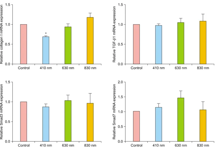
Fig. 2. Only expression of collagen type I showed a significant decrease in cell irradiate with 410 nm light compared to non-irradiated control after 48 hours. The results are represented as mean±standard error from five independent experiments. TGF-β1: transforming growth factor β1. *p<0.05 vs. non-irradiated control.
Fig. 1. Viable keloid fibroblasts had tendency to decrease after irradiation with all wavelengths compared to non-irradiated keloid control. However, there was no statistically significant decrease compared to the control. The results are represented mean±standard error from two independent experiments with two set of fibroblasts.
J/cm2 fluences of the three wavelengths of 410-, 630-, and 830-nm as preliminary experiment. Tryptan blue assay ex- hibited proliferations rates of keloid fibroblasts tend to de- crease only at 10 and 30 J/cm2, showing rather increased proliferation rate at 3 J/cm2, in all wavelengths, although it was not significant (Appendix). In the condition of similar proliferation rate, using lower dose is more suitable and convenient for conducting experiment. Therefore, we de- termined to irradiate 10 J/cm2 for keloid fibroblasts.
Cell viability assay
Viable keloid fibroblasts decreased with irradiation with all wavelengths compared to non-irradiated control.
However, it was not statistically significant (Fig. 1).
mRNA expression of TGF-β/Smad signaling components Collagen type I expression was significantly decreased with irradiation at 410 nm in 48 hours (p<0.005). We al- so observed lower Smad3 expression after irradiation at 410 nm in 48 hours, but it was not statistically significant (p=0.065). There were no differences between irradiated
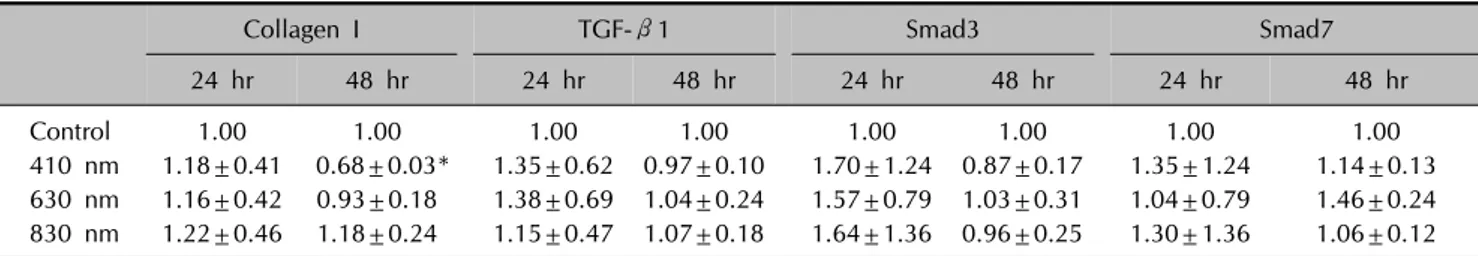
Table 1. Relative mRNA level of collagen type I, TGF-β1, Smad3, and Smad7 after LLLT at 10 J/cm2 in keloid fibroblasts
Collagen I TGF-β1 Smad3 Smad7
24 hr 48 hr 24 hr 48 hr 24 hr 48 hr 24 hr 48 hr
Control 1.00 1.00 1.00 1.00 1.00 1.00 1.00 1.00
410 nm 1.18±0.41 0.68±0.03* 1.35±0.62 0.97±0.10 1.70±1.24 0.87±0.17 1.35±1.24 1.14±0.13 630 nm 1.16±0.42 0.93±0.18 1.38±0.69 1.04±0.24 1.57±0.79 1.03±0.31 1.04±0.79 1.46±0.24 830 nm 1.22±0.46 1.18±0.24 1.15±0.47 1.07±0.18 1.64±1.36 0.96±0.25 1.30±1.36 1.06±0.12 Values are presented as mean only or mean±standard error. TGF-β1: transforming growth factor ß1, LLLT: low-level light therapy.
*p<0.05 vs. non-irradiated control.
Fig. 3. Low-level light therapy suppressed the expression of collagen type I in keloid fibroblasts irradiated with 410 nm light.
TGF-β1: transforming growth factor β1.
samples in 24 hours and non-irradiated controls (Fig. 2, Table 1).
Protein levels of TGF-β/Smad signaling components Irradiation at 410 nm reduced the protein levels of colla- gen type I in 48 hours. However, we observed no differ- ences between irradiated samples and the control for TGF- β1, Smad3, and Smad7 (Fig. 3).
DISCUSSION
In the present study, we investigated whether LLLT could affect the collagen synthesis in keloid fibroblasts. Irradiation with 410 nm decreased collagen type I expression in ke- loid fibroblasts.
In normal fibroblasts, the effect of LLLT can be explained by photobiomodulation. Wavelengths above 500 nm are widely used to stimulate wound healing9. Their energy is absorbed by the cytochrome C oxidase in the mitochon- drial membrane and activates various cellular signaling pathways through inducing the production of nitrogen ox-
ide, adenosine triphosphate and reactive oxygen species (ROS)10. It can induce the transcription of various growth factors including platelet-derived growth factor, TGF-β, and fibroblast growth factor, and cytokines such as inter- leukins and tumor necrosis factor-alpha. On the other hand, blue light induces a higher production of intra- cellular ROS in a dose-dependent manner, which might act as a mediator of cellular effects of blue light11. Because of their relative close wavelengths, it is possible that blue and ultraviolet A light has similar biological effect such as ROS generation and immunomodulation11. Flavins are photo-acceptors and involved in the generation of ROS.
Flavins are small, water-soluble molecules that absorb wavelengths below 500 nm and initiate free radical re- actions when excited by light. Flavin-mediated photo- sensitization generates cytotoxic free radicals and is re- sponsible for significant biological effects such as muta- genesis12,13.
Previous reports indicate that 410 nm LED irradiation is cytotoxic for normal fibroblasts. Opländer et al.14 demon- strated that single irradiation with 410 or 420 nm LED at 5∼10 J/cm2 inhibits the proliferation of dermal fibroblasts.
Bonatti et al.15 also reported a significant reduction in the number of normal fibroblasts irradiated with 470 nm light at 18 J/cm2. According to Seo et al.16, red (630 nm, 9.5 J/cm2) and green (530 nm, 9.8 J/cm2) LED-irradiated cells were significantly more proliferated than cells irradiated with blue light (460 nm, 27 J/cm2). Also, blue light did not increase the expression of collagen type I and TGF-β1 in the dermis compared to the control, whereas red and green light enhance the expression of both genes.
In contrast to studies on normal fibroblasts, reports on the effect of LLLT on keloid fibroblasts are extremely rare and disappointing. Bonatti et al.15 showed that a single irradi- ation with 470 nm LED at 6, 12, and 18 J/cm2 does not in- duce significant differences in the number of keloid fibroblasts. To our knowledge, no study reports that LLLT affects the molecules investigated here in keloid fibroblasts.
Our results showed that collagen mRNA levels decreased
after irradiation in keloid fibroblasts, which means that LLLT inhibited the transcription of this gene. Unfortunately, it is uncertain whether the TGF-β/Smad signaling path- way is inhibited in keloid fibroblasts, because other down- stream signaling components, except collagen type I, did not show any significant changes in their expression after LLLT. It should be considered that LLLT also affects other signaling pathways in keloid, such as the mitogen-ac- tivated protein kinase (MAPK), which is suggested to me- diate TGF-β⁄Smad signal transduction17,18. The influence of MAPK on keloid fibroblasts was evaluated after pulsed dye laser (PDL) treatment in a few reports. Activation of extracellular signal-regulated kinase and the p38 signal transduction pathway is involved in the suppression of ke- loid fibroblast proliferation and the induction of apoptosis after PDL treatment19,20. Recently, possible role of wing- less type (Wnt)/β-catenin signaling pathway in keloid pathogenesis has been reported, which was known to play a key role in cellular proliferation. Wnt5a and β-catenin, its effector, were highly expressed in keloid fibroblasts and mediate the effect of TGF-β in wound healing process21. In addition, unlike normal fibroblasts, abnormal TGF-β signaling and autocrine loop are important in keloid fibro- blasts1,4. For example, keloid fibroblasts show marked sen- sitivity to TGF-β, causing an accelerated increase in colla- gen and fibronectin synthesis compared to normal fibro- blast22,23. Therefore, the peak time of the TGF-β/Smad sig- naling components in keloid fibroblasts might be different from that of normal fibroblasts and it might be difficult to predict their expression pattern1,4.
This study has significance, in that it demonstrated that LLLT with 410 nm light inhibited collagen synthesis in ke- loid fibroblasts in vitro. Considering the initial pathophysi- ology of keloid, in which the expression of the TGF-β1 activates the adjacent fibroblasts to produce collagen, LLLT might be effective in preventing keloid formation at the initial stage. Corresponding clinical studies with blue LED and further investigative studies are needed to dem- onstrate the signaling mechanisms in keloid fibroblasts af- ter LLLT. We hope this study can be a starting point for clinical application of LLLT to patients with keloid scars.
ACKNOWLEDGMENT
This study was supported by Basic Science Research Program through the National Research Foundation of Korea (NRK) (Grant no. 2013R1A1A2006944). This work was supported by the GRRC program of Gyeonggi prov- ince (GRRC AJOU 2016B04, Photonics-Medical Convert- gence Technology Research Center).
CONFLICTS OF INTEREST
The authors have nothing to disclose.
REFERENCES
1. Tsujita-Kyutoku M, Uehara N, Matsuoka Y, Kyutoku S, Ogawa Y, Tsubura A. Comparison of transforming growth factor-beta/Smad signaling between normal dermal fibroblasts and fibroblasts derived from central and peripheral areas of keloid lesions. In Vivo 2005;19:959- 963.
2. Park SY, Park JY, Kim CH, Kang SU, Kim JH, Bark KM, et al.
Effects of Xanthium stramarium and Psoralea corylifolia extracts combined with UVA1 irradiation on the cell proliferation and TGF-β1 expression of keloid fibroblasts.
Ann Dermatol 2013;25:304-309.
3. Chin GS, Liu W, Peled Z, Lee TY, Steinbrech DS, Hsu M, et al. Differential expression of transforming growth factor- beta receptors I and II and activation of Smad 3 in keloid fibroblasts. Plast Reconstr Surg 2001;108:423-429.
4. Jagadeesan J, Bayat A. Transforming growth factor beta (TGFbeta) and keloid disease. Int J Surg 2007;5:278-285.
5. Phan TT, Lim IJ, Aalami O, Lorget F, Khoo A, Tan EK, et al.
Smad3 signalling plays an important role in keloid pa- thogenesis via epithelial-mesenchymal interactions. J Pathol 2005;207:232-242.
6. Mamalis AD, Lev-Tov H, Nguyen DH, Jagdeo JR. Laser and light-based treatment of Keloids--a review. J Eur Acad Dermatol Venereol 2014;28:689-699.
7. Barolet D, Boucher A. Prophylactic low-level light therapy for the treatment of hypertrophic scars and keloids: a case series. Lasers Surg Med 2010;42:597-601.
8. Carvalho RL, Alcântara PS, Kamamoto F, Cressoni MD, Casarotto RA. Effects of low-level laser therapy on pain and scar formation after inguinal herniation surgery: a randomized controlled single-blind study. Photomed Laser Surg 2010;
28:417-422.
9. da Silva JP, da Silva MA, Almeida AP, Lombardi Junior I, Matos AP. Laser therapy in the tissue repair process: a literature review. Photomed Laser Surg 2010;28:17-21.
10. Hamblin MR, Demidova T. Mechanisms of low level light therapy. Proc of SPIE 2006;6140:614001-12.
11. Mamalis A, Garcha M, Jagdeo J. Light emitting diode- generated blue light modulates fibrosis characteristics:
fibroblast proliferation, migration speed, and reactive oxygen species generation. Lasers Surg Med 2015;47:210-215.
12. Gorgidze LA, Oshemkova SA, Vorobjev IA. Blue light inhibits mitosis in tissue culture cells. Biosci Rep 1998;
18:215-224.
13. Eichler M, Lavi R, Shainberg A, Lubart R. Flavins are source of visible-light-induced free radical formation in cells.
Lasers Surg Med 2005;37:314-319.
14. Opländer C, Hidding S, Werners FB, Born M, Pallua N, Suschek CV. Effects of blue light irradiation on human dermal fibroblasts. J Photochem Photobiol B 2011;103:
118-125.
15. Bonatti S, Hochman B, Tucci-Viegas VM, Furtado F, Pinfildi CE, Pedro AC, et al. In vitro effect of 470 nm LED (Light Emitting Diode) in keloid fibroblasts. Acta Cir Bras 2011;
26:25-30.
16. Seo YK, Park JK, Song C, Kwon SY. Comparison of light-emitting diode wavelength on activity and migration of rabbit ACL cells. Lasers Med Sci 2014;29:245-255.
17. He S, Liu X, Yang Y, Huang W, Xu S, Yang S, et al.
Mechanisms of transforming growth factor beta(1)/Smad signalling mediated by mitogen-activated protein kinase pathways in keloid fibroblasts. Br J Dermatol 2010;162:
538-546.
18. Lim IJ, Phan TT, Tan EK, Nguyen TT, Tran E, Longaker MT, et al. Synchronous activation of ERK and phosphatidy- linositol 3-kinase pathways is required for collagen and extracellular matrix production in keloids. J Biol Chem 2003;278:40851-40858.
19. Kuo YR, Wu WS, Jeng SF, Huang HC, Yang KD, Sacks JM,
et al. Activation of ERK and p38 kinase mediated keloid fibroblast apoptosis after flashlamp pulsed-dye laser treat- ment. Lasers Surg Med 2005;36:31-37.
20. Kuo YR, Wu WS, Wang FS. Flashlamp pulsed-dye laser suppressed TGF-beta1 expression and proliferation in cultured keloid fibroblasts is mediated by MAPK pathway.
Lasers Surg Med 2007;39:358-364.
21. Igota S, Tosa M, Murakami M, Egawa S, Shimizu H, Hya- kusoku H, et al. Identification and characterization of Wnt signaling pathway in keloid pathogenesis. Int J Med Sci 2013;10:344-354.
22. Babu M, Diegelmann R, Oliver N. Keloid fibroblasts exhibit an altered response to TGF-beta. J Invest Dermatol 1992;
99:650-655.
23. Younai S, Nichter LS, Wellisz T, Reinisch J, Nimni ME, Tuan TL. Modulation of collagen synthesis by transforming growth factor-beta in keloid and hypertrophic scar fibro- blasts. Ann Plast Surg 1994;33:148-151.
Appendix. Proliferation rate of keloid fibroblasts irradiated at 3 J/cm2 was higher than control, while proliferation rate at 10 and 30 J/cm2 is lower than control.