Article https://doi.org/10.14478/ace.2021.1006
1. Introduction
1)Recently, people worldwide have been affected by COVID 19, which is caused by a type of coronavirus. Body temperature checks were used at most entrances, such as at hospitals and health centers, as a preliminary means to isolate people with fever. To date, many types of systems have been developed for checking body temperature.
Several researchers have developed sensors for checking body temper- ature using chemical materials[1-6]. However, it is necessary to devel- op a sensor for checking body temperature that is convenient to use.
In particular, a sensor that can show changes in body temperature through color changes is attractive.
Polydiacetylene (PDA) is a polymer with alternating double and tri- ple bonds. Therefore, PDA has multiple conjugate bonds that can dis- play specific colors. Because the color of PDA can be changed by ex- ternal stimuli, many PDAs can be applied to develop detecting sen- sors[7-10]. The outer stimuli may be changes in heat, pH, mechanical stress, and other changes in the surrounding environment. Among many
† Corresponding Author: Semyung University,
Department of Cosmetics and Biotechnology, Jecheon 27136, Korea Tel: +82-43-649-1619 e-mail: [email protected]
pISSN: 1225-0112eISSN: 2288-4505 @ 2021 The Korean Society of Industrial and Engineering Chemistry. All rights reserved.
previous reports, the research results about PDA color change at low temperature were reported[11-13]. We have also conducted several studies on PDA in our lab, including color change during drug per- meation[14-16], solvent effect in PDA color change[17,18], color change by newly synthesized PDA[19-21], color change for special chemical materials[22-24], and color change related to the molecular imprinting system[25,26].
Generally, almost all PDAs are blue, which changes to red when ex- posed to outer stimuli. When PDA was blue, the UV spectrum showed maximum absorption near 640 nm. When PDA was red, it showed maximum absorption near 540 nm. There have been many studies on PDA; however, the mechanism behind the color change from blue to red has not been clearly understood. The color change from blue to red indicates that the conjugation length in the conjugated multiple bonds is reduced. However, we do not know yet why the changed color is always red or a similar shade.
In this study, an easy body temperature checking system was fab- ricated using a color-changing PDA vesicle system which was formed with 10,12-pentacosadiynoic acid (PCDA). This system showed color changes at temperatures higher than the body temperature.
Polydiacetylene을 이용한 체온 측정 물질의 제조
김희선⋅허은진⋅신민재†
세명대학교 화장품생명공학과
(2021년 1월 19일 접수, 2021년 3월 12일 수정, 2021년 3월 19일 채택)
Preparing a Body Temperature Checking Material Using Polydiacetylene
Huiseon Kim, Eunjin Heo and Min Jae Shin†
Department of Cosmetics and Biotechnology, Semyung University, Jecheon 27136, Korea (Received February 19, 2021; Revised March 12, 2021; Accepted March 19, 2021)
Abstract
Considering the current COVID 19 pandemic, herein, we developed a material that can be used to fabricate a device for checking the body temperature of a person who has been exposed to influenza or corona virus. This material was formed by mixing pluronic F127 (F127) with a polydiacetylene (PDA) vesicle, which was formed with 10,12-pentacosadiynoic acid.
The color of the system started to change from blue to light purple at 37 ℃, finally turning reddish at 40 ℃. Thus, the developed material can be used to detect changes in body temperature, and thus, detect signs of fever. The mixing ratio of the PDA vesicle and F127 was an important factor for controlling the temperature at which the color change started. The results showed that the color change accompanied by the separation of the PDA vesicle with F127. We believe that this phe- nomenon plays an important role in reducing the conjugation length in the double and triple bond of PDA.
Keywords: Functional polymer, Sensor, Color change, Body temperature checking, Polydiacetylene
2. Experimental
2.1. Materials and instruments
PCDA and Pluronic F127 (F127) were purchased from Aldrich (Merck, Darmstadt, Germany). Sonication was conducted using a VC505 Ultrasonic Processor (Sonics Inc., CT, USA). UV spectra were obtained using a UV-1800 spectrometer (Shimadzu Crop., Kyoto, Japan), and particle size distribution was obtained using a DLS 9900 nanoparticle size analyzer (K-One Nano Ltd., Seoul, Korea).
2.2. Preparation of PDA vesicle solution
PCDA (3.75 mg, 0.010 mmol) was dissolved in chloroform (2.5 mL). This solution was evaporated to retain a thin PCDA film at the bottom of the flask. Distilled water (10 mL) was added to the flask and sonicated for 10 min. Thereafter, distilled water (5 mL) was added and sonicated for 5 min and this process was repeated one more time.
The total sonication time was 20 min, and the total solution volume was 20 mL, and the final product was the PCDA vesicle solution. The sonicated vesicle solution was stored at 4 ℃ in a refrigerator for 24 h before use. The polymerization of the vesicle solution was conducted by irradiation with 254 nm UV light for 1 min. Finally, a PDA vesicle solution was formed.
2.3. Formation of body temperature checking material
The mixed vesicle system with PDA vesicles and F127 was formed as follows. The thin membrane at the bottom of the flask was formed with PCDA using the method described in section 2.2. A specific amount of F127 was added to this flask, and then, water was added and the sonication process was conducted using the aforementioned method. The total volume of the resulting mixed vesicle system was 20 mL. The sonicated mixed vesicle solution was refrigerated at 4 ℃ for 24 h before use. The polymerization of the mixed vesicle solution was conducted by irradiation with 254 nm UV light for 1 min. Finally, a mixed vesicle solution with PDA and F127 was formed.
3. Results and Discussion
3.1. Preparation of body checking material
This study aimed to develop a material system to detect when the body temperature changes beyond normal conditions. Therefore, this system does not change color at 36~37 ℃, that is, the color of the sys- tem changes when the body has a fever caused by an influenza or a corona virus. When the PDA vesicle system was formed using only PCDA, the color of the system was blue after formation and did not change at temperatures below 40 ℃. At temperatures above 60 ℃, the color changed from blue to red. Thus, we attempted to devise a system in which the color change temperature can be reduced to 37 ℃ by mixing other surfactants in our PDA system. We experimented with more than 20 surfactants to achieve this goal; however, only one sur- factant, F127, showed a clear color change between 37 and 40 ℃.
The original name of F127 is poloxamer 407, but it is more known as Pluronic F-127, which is the trade name of BASF. F127 is a tri-
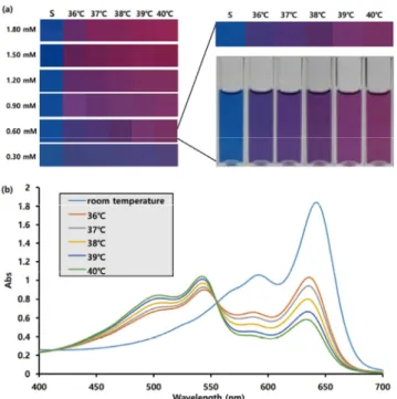
Figure 1. Temperature-dependent color change of vesicles formed with PCDA and F127. (a) Real color change depending on F127 concen- tration, S: room temperature. (b) UV spectrum of the mixed vesicle in case of 0.60 mM of F127.
block copolymer, poly(ethylene glycol)-poly(propylene glycol)-poly (ethylene glycol) [PEG-PPG-PEG]. The chemical structure is PEG101- PPG56-PEG101, and its average molecular weight is 12600. PEG is hydrophilic and PPG is hydrophobic; therefore, the hydrophilic part is more dominant than the hydrophobic part.
Photopolymerization was conducted after the mixed vesicle system with PCDA and F127 was obtained. When the temperature changed, there was a color change. The F127 concentration was controlled be- tween 0.30 and 1.80 mM, and the temperature was varied between 36 and 40 ℃. The results are shown in Figure 1(a).
Figure 1(a) shows that for 0.30 mM of F127, the color does not change when temperature increases from 36 to 40 ℃, and the blue col- or is retained. However, for 0.60 mM of F127, the color changed when temperature increased from 36 to 40 ℃, and the blue color gradually changed to red. For 0.90 mM of F127, the color rapidly changed to red. Thus, we concluded that 0.60 mM of F127 is the optimal concen- tration for our purpose. The UV spectra for 0.60 mM of F127 are shown in Figure 1(b). Figure 1(b) shows that when 0.60 mM of F127 was used, the absorption peaks of the blue color state at room temper- ature were 642 and 592 nm. When the temperature was increased to 36 ℃, a blue shift occurred. The blue color state peaks at 642 and 592 nm moved to slightly shorter wavelengths, that is, 636 and 587 nm.
At the same time, the red color state peaks also appeared at 544 and 505 nm. However, the real color was still almost blue because the ab- sorption of the blue color state was larger than that of the red color state. When the temperature was gradually increased from 36 to 40 ℃, the blue color state peaks reduced gradually, and the red color state peaks increased gradually. These results were repeated more than three
times, and same results were obtained. The color reproducibility of the results was well maintained. The real color also changed gradually from blue to reddish. Figure 1(b) shows that all UV spectra intersected at only one. This means that our vesicle system has only two states, those that are in a blue color state and those in a red color state. At 40 ℃, the blue color state peaks were shown at shorter wavelengths, 633 and 582 nm. Thus, increasing the temperature reduces the con- jugation length of multiple bonds in the blue color state as well as in the red color state.
3.2. Vesicle size change in increasing temperature
The vesicle sizes during the increasing temperature were recorded to explain the color change mechanism. The results are shown in Figure 2.
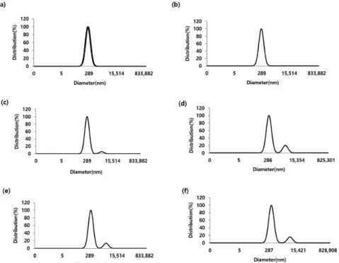
Figure 2 shows that the average size of the mixed PDA vesicle was 247 nm at room temperature and increased to 271 nm at 36 ℃. As the temperature was increased, the average size gradually increased to 289, 329, 354, and 405 nm at 37, 38, 39, and 40 ℃, respectively. At room temperature and at 36 ℃, only one peak was observed, but above 37 ℃, two peaks were observed. The second peaks were at 2825, 3119, 3652, and 5006 nm at 37, 38, 39, and 40 ℃, respectively.
The amount of second particles was smaller than the amount of the main vesicle. When the temperature was increased, the size of the sec- ond particles also increased. We believe that the color change is close- ly related to the phase separation process. It was assumed that F127 was the main component of the big second particle because this large particle did not form under the same conditions when only PCDA was used. A schematic of this phenomenon is shown in Figure 3.
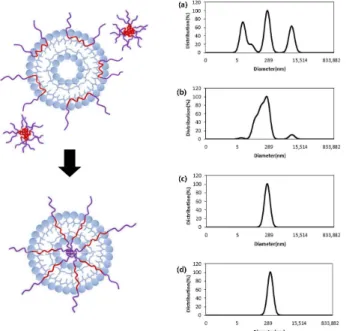
Figure 3. Schematic of the mixed vesicle structure with PDA and F127 depending on temperature increase.
3.3. Vesicle size change during stay in refrigerator
When we prepared the mixed vesicle with PDA and F127, the stabi- lization was conducted by storing in the refrigerator at 4 ℃ for 24 h.
So far, we have used this stabilization method for all the vesicle sys- tems that we have studied. In this study, we checked the size dis- tribution change during the stabilization process. The results are shown in Figure 4.
Figure 4(a) shows that when the mixed vesicle was formed with PDA and F127, the size distribution was 11, 243, and 5278 nm before stabilization. Among these values, 243 nm is attributed to the mixed vesicle of PDA and F127 because the size of the mixed vesicle was 247 nm after stabilization. We assumed that F127 is the main compo- nent in the particles of 11 and 5278 nm. We think that 11 nm is the micelle with F127 and that 5278 nm is the entanglement with F127.
The size distribution after storage in the refrigerator at 4 ℃ for 12 h is shown in Figure 4(b), which shows that the peaks at 11 and 5278 nm were almost reduced. The size distribution after storage in the re- frigerator for 24 h is shown in Figure 4(c). The results showed that the peaks at 11 and 5278 nm were removed perfectly and that only Figure 2. Size distribution profiles of the mixed vesicles with PDA/F127 (a) room temperature, (b) 36 ℃, (c) 37 ℃, (d) 38 ℃, (e) 39 ℃, and (f) 40 ℃.
Figure 4. Size changes of the mixed vesicles during the formation of vesicles, after (a) 0 h, (b) 12 h, (c) 24 h, and (d) 48 h.
Figure 5. Color change stability of the mixed vesicle depending on duration.
one kind of mixture was formed. The average particle size was 247 nm. The size distribution after storage for 48 h is shown in Figure 4(d), which shows almost similar results as those after 24 h. The aver- age particle size was 249 nm. In conclusion, storage in the refrigerator at 4 ℃ for 24 h is the optimal duration for stabilizing the vesicle system.
3.4. Long-term stability of the system
The long-term stability of our system was checked, and the results are shown in Figure 5.
The results showed that the system showed the same color change after 4 days.
4. Conclusions
In this study, a color-changing body temperature checking system was fabricated using PDA and F127. The system showed a gradual color change from 36 to 40 ℃. Using this material, a person with a fever can be easily identified. We revealed that the color change was
accompanied by phase separation. When the vesicle system was formed, stabilization was achieved after storing the vesicle solution in a re- frigerator at 4 ℃ for 24 h. We also revealed that one type of vesicle was formed during the stabilization process. Therefore, stabilization is necessary for obtaining optimal results in the research on the PDA vesicle system.
Acknowledgement
This study was supported by Semyung University’s University Inno- vation Support Project in 2020.
References
1. M. Jung, S. Jeon, and J. Bae, Scalable and facile synthesis of stretchable thermoelectric fabric for wearable self-powered temper- ature sensors, RSC Adv., 8, 39992-39999 (2018).
2. Y.-F. Wang, T. Sekine, Y. Takeda, K. Yokosawa, H. Matsui, D.
Kumaki, T. Shiba, T. Nishikawa, and S. Tokito, Fully printed PEDOT:PSS-based temperature sensor with high humidity stability for wireless healthcare monitoring, Sci. Rep., 10, 2467 (2020).
3. C. Xu, Y. Yang, and W. Gao, Skin-interfaced sensors in digital medicine: From materials to applications, Matter, 2, 1414-1445 (2020).
4. X. Gong, L. Zhang, Y. Huang, S. Wang, G. Pan, and L. Li, Directly writing flexible temperature sensor with graphene nanoribbons for disposable healthcare devices, RSC Adv., 10, 22222-22229 (2020).
5. H. Ota, M. Chao, Y. Gao, E. Wu, L.-C. Tai, K. Chen, Y. Matsuoka, K. Iwai, H. M. Fahad, W. Gao, H. Y. Y. Nyein, L. Lin, and A.
Javey, 3D printed “earable” smart devices for real-time detection of core body temperature, ACS Sens., 2, 990-997 (2017).
6. S. Majumder, T. Mondal, and M. J. Deen, Wearable sensors for remote health monitoring, Sensors, 17, 130 (2017).
7. X. Chen, G. Zhou, X. Peng, and J. Yoon, Biosensors and chemo- sensors based on the optical responses of polydiacetylenes, Chem.
Soc. Rev., 41, 4610-4630 (2012).
8. S. Lee, J.-Y. Kim, X. Chen, and J. Yoon, Recent progress in stim- uli-induced polydiacetylenes for sensing temperature, chemical and biological targets, Chem. Commun., 52, 9178-9196 (2016).
9. J. Seo, C. Kantha, J. F. Joung, S. Park, R. Jelinek, and J.-M. Kim, Covalently linked perylene diimide-polydiacetylene nanofibers dis- play enhanced stability and photocurrent with reversible FRET phenomenon, Small, 15, 1901342 (2019).
10. D.-H. Park, J. M. Heo, W. Jeong, Y. H. Yoo, B. J. Park, and J.-M.
Kim, Smartphone-based VOC sensor using colorimetric polydiace- tylenes, ACS Appl. Mater. Interfaces, 10, 5014-5021 (2018).
11. X. Chen and J. Yoon, A thermally reversible temperature sensor based on poydiacetylene: Synthesis and thermochromic properties, Dyes Pigm., 89, 194 (2011).
12. I. S. Park, H. J. Park, W. Jeong, J. Nam, Y. Kang, K. Shin, H.
Chung, and J.-M. Kim, Low temperature thermochromic polydiace- tylenes: Design, colorimetric properties, and nanofiber formation, Macromolecules, 49, 1270 (2016).
13. A. Saenjaiban, T. Singtisan, P. Suppakul, K. Jantanasakulwong, W Punyodom, and P. Rachtanapun, Novel color change film as a time-temperature indicator using polydiacetylene/silver nanoparticles
embedded in carboxymethyl cellulose, Polymers, 12, 2306 (2020).
14. M. J. Shin and J. S. Shin, Chromatic response of cationic poly- diacetylene vesicles induced by permeation of target compound, J.
Appl. Polym. Sci., 137, 49355 (2020).
15. Y. J. Shin, M. J. Shin, and J. S. Shin, Permeation-induced chro- matic change of a polydiacetylene vesicle with nonionic surfactant, Colloid Surf. A, 520, 459-466 (2017).
16. M. J. Shin, Y. J. Kim, and J.-D. Kim, Chromatic response of poly- diacetylene vesicle induced by the permeation of methotrexate, Soft Matter, 11, 5037-5043 (2015).
17. M. J. Shin and J.-D. Kim, Reversible chromatic response of poly- diacetylene derivative vesicles in D2O solvent, Langmuir, 32, 882-888 (2016).
18. M. J. Shin and J. S. Shin, Effect of ethanol on the color transition of the polydiacetylene vesicle of 10,12-pentacosadiynoic acid for butylamine detection, J. Appl. Polym. Sci., 136, 47688 (2019).
19. N. Han, H. J. Woo, S. E. Kim, S. Jung, M. J. Shin, M. Kim, and J. S. Shin, Systemized organic functional group controls in poly- diacetylenes and their effects on color changes, J. Appl. Polym.
Sci., 134, 45011 (2017).
20. K. Yoo, S. Kim, N. Han, G. E. Kim, M. J. Shin, J. S. Shin, and M. Kim, Stepwise blue-red-yellow color change of a polydiace- ty- lene sensor through internal and external transitions, Dyes Pigm., 149, 242-245 (2018).
21. M. J. Shin and J.-D. Kim, Chromatic reversibility of multilayered polydiacetylene cast film, J. Ind. Eng. Chem., 35, 211-216 (2016).
22. M. J. Shin, D. H. Byun, and J.-D. Kim, Sensitivity limitation of the sensor fabricated with polydiacetylene, J. Ind. Eng. Chem., 23, 279-284 (2015).
23. M. Kim, Y. J. Shin, M. J. Shin, and J. S. Shin, Gas-sensor fabrica- tion by a layer‐by‐layer technique using polydiacetylene, J. Appl.
Polym. Sci., 134, 44997 (2017).
24. M. Kim, Y. J. Shin, S. W. Hwang, M. J. Shin, and J. S. Shin, Chromatic detection of glucose using polymerization of diacetylene vesicle, J. Appl. Polym. Sci., 135, 46394 (2018).
25. M. Kim, N. Han, M. J. Shin, M. Kim, and J. S. Shin, Effect of head structure on ATP detection in polydiacetylene systems, Macromol.
Res., 28, 62-66 (2020).
26. M. J. Shin and J. S. Shin, A molecularly imprinted polymer under- going a color change depending on the concentration of bisphenol A, Microchim. Acta, 187, 44 (2020).
Authors
Huiseon Kim; Master Student, Department of Cosmetics and Biotechnology, Semyung University, Jecheon 27136, Korea;
huiseon0108 @gmail.com
Eunjin Heo; Undergraduate Student, Department of Cosmetics and Biotechnology, Semyung University, Jecheon 27136, Korea; ejhur97
@ naver.com
Min Jae Shin; Professor, Department of Cosmetics and Biotechnology, Semyung University, Jecheon 27136, Korea; [email protected]