I. 서론
치주질환에 의해 상실된 치주조직을 회복시키거나 임플란트를 식립할 부위에서 소실된 골조직을 재건 시키기 위하여 치료계획을 세울 경우에 임상의는 어 떠한 방법에 의해 이러한 결손부를 회복시킬 것인지 를 결정하는 것은 무척 어려운 일이다. 그렇지만, 이 러한 목적을 성취하기 위하여 자가골을 비롯하여 다 양한 이식재들이 개발되어 그 임상결과들이 보고되 고 있다.20)1)
수 많은 이식재들 중에서 자가골을 이용하는 방법 이 가장 좋은 것으로 보고되고 있으나 치근흡수, 골 채취의 어려움, 많은 양을 채득할 수 없다는 점, 환 자의 불편감, 예측할 수 없는 치료결과 등이 문제점 으로 대두되었다. 따라서, 동종골, 이종골 그리고 합 성골 등이 개발되어 사용되고 있는데, 동종골은 공 여자의 골상태에 좌우되어 골형성 효과가 좌우된다
고 보고되었다. 한편 주로 어린 송아지 뼈를 이용하 는 이종골은 특이질환에 감염될 수 있다는 위험성 때문에 환자들이 사용을 꺼리는 경향이 나타났고 치 주결손부에서 동종골과 합성골을 사용하여 비교연구 한 결과 그 효과는 비슷하다고 보고된 바 현재에는 무한정 이용될 수 있는 합성골에 관한 연구가 진행 되고 있다.22)
현재 합성골로서 bioactive glass4), hydrox- yapatite (HA)18), ß-tricalcium phosphate (ß-TCP)23,26), 그리고 coralline calcium carbo- nate12) 제재가 판매되고 있는데, 사용하기 쉽다는 점, 재료공급이 무한정하다는 점, 이식재의 조기 흡 수, 그리고 골형성에 있어서 낮은 예측성 등이 보고 되어 이 재료들 또한 장․단점을 가지고 있다.1)
이 연구에 이용된 HA와 ß-TCP 같은 calcium phosphate ceramics는 골대체재로 사용하기 위하 여 오래전부터 연구되어 왔다.17) 이러한 이식재들을 단독으로 사용하여 이루어진 1980년대의 연구를 살
* This study was supported by a grant of the Korea Health 21 R & D Project, Ministry of Health &
Welfare, Republic of Korea (03-PJ1-PG1-CH08-0001).
* 교신저자:김병옥, 광주광역시 동구 서석동 421번지 조선대학교 치과대학 치주과학교실, 우편번호 501-759, E-mail:[email protected]
대한치주과학회지 : Vol. 36, No. 1, 2006
토끼의 두개골내에 형성된 골결손부에서 HA/ß-TCP composite powders의
골형성에 관한 조직학적 연구
이광호1, 장현선1,4, 박주철2,4, 김흥중3,4, 김종관5, 김병옥1,4*
1조선대학교 치과대학 치주과학 교실, 2조선대학교 치과대학 구강조직학 교실
3조선대학교 치과대학 구강해부학 교실, 4조선대학교 치과대학 구강생물학연구소
5연세대학교 치과대학 치주과학 교실
펴보면, 비계역할에 대해 각각 상반된 결과가 도출 되었다. HA는 새로운 골을 형성하기 위해서 비계로 서 이용될 수 있다고 보고되었는데, Rabalais 등21) 은 치주결손부를 제거하는데 있어서 단순히 치주판 막술을 시행했을 때 보다는 HA를 사용했을 때 결손 부 충전효과가 더 좋았다고 하였고, Froum 등8)은 염증소견은 최소이거나 전혀 없었으며 결합조직내에 서 잘 적응된 이물반응 (well-tolerated foreign material response)을 나타냈다고 하였으며, Moskow와 Bubarr18)는 사람의 치주조직 결손부를 치료하기 위하여 순수한 HA (Durapatite)를 이용 한 연구에서 새로운 치주조직의 부착이나 골형성, 또는 백악질 형성과 같은 소견은 관찰되지 않았다고 한 반면, Yukna 등27)은 13명의 치주환자에 HA를 사용하여 수술하고 12개월 후에 재수술을 하여 결손 부에 대해 검사를 시행한 결과 경조직반응에 대해서 치주판막술만 시행한 경우보다 장점은 있으나 항상 좋은 결과를 나타내지는 않았다고 보고하였다.
반면, ß-TCP에 관련된 연구를 살펴보면, Jarcho11)는 생체액과 다양한 용매내에서 예측할 수 없을 정도로 흡수된다고 하였고, Levin 등16)은 치 주결손부내에서 골이 형성되었나 새로운 치주조직의 부착에 대해서는 의문시 된다고 하였으며, 체액에 너무 빨리 흡수된다고 알려져 비계로서 빈번하게 이 용되지는 않았다. 그러나, Lee 등14)은 ß-TCP와 chitosan을 스폰지 형태로 만들어 골아세포를 3-차 원적으로 배양하여 골형성을 위한 비계로서 이용할 수 있다고 하였으며, Lee 등15)은 chitosan/ß-TCP 스폰지에 성장인자와 함께 사용하여 골형성효과가 있음을 보고하여 골재생을 위해 비계물질로서 사용 할 수 있다고 하였다.
한편, biphasic calcium phosphate ceramic (BCPC) 방법이 개발되어 HA와 ß-TCP의 장점들 을 이용할 수 있게 되었는데, 대략적으로 60%의 HA와 40%의 ß-TCP의 비율(60HA/40ß-TCP)로 연구한 결과 골전도 성질이 관찰되었다.6,7,9,10)
이 연구는 국내에서 개발된 BCPC 이식재를 생리 식염수 또는 정맥혈과 함께 각각 혼합하여 토기의
두개골에 형성된 골결손부에 충전한 후 시간경과에 따른 골형성 효과를 평가하는 것이다.
II. 연구재료 및 방법
A. 연구재료
이 연구는 토끼의 두개골에 형성된 골결손에 새로 이 개발된 BCPC (60HA/40ß-TCP) (Dentium, Korea)의 골형성효과를 평가하기 위하여 시행되었 다.
B. 연구방법
이 연구는 조선대학 동물연구위원회 (Animal Research Committee of Chosun University)의 허락을 받아 시행되었다. 이 연구에서는 10마리의 가토 (무게: 2.5 ㎏에서 3.0 ㎏)를 이용하였는데 Ketamine-HCl (5 ㎎/㎏, Yuhan Cor. Korea)과 Xylazine-HCl (1.5 ㎖/㎏, Yuhan Cor. Korea) 로 마취하였으며, 두개골 부위를 조심스럽게 털을 제거한 후 소독된 상태에서 두개골을 노출시켰다.
두개골에 6 ㎜ trephine bur (외경 8 ㎜, 3i, USA)를 이용하여 뇌경막에 손상을 주지 않도록 하 면서 4개의 결손부를 형성하였는데 각각의 결손부를 다음과 같이 구분하였다 (Figure 1). (1) 대조군:
이식재를 넣지 않은 군, (2) 실험 1군: 생리식염수 에 적신 이식재를 넣은 군, (3) 실험 2군: 정맥혈에 적신 이식재를 넣은 군, (4) 실험 3군: 이식재만 넣 은 군. 실험을 시행한 후 흡수성 봉합사로 일차봉합 을 시행하였고 항생제 Gentamicin (0.1 ㎎/㎏, Dae Sung MIcrob. Korea)를 근육내로 주사하였 다. 수술 1주, 2주, 4주, 6주, 그리고 8주후에 phentobarbital (100 ㎎/㎏)을 정맥내 주사하여 각각 희생시키고 통상적인 방법에 의해 조직 표본을 제작하여 H-E 염색을 시행한 후 광학현미경으로 관 찰하였다.
III. 연구결과
1. 대조군 (No HA/TCP)
골 결손 형성 후 1주 소견에서 결손부는 만성염증 세포가 침윤되고 사이사이로 모세혈관이 증식되어 결체조직으로 채워져 있었으나 새로운 골형성은 관 찰되지 않았다 (Figure 2). 2주 후 소견에서 결손 부는 새로운 골 형성은 관찰되지 않았으며 소성결체 조직으로 구성되어 있었다 (Figure 3). 4주, 6주 후 소견에서도 골 결손부의 중앙부위에서는 골형성 이 관찰되지 않았으며 기존골과 골 결손 형성의 경 계부의 골로부터 일부의 골형성 소견이 관찰 되었다 (Figure 4). 8주 후에도 골 결손부의 중앙부위에서 는 골형성이 관찰되지 않았다.
2. 실험 1군(Saline + HA/TCP)
1주 소견에서 이식재의 상부와 이식재 주위에 일 부 만성염증세포의 침윤이 있었으며 (Figure 5), 2 주 후에는 이식재 주위에 세포의 분화와 더불어 소 량의 신생골 형성도 관찰 되었다. 4주후에는 골 결 손부 전체에서 신생골 형성이 관찰 되었으며 (Figure 6), 6주, 8주로 경과 할수록 골 양의 증가 와 더불어 보다 더 성숙된 소견을 보였다 (Figure 7).
3. 실험 2군 (Blood + HA/TCP)
1주 소견에서 이식재 주위에 염증 양상이 없이 다 양한 세포들이 모여드는 소견을 보였으며(Figure 8), 2주 후에는 이식재 주위에 골모세포의 분화와 더불어 새로 형성된 신생골이 관찰 되었다 (Figure 9). 4주 후에는 골 결손부 전체에 신생골 형성이 관 찰 되었으며 (Figure 10), 6주, 8주로 경과 할수록 형성된 골의 양이 많아지고 골이 성숙이 관찰 되었 다 (Figure 11).
4. 실험 3군 (HA/TCP only)
골 결손 형성 후 결손부를 HA와 TCP 처리한 후 1주 소견에서 이식재가 주위 결합조직과 일부 만성 염증세포에 의하여 둘러싸여 있었으며 (Figure 12), 2주 후에는 이식재 주위를 1-2층의 입방형 또는 타 원형의 세포가 둘러싸고 있었으며 일부 이식재 주위 에 소량의 신생골 형성이 관찰 되었다 (Figure 13).
4주후에는 이식재 주위에 골이 형성되기 시작하여 전체적으로 골 결손부를 모두 수복할 정도의 신생골 형성이 관찰 되었으며 (Figure 14), 6주, 8주로 경 과 할수록 형성된 골의 양이 많아지고 골의 층판이 형성되고 골수강이 형성되는 것 같은 양상을 나타내 는 골이 성숙되는 양상을 보였다 (Figure 15, 16).
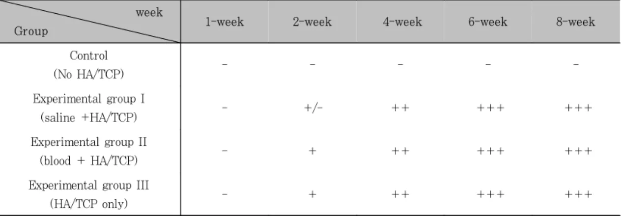
Table 1. Degree of new bone formation according to time lapse in rabbit calvarial defect
weekGroup 1-week 2-week 4-week 6-week 8-week
Control
(No HA/TCP) - - - - -
Experimental group I
(saline +HA/TCP) - +/- ++ +++ +++
Experimental group II
(blood + HA/TCP) - + ++ +++ +++
Experimental group III
(HA/TCP only) - + ++ +++ +++
-: no bone formation, +: mild bone formation, ++: moderate bone formation, +++: abundant bone formation Degree of new bone formation in experimental groups was evlauated compared to control.
IV. 고 찰
Calcium phosphate ceramics 제재로 널리 사 용되고 있는 ceramics는 HA와 ß-TCP이다. 이들 의 용해도는 구성비율, 결정도, 용액의 pH 등에 좌 우되지만5), HA는 이식 후 체액내에서 안정하여 용 해율이 너무 낮으며 ß-TCP는 용해율이 너무 높아 적절한 결과를 얻을 수 없다고 알려져 있다.
그렇지만, 이러한 재료들이 적절하게 흡수될 수 있도록 BCPC에 관한 연구에 진행되어 왔는데 현저 한 생체 적합성, 생체활성 그리고 골전도 (osteoconduction) 성질 때문에 골이식 대체재로 서 연구되고 있다. Wullstein이 1981년에 최초로 mastoid cavity를 충전시키기 위하여 과립과 fi- brin sealant를 동시에 사용하였다.3) BCPC의 흡 수성은 HA/TCP ratio에 의해 결정된다고 보고되 었는데24), Kwon 등13)은 ß-TCP와 biphasic HA/ß-TCP composite powders의 용해에 대한 연구를 시행하여 TCP는 Ringer 용액에 가장 높은 용해율을 나타냈으며 biphasic HA/ß-TCP com- posite powders는 HA와 ß-TCP 사이의 중간정도 의 용해율을 나타냈다고 하였다. 또한, Trecant 등
25)은 원통형의 calcium phosphate ceramics를 토끼와 개의 대퇴골에 이식한 후에 물리-화학적 변 화에 대해 연구를 하였는데 시간이 경과됨에 따라 압축강도의 증가와 새로운 성분 (콜라젠 기질과 생
물학적인 apatite와 물)이 나타났다고 하였다.
BCPC 제품은 자가골 또는 동종골과 전혈 (whole blood)을 함께 사용하도록 추천하고 있는 데, Bagot D'arc와 Daculsi3)는 1986년부터 만성 중이염 수술에 BCPC와 fibrin sealant를 사용한 후 15년간의 결과를 보고하였는데, ceramic 과립들 은 기계적인 안정성을 부여하였으며 fibrin sealant 가 ceramic 과립들을 강력하게 결합시켜 골유도가 촉진되었으며 창상치유과정이 향상되었다고 하였다.
또한, Gauthier 등9)은 골이 과립내로 유입되기 위 해서는 공극률보다는 세공의 직경이 더 중요하다고 하였는데, 균일하고 많은 골이 유입되기 위해서는 세공의 직경이 565 ㎛이며 40%의 공극률을 가진 이식재가 적절하다고 하였다.
치과영역에서 BCPC를 이용한 연구를 살펴보면, Nery 등19)은 개에 치주결손부를 형성하고 다양한 비율로 혼합된 HA와 ß-TCP를 충전한 후 조직학적 인 소견을 보고하였는데, ß-TCP보다는 HA가 많은 군 (65HA/35ß-TCP, 85HA/15ß-TCP)에서 부착 수준과 골재생이 훨씬 더 많았다고 하였다.
Gauthier 등10)은 직경 BCPC (60HA/40ß-TCP) 와 polymeric carrier로서 cellulous derivative (methyl-hydroxy-prophyl-cellulous)를 이용하 여 결손부에 주입할 수 있는 형태로 제작한 다음 개 의 발치와에 충전시키고 3개월 후에 조직학적으로 평가하였다. 이들의 연구에 의하면 모든 발치와면에
Table 2. Severity of inflammation according to time lapse in defect area
week
Group 1-week 2-week 4-week 6-week 8-week
Control
(HA/TCP only) + + - - -
Experimental group I
(saline + HA/TCP) + + - - -
Experimental group II
(blood + HA/TCP) - - - - -
Experimental group III
(HA/TCP only) + - - - -
-:inflammation-free, +:inflammation-present
The severity was evaluated by aucte and chronic inflammatory cell infiltration and blood cell numbers.
서 광화가 잘 되었으며 새로이 형성된 층판골이 광 범위하게 놓여 있었으며 치조제가 흡수되는 것을 예 방하는 것처럼 보였다고 하였다. Saffar 등23)은 사 람의 골연하낭에 ß-TCP로 이식하고 16개월 에서 40개월 후에 조직학적 검사를 시행한 결과, 미성숙 한 표본에서의 이식재들은 고도로 섬유성이며 세포 성이나 맥관조직이 별로 없는 결체조직으로 둘러싸 여 있었으며 이식재의 표면에서는 Howship's la- cuna와 유사한 형태의 소와가 존재했으며, 성숙한 표본에서의 이식재들은 골수강에서 부터 과립으로 향하여 광화되고 있는 무세포성의 섬유조직내에 묻 어 있었는데 골형성능 (osteogenic potential)을 가지고 있다고 보고하면서 위와 같은 소견들은 사람 의 골결손부에서 오랫동안 지속된다고 하였으며, Wiltfang 등26)도 상악동거상술을 시행하고 6개월 후에 조직학적으로 검사한 결과 새로운 골이 형성되 었으며 과립을 둘러싸고 있는 연조직과 foreign body giant cell이 존재하였으나 흡수가 빨리 일어 나지 않았다고 하였다.
이 연구는 국내에서 개발된 BCPC의 골형성 효과 를 평가한 것으로 토끼의 두개골에 결손부를 형성한 후 이 골대체재로 충전하였다. 골 결손부 형성 후 아 무것도 처리하지 않은 대조군은 1주후에 결손부와 주위에 소수의 염증세포를 포함하는 소성 결합조직 이 존재하다가 2주, 4주로 갈수록 조직이 소실되었 는데, 결과적으로 8주가 경과되어도 골 결손부에서 골이 형성된 소견은 관찰되지 않았다. 실험 1군 (HA와 TCP 골이식재 + 생리식염수), 실험 2군 (HA와 TCP 골이식재 + 정맥혈), 그리고 실험 3 군 (HA와 TCP만 사용한 군)에서는 이식 1주 후에 는 이식재 주위에 골모세포와 골모세포로 분화할 세 포들이 모여드는 소견이 관찰되었으며, 이식 2주 후 에는 이식재 주위에서 분화한 골모세포에 의하여 골 형성이 시작된 소견이 관찰되었다. 그리고, 4주 후 에는 신생 미성숙골이 골 결손부의 대부분을 채우고 있었으며, 6주와 8주로 갈수록 미성숙골이 층판을 형성하는 것처럼 골이 성숙된 소견을 나타냈다. 실 험 군들 사이에 4주, 6주, 8주 경과하면서 결과적인
골형성 양상에서 뚜렷한 차이가 관찰되지 않았다.
그러나 이식재를 정맥혈로 처리한 실험 2군에서는 다른 군에 비하여 이식 1주 후에 이식재 주위에 많 은 세포들이 모여들고 2주 후에는 다른 군에 비하여 신생골 형성이 다소 빠르게 진행되는 소견을 보였다.
Gauthier 등9)의 연구는 발치와를 이용하였지만 수 술 3개월째의 조직학적 소견을 보고하여 BCPC의 초기치유에 관한 소견을 알 수 없었으나 이 연구에 의하면 6주부터 층판골이 형성되는 것을 관찰할 수 있었다. 또한 정맥혈과 섞어서 이식했을 경우에 다 소나마 골형성이 빨랐던 것을 관찰할 수 있어 이 이 식재를 사용할 경우 정맥혈의 필요성을 확인할 수 있었다.
한편, 실험기간동안 염증세포의 발현에 관련되어 대조군, 실험 I군 그리고 III군과 비교시 실험 II군 에서만 염증세포가 관찰되지 않은 점을 감안했을 경 우, 이미 판매되고 있는 이런 형태의 이식재들을 사 용할 때와 마찬가지로 이 이식재를 임상적으로 사용 할 때는 정맥혈과 동시에 사용해야 할 것으로 생각 되었다.
이 연구는 토끼의 두개골을 이용하여 국내에서 개 발된 BCPC의 골형성 효과를 평가하였는데 이 제품 의 효능을 보다 더 정확하게 평가하기 위해서는 사 람에게서 자가골 또는 동종골과 함께 정맥혈 또는 혈소판풍부혈장을 사용한 후 조직계측학적인 연구가 추가적으로 필요하리라 생각되었다.
V. 결 론
이 연구는 국내에서 개발된 이식재인 biphasic calcium phosphat ceramics (60HA/40ß-TCP) 의 시간경과에 따른 골형성 효과를 평가하는 것이다.
10마리의 가토 (무게: 2.5 ㎏에서 3.0 ㎏)의 두 개골에 6 mm trephine bur (외경 8 mm)를 이용 하여 경뇌막의 손상을 주지 않도록 하면서 4개의 결 손부를 형성한 후 각각의 결손부는 (1) 대조군: 이 식재를 넣지 않은 군, (2) 실험 1군: 생리식염수에 적신 이식재를 넣은 군, (3) 실험 2군: 정맥혈에 적
신 이식재를 넣은 군, (4) 실험 3군: 이식재만 넣은 군으로 구분하였다. 수술 1주, 2주, 4주, 6주, 그리 고 8주후에 동물을 희생시키고 결손부를 제거한 후 통상적인 방법에 의해 조직 표본을 제작하였다.
H-E 염색을 시행한 후 광학현미경으로 관찰하여 다 음과 같은 결과를 얻었다.
1. 실험군에서는 이식 후 2주부터 신생골 형성이 관찰되어 시작되어 시간이 경과할수록 신생골 의 양이 많아지고 성숙되는 소견이 관찰되었 다.
2. 실험군들 사이에 골형성 차이는 조직학적으로 식별 될 수 없었다. 그러나 실험 2군은 1주에 이식재 주위에 더 많은 세포들이 모여들고 2 주 후에 신생골 형성이 다소 빠르고 왕성하게 진행되는 양상을 보였다. 또한 실험 1군은 1 주와 2주에서 이식재 주위에 다소의 염증 소 견이 관찰 되었다.
3. 대조군에서는 골형성이 관찰되지 않았다.
이상의 조직학적인 연구를 토대로 보았을 때, 국 내에서 개발된 새로운 HA/ß-TCP composite powders도 골결손부의 재생을 위한 이식재로 사용 될 수 있음을 시사하였다.
참고문헌
1. 김병옥. 골이식을 위한 치주치료의 최신지견.
『대한치과의사협회지』. 2005, 43(12):768-773 2. 김현수. BBP가 치주 골내낭 치유에 미치는 영향. 『대한치주과학회지』2002, 40(1):213-223 3. Bagot D'arc M,, Daculsi G. Micro
macroporous biphasic ceramics and fibrin sealant as moldable material for bone reconstruction in chronic otitis media surgery. A 15 years experience.
Kluwer Academc Publishers, 2003.
4. Cancian DCJ, Hochuli-Veira E, Marcantonio
RAC, et al. Utilization of autogenous bone, bioactive glasses, and calcium phosphate cement in surgical mandibular bone defects in cebus apella monkeys. INT J ORAL MAXILLOFAC IMPLANTS, 2004, 19:73-79
5. de Groot K. Clinical applications of calcium phosphate biomaterials: a review.
Ceramics International, 1993, 19:363-366 6. Dupraz A, Nguyen TP, Richar M, et al.
Influence of a cellulosic ether carrier on the structure of biphasic calcium phosphate ceramic particles in an injectable composite material. Biomaterials, 1999, 20:663-673
7. Ellinger RF, Nery EB, Lynch KL.
Histological assessment of periodontal osseous defects following implantation of hydroxyapatite and biphasic calcium phosphate ceramic. A case report. Int J Periodontics Restorative Dent, 1986, 6(3):22-33
8. Froum SJ, Kushner L, Scopp IW, et al.
Human clinical and histologic responses to durapatite implants in intraosseous lesions. J Periodontol, 1982, 53(12): 719-725 9. Gauthier O, Bouler JM, Aguado E, et
al. Macroporous biphasic calcium phosphate ceramics: influence of macropore diameter and macroporosity percentage on bone ingrowth. Biomaterials. 1998, 19:133-139
10. Gauthier O, Biox D, Grimanoi G. et al.
A new injectable calcium pphosphate biomaterial for immediate bone filling of extraction sockets: A preliminary study in dogs. J Periodontol, 1999, 70:375-383 11. Jarcho M. Biomaterial aspect of calcium
phosphates (properties and applicaions).
Dent Clin Nor Am, 1986, 30:25-47 12. Kim Ck, Choi EJ, Cho KS et al.
Periodontal repair in intrabony defects treated with a calcium carbonate implant and guided tissue regeneration.
J Periodontol 1996, 67: 1301-1306.
13. Kwon SH, Jun YK, Hong SH et al.
Synthesis and dissolution behavior of ß-TCP and HA/ß-TCP composite powders.
J. Euro. Ceram. Soc., 2003, 23:1039-1045.
14. Lee YM, Park YJ, Lee SJ, et al. Tissue engineered bone formation using chitosan/tricalcium phosphate sponges.
J Periodontol, 2000, 71:410-417.
15. Lee YM, Park YJ, Lee SJ, et al. The bone regenerative effect of platelet- derived growth factor-BB delivered with a chitosan/tricalcium phosphate sponge carrier. J Periodontol, 2000, 71:418-424.
16. Levin MP Getter L, Adrian J et al.
Healing of periodontal defects with ceramic implants. J Clin Periodontol, 1974, 1:197-205.
17. Monroe EA, Votava W, Bass DB et al.
New calcium phosphate ceramic material for bone and tooth implants. J Dent Res, 1971, 50:860-861.
18. Moskow BS, Bubarr A. Histological assessment of human periodontal defects after durapatite ceramic implants. J Periodontol, 1983, 54:455-462.
19. Nery EB, LeGeros RZ, Lynch KL, et al
"Tissue response to biphasic calcium phosphate ceramic with different ratios of HA/ß-TCP in periodontal osseous defects, J Periodontol, 1992, 63:729-735.
20. Position paper. Periodontal regeneration.
J Periodontol, 2005. 75:1601-1622.
21. Rabalais ML, Yukna TA, Mayer ET, Evaluation of durapatite ceramic as an alloplastic implant in periodontal osseous defects. I Initial six month results. J Periodontol, 1981. 52(11): 680-689.
22. Reynolds MA, Aichelmann-Reudt ME, Branch-Mays GL, et al. The efficacy of bone replacement grafts in the treatment of periodontal osseous defects.
A systematic review. Ann Periodontol, 2004, 227-265.
23. Saffar JL, Colombier ML, Detienville R.
Bone formation in tricalcium phosphate -filled periodontal intrabony lesion.
Histological observations in humans. J Periodontol, 1990, 61:209-216.
24. Takeishi A, Hayashi H, Kamatsubara H, et al. Implant of calcium phosphate ceramics altering Ca/P ratio in bone. J Dent Res, 1989, 68:680.
25. Trecant M, Delecrin T. Mechanical changes in macroporous calcium phosphate ceramics after implantation in bone.
Clinical Materials, 15:233-240, 1994.
26. Wiltfang J, Schlegel KA, Schultze- Mosgau S, et al. Sinus floor augmentation with ß-tricalcium phosphate (ß-TCP):
does platelet-rich plasma promote its osseous integration and degradation?.
Clin Oral Impl Res, 14:213-218, 2003.
27. Yukna RA, Harrison BG, Caudill RF, et al. Evaluation of durapatite ceramic as an alloproastic implant in periodontal osseous defects. II. Twelve month reentry results. J Periodontol, 56(9): 540-547, 1985.
사진 부도 설명(Ⅰ)
Figure 1. Photographs of the surgical sites.
Figure 2. Control group after 1-week of surgery. Microphotograph shows that new bone formation is not evident. The defect is infiltrated with chronic in- flammatory cells, capillaries and connective tissues. (magnification×40). DF:
artificial defect.
Figure 3. Control group after 2-week of surgery. Microphotograph shows that the de- fect is filled with loose connective tissue. Any new bone formation is not evident. (magnification×40). DF: artificial defect.
Figure 4. Control group after 4-week of surgery. Microphotograph shows that the center of the defect is not filled with new bone. New bone formation is evi- dent in the boundaries between the native bone and the defect.
(magnification×100). DF: artificial defect.
Figure 5. Experimental group I after 1-week of surgery. Microphotograph shows that new bone formation is evident in the entire defect area. (magnification×200).
Figure 6. Experimental group I after 4-week of surgery. Microphotograph shows that osteoblastic-like cells are differentiated around the graft particles, and a minimum of new bone are formed. (magnification×100).
Figure 7. Experimental group I after 6-week of surgery. Microphotograph shows that lots of new bone formation are evident and mature bone compared to 4-week after of surgery. (magnification×100).
Figure 8. Experimental group II after 1-week of surgery. Microphotograph shows that inflammation is absent and various cells are present around bone particles.
(magnification×200). Gr: Graft material.
Figure 9. Experimental group II after 2-week of surgery. Microphotograph shows the differentiation of osteoblastic-like cell and new bone formation around bone particles. (magnification×100). Gr: Graft material.
Figure 10. Experimental group II after 4-week of surgery. New bone formation is evi- dent in the entire defect area. (magnification×100).
Figure 11. Experimental group II after 8-week of surgery. Microphotograph shows bone remodeling and mature bone. (magnification×100)
Figure 12. Experimental group III after 1-week of surgery. The graft particles are sur- rounded by connective tissue and chronic inflammatory cells.
(magnification×100). DF: artificial defect
사진 부도 설명(Ⅱ)
Figure 13. Experimental group III after 2-week of surgery. The graft particles are sur- rounded by 1 or 2 layers of cuboidal or ovoidal cells, and a small amount of newly formed bone. (magnification×100). Gr: Graft material.
Figure 14. Experimental group III after 4-week of surgery. Microphotograph shows that new bone is formed over the entire defect area. (magnification×100)
Figure 15. Experimental group III after 6-week of surgery. Microphotograph shows that mature bone is filled in defect area. (magnification×100).
Figure 16. Experimental group III after 8-week of surgery. There is mature new bone in defect area. (magnification×100).
사진 부도 설명(Ⅰ)
Figure 1.
Figure 2.
figure 3.
Figure 4.
Figure 5.
Figure 6
사진 부도 설명(Ⅱ)
Figure 7.
Figure 8.
Figure 9.
Figure 10.
Figure 11.
Figure 12.
사진 부도 설명(Ⅲ)
Figure 13.
Figure 14.
Figure 15.
Figure 16.
-Abstract-
Bone formation e ffe ct of HA/ß-TCP composite pow- ders in rabbit calvarial bone defects: Histologic study
Kwang-Ho Lee1,4, Hyun-Seon Jang1,4, Joo-Cheol Park2,4, Heung-Joong Kim3,4, Chong-Kwan Kim5, Byung-Ock Kim1,4,*
1Department of Periodontology, College of Dentistry, Chosun University, 2Department of Oral
Histology, College of Dentistry, Chosun University, 3Department of Oral Anatomy, College of Dentistry, Chosun University 4Oral Biology Research Institute, Chosun University,
5Department of Periodontology, College of Dentistry, Yonsei University
The purpose of the present study was to evaluate the histologic results of bone cavities that were surgically created in the calvaria of rabbit and filled with HA/ß-TCP composite pow- ders, which had been developed in Korea (Dentium, Korea).
Ten young adult rabbits were used. Four defects were surgically produced in calvaria of each rabbit. Each rabbit was anesthetized with Ketamine-HCl (5 ㎎/㎏, Yuhan Cor. Korea) and Xylazine-HCl (1.5 ㎖/㎏, Yuhan Cor. Korea)). An incision was made to the bony cranium and the periosteum was reflected. Using a trephine bur (external diameter: 8 mm, 3i, USA), 4 'through-and-through' bone defects were created with copious irrigation, and classified into 4 groups: control group: no graft materials, experimental group I: normal saline + graft mate- rials: experimental group II: venous blood + graft materials: experimental group III: graft materials only. The defects were randomly filled with graft materials. The defects were closed with resorbable suture material. At the end of the surgical procedure, all animals received a single intramuscular injection of antibiotics Gentamicin (0.1 ㎎/㎏, Dae Sung Microb. Korea).
Rabbits were sacrificed with phentobarbital (100 ㎎/㎏) intravenously at 1-, 2-, 4-, 6- and 8-week after. Specimens were treated with hydrochloric acid decalcifying solution (Fisher Scientific, Tustin, CA) and sectioned by bisecting the 8 mm diameter defects. The histologic specimens were prepared in the general method with H & E staining at 6 ㎛ in thickness. The results were as follows;
1. New bone formation showed from after 2-week of surgery in defect area. As time lapsed, lots of new bone formation and mature bones showed.
2. Histologically, degree of new bone formation could not be discerned among the ex- perimental groups. But, for experimental group II, lots of cells gathered around graft materials after 1-week of surgery, new bone formed slightly faster and than the others at 1-week after. For experimental group I, a few inflammatory finding showed around graft material at after 1-week and after 2-week of surgery.
3. No bone formation did show for control group.
Based on histologic results, the new HA/ß-TCP composite powders appeared to act as a scaffolding material for regeneration of osseous defects.2)
Key words:HA/ß-TCP composite powders, Bone formation effect, Bone defeat