Received:
Revised:
Accepted:
June 25, 2015 August 21, 2015 September 22, 2015
Corresponding Author: Kyung-Jin Yun, Division of Endocrinology and Metabolism, Department of Internal Medicine, Yeouido St. Mary’s Hospital, 62 Yeouido-dong Youngdeungpo-gu Seoul 150-713, Korea
Tel: +82-2-3779-2214, Fax: +82-2-786-1471, Email: [email protected]
This is an Open Access article distributed under the terms of the Creative Commons Attribution Non-Commercial License (http://creativecommons.org/licenses/by-nc/3.0) which permits unrestricted non-commercial use, distribution, and reproduction in any medium, provided the original work is properly cited.
http://dx.doi.org/10.12997/jla.2016.5.1.11
pISSN 2287-2892 • eISSN 2288-2561
JLA
Effects of Small Dense LDL in Diabetic Nephropathy in Females with Type 2 Diabetes Mellitus
Seongyul Ryu, Youngwoo Kim, Mee Kyoung Kim, Hyuk-Sang Kwon, Ki-Hyun Baek, Ki-Ho Song, Kyung-Jin Yun
Department of Internal Medicine, the Catholic University of Korea, Seoul, Korea
Objective: This study investigated the association between small-dense low-density lipoprotein (sdLDL) and diabetic nephropathy (DN) in type 2 diabetic patients.
Methods: A total of 172 type 2 diabetic patients (95 men and 77 women) who had not taken lipid-lowering agents were enrolled in this study. Measured LDL cholesterol fractionates into seven parts (LDL1 through 7) according to the size and the extent of charge. Using this system, we analyzed mean LDL particle size and the proportion of sdLDL (the percent of LDL3 through 7 over whole LDL). DN was defined as the albumin-to-creatinine ratio (ACR) ≥30 mg/g after excluding other causes of proteinuria.
Results: The mean LDL cholesterol, LDL cholesterol size, proportion of sdLDL and ACR did not differ significantly between males and females. The presence of DN was negatively correlated with mean LDL size (r=-0.33, p value=0.02) and positively correlated with the proportion of sdLDL (r=0.34, p value=0.01) in females but not in males. After adjusting for other confounding factors related to DN, mean LDL size and proportion of sdLDL remained independent risk factors for DN in females [for mean LDL size, Odds ratio (OR)=0.86, 95% Confidence interval (CI)=0.77-0.96, p=0.01; for proportion of sdLDL, OR=1.07, 95% CI: 1.10-1.12, p=0.01], but not in males.
Conclusion: sdLDL is closely related to DN in female type 2 diabetic patients. Further studies are necessary to clarify the association of sdLDL and DN with gender. (J Lipid Atheroscler 2016 June;5(1):11-19)
Key Words: LDL Cholesterol, Type 2 Diabetes Mellitus, Diabetic Nephropathies
INTRODUCTION
Diabetes is one of the most common chronic diseases in the world, and its prevalence has been increasing continuously.1,2 According to research sponsored by the International diabetes federation (IDF) in 2013, there were 382 million people with diabetes throughout the world, and this figure is estimated to increase by approximately 592 million by 2035.2 Diabetes is important because it
causes several complications. In particular, diabetic nephropathy (DN), one of the microvascular complications of diabetes, is the most common cause of end-stage renal disease, which reduces the quality of life and increases mortality.3,4
Low-density lipoprotein (LDL) cholesterol consists of a heterogeneous spectrum of particles in terms of size, density, and lipid composition.5 Austin et al.6 classified LDL particles into two distinct phenotypes: large, more
buoyant LDL particles and small-dense LDL (sdLDL) parti- cles. Several studies have shown that sdLDL cholesterol levels are associated with the incidence of microvascular complications, such as nephropathy, retinopathy, and neuropathy in diabetic patients.4,5,7-13 In addition, some studies showed that sdLDL was a significant risk factor for albuminuria in type 2 diabetic patients.4,5 A previous study that included 40 patients with type 2 diabetes found a significant correlation between sdLDL and albuminuria.4 Another study, conducted with 23 patients with type 2 diabetes, reported that sdLDL is a risk factor for micro- albuminuria.5
The mechanism underpinning the association between sdLDL and microvascular complications in diabetic patients has been described previously. First, due to its small size, sdLDL penetrates the intima more easily than large LDL.7 Second, sdLDL was shown to be more susceptible to oxidative modification than large LDL particles. Therefore sdLDL is considered to contribute more to foam cell formation via macrophage scavenger receptors.14 Third, sdLDL generates a structural change in apolipoprotein-B and lower affinity for the LDL receptor, and it decreases hepatic clearance by the LDL receptor.15
Several previous studies conducted with patients with type 1 diabetes mellitus found that increased sdLDL was a risk factor for microvascular complications.11-13 However, the relationship between sdLDL and microvascular compli- cations, especially DN in type 2 diabetic patients, remains unclear. Therefore, in this study we assessed the relationship between sdLDL and DN in type 2 diabetic patients.
METHOD
1. SubjectsThe present study was conducted at Yeouido St. Mary’s Hospital in Seoul, Korea from September 2010 to October 2011. Subject inclusion criteria consisted of the following:
1) previous diagnosis of type 2 diabetes according to the
diagnostic criteria in the American Diabetes Association (ADA) guidelines,16 with no evidence of type 1 diabetes characterized low c-peptide levels or other secondary causes of diabetes mellitus, such as pancreatitis; 2) aged older than 20 years; and 3) no history of using lipid- lowering agents (fibrates, statin, nicotinic acid or ezeti- mibe). Patients who had taken corticosteroids, PPAR gamma agonists (pioglitazone, rosiglitazone), doxazosin, or antioxidants within 3 months before screening were excluded. Finally, a total 172 type 2 diabetes mellitus subjects (95 male and 77 female) were consecutively enrolled in this study. The study was approved by the institutional review board of Yeouido St. Mary’s Hospital.
2. Measurement and Classification of Variables
Clinical information regarding age, duration of diabetes, alcohol consumption, smoking history and other health- related variables was obtained through a standardized questionnaire. Waist circumference was measured with a measuring tape placed directly on the patient’s skin at the top of the iliac crest. Body weight and height were measured with the participant barefoot and wearing light clothing, and the measurements were used to calculate body mass index (BMI) using the following formula: BMI
=weight (kg)÷height (m2). Blood pressure was measured using a mercury sphygmomanometer with the participant in a seated position after a 5-min rest.
Venous blood sampling was performed in the morning after a minimum fasting time of 8hr. Hemoglobin A1c (HbA1c) levels were measured using high-performance liquid chromatography with an HLC-723 G7 analyzer (Tosoh Corp., Tokyo, Japan). Fasting plasma glucose concentrations were measured in sodium fluoride tubes using the hexokinase method (Johnson & Johnson LifeScan, Milpitas, CA, USA). Total cholesterol was measured using an enzymatic method (Pureauto S CHO-N kit; Sekisui Medical, Osaka, Japan), as were triglycerides (Pureauto S TG-N kit). High-density lipoprotein (HDL) and LDL
cholesterol levels were measured using direct enzymatic methods (Cholestest N HDL kit; Sekisui Chemical, Osaka, Japan). Apolipoprotein A-I and apolipoprotein B levels were determined using immunoturbidometry a Hitachi- 7600 analyser (Sekisui Chemical).
Estimated Glomerular Filtration Rate (eGFR), according to Modified Diet in Renal Disease (MDRD), was calculated using the following formula=eGFR (mL/min/1.73m2) : 186
×[serum creatinine (mg/dL)−1.154×age (years)−0.203× (0.742 if female)].17 Urine albumin excretion was determined by measuring the urine albumin-to-creatinine ratio (ACR) in spot urine samples. DN was defined as ACR ≥ 30 mg/g after excluding other causes of proteinuria.
3. Low-density lipoprotein subfraction analysis
Mean LDL-cholesterol particle size was measured using polyacrylamide tube gel electrophoresis (Quantimetrix Lipoprint TM; LDL System, Redondo Beach, CA, USA), and dividing it into seven subfractions: LDL 1 and 2 were predominantly large and buoyant LDL cholesterol, defined as large or intermediate LDL; and LDL 3 to 7 were predominantly small and dense LDL cholesterol, defined as sdLDL. Subsequently, we calculated the proportion of sdLDL using the following formula: proportion of sdLDL
=(LDL3+LDL4+LDL5+LDL6+LDL7)/(LDL1+LDL2+LDL3+
LDL4+LDL5+LDL6+LDL7)*100.
STATISTICAL ANALYSIS
All statistical analyses were performed using SAS version 9.3 (SAS Institute Inc.; Cary, NC, USA). T-tests or chi-square tests were performed to identify differences between males and females in baseline clinical characteristics. Data are presented as means±standard deviation (SD) for continuous variables except for triglyceride, ACR and proportion of sdLDL, which are presented as median (interquartile ranges), and as proportions for categorical variables. Figure 1 shows the results of the t-test for LDL
size and wilcoxon signed-rank test for proportions of sdLDL according to the presence of DN in males and females.
Spearman’s correlation analysis was used to examine the relationships between DN and other clinical factors in males and females. Values were adjusted for age, BMI, and duration of diabetes. In a multivariate logistic regre- ssion analysis, odds ratios (OR) and 95% confidence intervals (95% CIs) were used to identify associations between DN and LDL size or proportion of sdLDL. The results are shown both before and after adjusting for age, BMI, duration of diabetes mellitus, systolic blood pressure (SBP), HbA1c, and eGFR. A p value <0.05 was considered to indicate statistical significance.
RESULTS
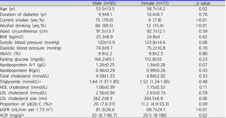
The baseline clinical characteristics of the study popu- lation are shown in Table 1. All subjects were subdivided into two groups according to gender. Males were younger (mean age of men 54±14 years, mean age of women 59±14 years, p value=0.02), smoked more, and consumed more alcohol than females. However, there were no significant differences in lipid profiles, including LDL cholesterol, between males and females. In addition, the mean LDL cholesterol size and proportions of sdLDL and ACR did not significantly differ between the two groups.
Subjects with DN showed no significant differences in LDL size and proportion of sdLDL compared with subjects without DN. (Data not shown). However, there were significant differences in LDL size and proportion of sdLDL according to presence of DN in female group. Figure 1 presents a comparison of the proportions of sdLDL and LDL sizes according to the presence of DN in males and females. Female patients exhibited significantly smaller mean LDL size in DN group as compared with non DN group (266.0±6.4 vs. 262.7±7.2, p value=0.04).
Additionally, female patients with DN exhibited signifi- cantly larger proportion of sdLDL as compared with non
Table 1. Clinical and biochemical characteristics of the study population
ㅤ Male (n=95) Female (n=77) p value
Age (yr) 53.5±13.5 58.7±14.2 0.02
Duration of diabetes (yr) 9.9±8.1 10.4±8.7 0.70
Current smoker (yes,%) 75 (79.0) 6 (7.8) <0.01
Alcohol drinking (yes,%) 66 (69.5) 12 (15.4) <0.01
Waist circumference (cm) 91.5±13.7 92.7±12.1 0.59
BMI (kg/m2) 25.3±8.9 24.8±4 0.62
Systolic blood pressure (mmHg) 120±13.9 123.8±14.6 0.08
Diastolic blood pressure (mmHg) 74.6±9.7 75.2±10.8 0.70
HbA1c (%) 9.9±2.2 9.8±2.5 0.80
Fasting glucose (mg/dL) 164.2±65.1 152.8±55 0.23
Apolipoprotein A-1 (g/L) 1.26±0.25 1.34±0.28 0.07
Apolipoprotein B(g/L) 0.96±0.29 0.99±0.26 0.43
Total cholesterol (mmol/L) 4.58±1.33 4.84±2.02 0.33
Triglyceride (mmol/L)* 1.64 (1.37-1.85) 1.52 (1.24-1.85) 0.48
HDL cholesterol (mmol/L) 1.06±0.39 1.15±0.33 0.11
LDL cholesterol (mmol/L) 2.56±0.94 2.63±0.74 0.59
LDL cholesterol size (nm) 262.2±8.3 264.5±6.9 0.06
Proportion of sdLDL-C (%)* 20 (7.6-37) 11.2 (4.9-33.3) 0.09
eGFR (mL/min per 1.73 m2) 81.3±26.6 69.7±24.1 <0.01
ACR (mg/g)* 20 (6.7-86.7) 20.5 (8-186) 0.62
Data are presented as means±standard deviation (SD) for continuous variables and as proportions for categorical variables.
*Median (interquartile ranges)
BMI; body mass index, HbA1c; hemoglobin A1c, HDL; high-density lipoprotein, LDL; low-density lipoprotein, sdLDL; small dense LDL, eGFR; estimated glomerular filtration rate, ACR; albumin-to-creatinine ratio
DN group [7.3 (4.5-23.9) vs. 24.4 (6.4-34.5), p value=
0.04]. However, no significant relationship between ACR and mean LDL size or proportion of sdLDL was observed in males (for mean LDL size, p value =0.21; for proportion of sdLDL, p value=0.31).
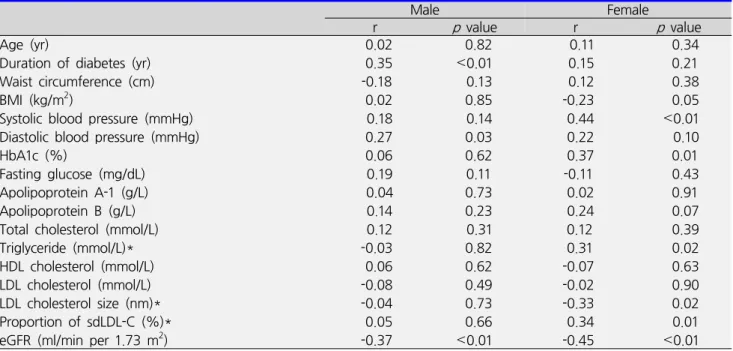
Table 2 shows the relationship between the presence of DN and other clinical factors in males and females using Spearman’s correlation analysis. All values were adjusted for age, BMI, and duration of diabetes mellitus. In males, there was a significant relationship between DN and diabetes duration, diastolic blood pressure (DBP), and eGFR. However, DN was not significantly associated with LDL size or the proportion of sdLDL in males. In females, there was a significant negative correlation between DN and LDL size (r=-0.33, p value=0.02), or positive corre- lation between DN and proportion of sdLDL (r=0.34, p value =0.01).
A multivariate logistic regression analysis was performed
to assess the association between presence of DN and mean LDL size or proportion of sdLDL. After adjusting for multiple risk factors for DN, sdLDL remained an independent risk factor for DN in females (for mean LDL size, OR=0.86, 95% CI: 0.77-0.96, p value=0.01; for proportion of sdLDL, OR=1.07, 95% CI: 1.10-1.12, p value
=0.01), but no significant relationship was found in males.
DISCUSSION
In the present study, both smaller mean LDL size and larger proportion of sdLDL were closely associated with the prevalence of DN in type 2 diabetic female patients.
The presence of DN was negatively correlated with mean LDL size (r=-0.33, p value=0.02) and positively correlated with proportion of sdLDL (r=0.34, p value=0.01) in females but not in males. In addition, multivariate logistic regression analysis showed that mean LDL size and
Fig. 1. Comparison of LDL size and the proportion of sdLDL according to presence of DN in females and males. A-1) Mean LDL size according to presence of DN in males (p value=0.21), A-2) Mean LDL size according to presence of DN in females (p value=0.04), B-1) Proportion of sdLDL according to presence of DN in males (p value=0.31), B-2) Proportion of sdLDL according to presence of DN in females (p value=0.04), LDL; low-density lipoprotein, sdLDL; small-dense LDL, DN; diabetic nephropathy
proportion of sdLDL were significant risk factors for DN in females. However, in males, the association between DN and mean LDL size or the proportion of sdLDL was not significant.
Type 2 diabetic patients do not always have higher LDL levels than non-diabetic individuals. However, their sdLDL particles are markedly increased.7,18 Due to the insulin resistance of type 2 diabetic patients, the release of free fatty acids in the muscle and fat tissue is increased, leading to increased production of very-low-density lipoprotein
(VLDL) in the liver. This interference leads to excessive exchange of triglycerides for cholesterol via the cholesteryl ester transfer protein (CETP) action between VLDL and HDL and between VLDL and LDL particles, resulting in the formation of small, relatively cholesterol-poor HDL and LDL particles.7,18 Additionally, these changes are affected by the specific drug used. Several studies have reported that treatment of diabetic dyslipidemia with statins or fibrates or the use of several diabetic drugs especially thiazolidinedione, can increase LDL particle
Table 3. Association between presence of diabetic nephropathy and mean LDL size or proportion of sdLDL using multivariate logistic regression analysis
Mean LDL size
Male Female
OR (95% CI) p value OR (95% CI) p value
*Model 1 1.03 (0.98-1.09) 0.42 0.93 (0.87-0.99) <0.01
†Model 2 1.02 (0.97-1.08) 0.09 0.88 (0.81-0.96) 0.01
‡Model 3 1.06 (0.99-1.13) 0.08 0.86 (0.77-0.96) 0.01
Proportion of sdLDL
Male Female
OR (95% CI) p value OR (95% CI) p value
*Model 1 0.99 (0.96-1.01) 0.47 1.03 (1.00-1.06) <0.01
†Model 2 0.99 (0.97-1.02) 0.10 1.06 (1.02-1.09) 0.01
‡Model 3 0.98 (0.95-1.00) 0.09 1.07 (1.102-1.12) 0.01
*Model 1: Non-adjusted
†Model 2: Adjustments for age and BMI
‡Model 3: Model 2 plus adjustments for duration of diabetes, systolic blood pressure, HbA1c and eGFR
LDL; low-density lipoprotein, sdLDL; small dense LDL, OR; odd ratio, Cl; confidence interval, BMI; body mass index, HbA1c;
hemoglobin A1c, eGFR; estimated glomerular filtration rate
Table 2. Spearman’s correlation coefficients for the relationship between the presence of diabetic nephropathy and other clinical factors in males and females
Male Female
ㅤ r p value r p value
Age (yr)
Duration of diabetes (yr) Waist circumference (cm) BMI (kg/m2)
Systolic blood pressure (mmHg) Diastolic blood pressure (mmHg) HbA1c (%)
Fasting glucose (mg/dL) Apolipoprotein A-1 (g/L) Apolipoprotein B (g/L) Total cholesterol (mmol/L) Triglyceride (mmol/L)*
HDL cholesterol (mmol/L) LDL cholesterol (mmol/L) LDL cholesterol size (nm)*
Proportion of sdLDL-C (%)*
eGFR (ml/min per 1.73 m2)
0.02 0.35 -0.18 0.02 0.18 0.27 0.06 0.19 0.04 0.14 0.12 -0.03 0.06 -0.08 -0.04 0.05 -0.37
0.82
<0.01 0.13 0.85 0.14 0.03 0.62 0.11 0.73 0.23 0.31 0.82 0.62 0.49 0.73 0.66
<0.01
0.11 0.15 0.12 -0.23 0.44 0.22 0.37 -0.11 0.02 0.24 0.12 0.31 -0.07 -0.02 -0.33 0.34 -0.45
0.34 0.21 0.38 0.05
<0.01 0.10 0.01 0.43 0.91 0.07 0.39 0.02 0.63 0.90 0.02 0.01
<0.01 Values are adjusted for age, BMI, and duration of diabetes.
*Median (interquartile ranges)
BMI; body mass index, HbA1c; hemoglobin A1c, HDL; high-density lipoprotein, LDL; low-density lipoprotein, sdLDL; small dense LDL, eGFR; estimated glomerular filtration rate
size.19,20 Yoshiho et al.19 demonstrated that treatment of diabeties with acarbose or troglitazone and treatment of hyperlipidemia with statins and fibrates was successful in increasing LDL size. Another study reported that
pioglitazone or rosiglitazone can increase LDL particle size,20 confirming why the patients who used those drugs were excluded from our study.
Several previous studies have examined the relationship
between sdLDL and macrovascular disease in type 2 diabetic patients.9,21-23 A previous prospective cohort study showed that an increase in the proportion of sdLDL was associated with an increase in the intima media thickness of type 2 diabetic patients.21 Another study reported that type 2 diabetic patients with coronary artery disease had significantly higher sdLDL levels than healthy controls.22 However, contradictory results regarding the relevance of sdLDL to diabetic microvascular complications have been observed.4,5,8,10,24,25 Although, similar to our research, the majority of studies have shown that sdLDL is associated with microvascular disease,4,5,8,10 a few have reported conflicting results.24,25 Specifically, with respect to the relationship between sdLDL and DN, Hirano et al.8 demonstrated that the average LDL size was smaller in type 2 diabetic patients with nephropathy than in control subjects or type 2 diabetic patients without nephropathy.
Matsui et al.5 also demonstrated that, in type 2 diabetic patients, smaller LDL cholesterol was a risk factor for microalbuminuria. Another study also reported a statis- tically significant correlation between sdLDL and albumin- uria.4 In contrast, one study suggested that the high prevalence of sdLDL in type 2 diabetic patients with nephropathy was not directly associated with kidney damage.24 However, several recent studies reported that sdLDL was a risk factor for microvascular complications in type 2 diabetic patients, which is in agreement with our study results.4,5,10 Additional well-designed, large prospective studies are necessary to clarify the relationship between sdLDL and DN in type 2 diabetic patients.
Unlike previous research, our study showed that results varied according to gender, which may explain the discrepant findings reported in previous studies. Baseline characteristics such as age, duration of disease, blood pressure, HbA1c, mean LDL size, and proportion of sdLDL, did not differ between males and females. Differences in smoking and drinking history were observed between males and females and may be related to gender
differences. Although the exact mechanisms have not yet been found, some studies reported that smoking and drinking history may affect the development of DN. Several studies reported that smoking is regarded as a risk factor for DN in type 2 diabetic patients.26,27 In addition, some studies showed that high alcohol consumption was significantly associated with albuminuria.28,29 In our study, however, there are no significant differences of ACR according to smoking or drinking status in both gender.
(Data not shown). Because this study was small sample based, cross-sectional study, it is difficult to define the effect of smoking and alcohol consumption with regard to gender.
Differences in sex hormones may offer a possible explanation for these results. For example, several previous studies30-32 reported that estrogen exerted renoprotective effects. However, this advantage no longer exists in a diabetic kidney due to increased expression of the estrogen receptor; however, estrogen replacement therapy can attenuate the progression of DN by compensating for the shortage of estrogen. However, we could not determine the relevance of sex hormones to DN because we did not measure sex hormones and divide the female group into premenopausal and postmenopausal. Additionally, this was not a prospective study, and issues related causation and underlying mechanisms could not be resolved due to the limitations of this approach. Therefore, further larger studies are necessary to confirm the effects of sdLDL in DN according to gender.
The present study had several limitations. First, this study was conducted at a single center and did not have a sufficient sample size. Second, this study was cross- sectional in design. Therefore, we could not address causality and were able to identify only an association between DN and sdLDL. Also, ACR was measured only once at each time point. Most guidelines recommended at least two of three random spot urine collections done in a three to six month period for accurate diagnosis of
albuminuria. Scheid et al.33, however, reported that current recommendations for repeated test may not improve diagnostic accuracy. And many other studies34-36 also used ACR results based on a single measurement at each time point. Third, clinical data including smoking or alcohol history was obtained through a standardized questionnaire. Thus, the accuracy of the data may have been compromised due to recall bias. Fourth, all study subjects were type 2 diabetic patients, and data from healthy controls and type 2 diabetic patients were not compared.
In conclusion, sdLDL is closely related to DN in female type 2 diabetic patients. Future studies are needed to clarify the association between sdLDL and DN according to gender.
REFERENCES
1. King H, Aubert RE, Herman WH. Global burden of diabetes, 1995-2025: prevalence, numerical estimates, and projections. Diabetes Care 1998;21:1414-1431.
2. International Diabetes Federation (BE). IDF diabetes atlas: 2014 update poster. 6th ed. Brussels: International Diabetes Federation; 2014.
3. Ahn JH, Yu JH, Ko SH, Kwon HS, Kim DJ, Kim JH, et al. Prevalence and determinants of diabetic nephropathy in Korea: Korea National Health and Nutrition Examination Survey. Diabetes Metab J 2014;38:109-119.
4. Abd-Allha E, Hassan BB, Abduo M, Omar SA, Sliem H.
Small dense low-density lipoprotein as a potential risk factor of nephropathy in type 2 diabetes mellitus. Indian J Endocrinol Metab 2014;18:94-98.
5. Matsui J, Tamasawa N, Tanabe J, Kasai N, Murakami H, Yamato K, et al. LDL particle size and lipid composition are risk factors for microalbuminuria in normotensive and normocholesterolemic patients with type 2 diabetes.
Diabetes Res Clin Pract 2004;66:229-236.
6. Austin MA, King MC, Vranizan KM, Krauss RM. Athero- genic lipoprotein phenotype. A proposed genetic marker for coronary heart disease risk. Circulation 1990;82:495- 506.
7. Jenkins AJ, Rowley KG, Lyons TJ, Best JD, Hill MA, Klein RL. Lipoproteins and diabetic microvascular compli- cations. Curr Pharm Des 2004;10:3395-3418.
8. Hirano T, Naito H, Kurokawa M, Ebara T, Nagano S, Adachi M, et al. High prevalence of small LDL particles in non-insulin-dependent diabetic patients with nephro- pathy. Atherosclerosis 1996;123:57-72.
9. Yoshino G, Hirano T, Kazumi T. Atherogenic lipoproteins and diabetes mellitus. J Diabetes Complications 2002;16:
29-34.
10. Jang EH, Park YM, Hur J, Kim MK, Ko SH, Baek KH, et al. Higher levels of small dense low-density lipoprotein (LDL) are associated with cardiac autonomic neuropathy in patients with type 2 diabetes. Diabet Med 2013;30:
694-701.
11. Sibley SD, Hokanson JE, Steffes MW, Purnell JQ, Marcovina SM, Cleary PA, et al. Increased small dense LDL and intermediate-density lipoprotein with albumin- uria in type 1 diabetes. Diabetes Care 1999;22:1165- 1170.
12. Chaturvedi N, Fuller JH, Taskinen MR; EURODIAB PCS Group. Differing associations of lipid and lipoprotein disturbances with the macrovascular and microvascular complications of type 1 diabetes. Diabetes Care 2001;24:
2071-2077.
13. Lyons TJ, Jenkins AJ, Zheng D, Lackland DT, McGee D, Garvey WT, et al. Diabetic retinopathy and serum lipoprotein subclasses in the DCCT/EDIC cohort. Invest Ophthalmol Vis Sci 2004;45:910-918.
14. de Graaf J, Hak-Lemmers HL, Hectors MP, Demacker PN, Hendriks JC, Stalenhoef AF. Enhanced susceptibility to in vitro oxidation of the dense low density lipoprotein subfraction in healthy subjects. Arterioscler Thromb 1991;11:298-306.
15. American Diabetes Association. Diagnosis and classifi- cation of diabetes mellitus. Diabetes Care 2014;37 Suppl 1:S81-S90.
16. Mula-Abed WA, Al Rasadi K, Al-Riyami D. Estimated glomerular filtration rate (eGFR): a serum creatinine- based test for the detection of chronic kidney disease and its impact on clinical practice. Oman Med J 2012;
27:108-113.
17. Wu L, Parhofer KG. Diabetic dyslipidemia. Metabolism 2014;63:1469-1479.
18. Yoshino G, Hirano T, Kazumi T. Treatment of small dense LDL. J Atheroscler Thromb 2002;9:266-275.
19. Deeg MA, Buse JB, Goldberg RB, Kendall DM, Zagar AJ, Jacober SJ, et al. Pioglitazone and rosiglitazone have different effects on serum lipoprotein particle concent- rations and sizes in patients with type 2 diabetes and dyslipidemia. Diabetes Care 2007;30:2458-2464.
20. Gerber PA, Thalhammer C, Schmied C, Spring S, Amann-Vesti B, Spinas GA, et al. Small, dense LDL particles predict changes in intima media thickness and insulin resistance in men with type 2 diabetes and prediabetes--a prospective cohort study. PLoS One 2013;8:e72763.
21. Huang YC, Chang PY, Hwang JS, Ning HC. Association of small dense lowdensity lipoprotein cholesterol in type 2 diabetics with coronary artery disease. Biomed J 2014;
37:375-379.
22. Berneis K, Jeanneret C, Muser J, Felix B, Miserez AR.
Low-density lipoprotein size and subclasses are markers of clinically apparent and non-apparent atherosclerosis in type 2 diabetes. Metabolism 2005;54:227-234.
23. Hirano T, Oi K, Sakai S, Kashiwazaki K, Adachi M, Yoshino G. High prevalence of small dense LDL in diabetic nephropathy is not directly associated with kidney damage: a possible role of postprandial lipemia.
Atherosclerosis 1998;141:77-85.
24. Suh S, Park HD, Kim SW, Bae JC, Tan AH, Chung HS, et al. Smaller mean LDL particle size and higher proportion of small dense LDL in Korean type 2 diabetic patients. Diabetes Metab J 2011;35:536-542.
25. Mehler PS, Jeffers BW, Biggerstaff SL, Schrier RW.
Smoking as a risk factor for nephropathy in non- insulin-dependent diabetics. J Gen Intern Med 1998;13:
842-845.
26. Chuahirun T, Khanna A, Kimball K, Wesson DE. Cigarette smoking and increased urine albumin excretion are interrelated predictors of nephropathy progression in type 2 diabetes. Am J Kidney Dis 2003;41:13-21.
27. Tapp RJ, Shaw JE, Zimmet PZ, Balkau B, Chadban SJ, Tonkin AM, et al. Albuminuria is evident in the early stages of diabetes onset: results from the Australian Diabetes, Obesity, and Lifestyle Study (AusDiab). Am J Kidney Dis 2004;44:792-798.
28. Klein R, Klein BE, Moss SE. Prevalence of microalbumin- uria in older-onset diabetes. Diabetes Care 1993;16:
1325-1330.
29. Wells CC, Riazi S, Mankhey RW, Bhatti F, Ecelbarger C, Maric C. Diabetic nephropathy is associated with decreased circulating estradiol levels and imbalance in the expression of renal estrogen receptors. Gend Med 2005;2:227-237.
30. Riazi S, Maric C, Ecelbarger CA. 17-beta Estradiol attenuates streptozotocin-induced diabetes and regulates the expression of renal sodium transporters. Kidney Int 2006;69:471-480.
31. Maric C, Sullivan S. Estrogens and the diabetic kidney.
Gend Med 2008;5 Suppl A:S103-S113.
32. Scheid DC, McCarthy LH, Lawler FH, Hamm RM, Reilly KE. Screening for microalbuminuria to prevent nephro- pathy in patients with diabetes: a systematic review of the evidence. J Fam Pract 2001;50:661-668.
33. Mann JF, Yi QL, Gerstein HC. Albuminuria as a predictor of cardiovascular and renal outcomes in people with known atherosclerotic cardiovascular disease. Kidney Int Suppl 2004:S59-S62.
34. Konta T, Kudo K, Sato H, Ichikawa K, Ikeda A, Suzuki K, et al. Albuminuria is an independent predictor of all-cause and cardiovascular mortality in the Japanese population: the Takahata study. Clin Exp Nephrol 2013;
17:805-810.
35. Friedman AN, Marrero D, Ma Y, Ackermann R, Narayan KM, Barrett-Connor E, et al. Value of urinary albumin-to- creatinine ratio as a predictor of type 2 diabetes in pre-diabetic individuals. Diabetes Care 2008;31:2344- 2348.