https://doi.org/10.4174/astr.2018.95.3.129 Annals of Surgical Treatment and Research
Wound healing and postsurgical complications in breast cancer surgery: a comparison between PEAK
PlasmaBlade and conventional electrosurgery – a preliminary report of a case series
Corrado Chiappa1, Anna Fachinetti1, Carlo Boeri1, Veronica Arlant1, Stefano Rausei1, Gianlorenzo Dionigi2, Francesca Rovera1
1First Division of Surgery - Senology Research Center, Department of Surgical and Morphological Sciences, University of Insubria, Varese, Italy
2Department of Human Pathology in Adulthood and Childhood ''G. Barresi'' University Hospital - Policlinico “G. Martino” - The University of Messina, Messina, Italy
INTRODUCTION
Over the last 30 years the surgical treatment for breast
cancer has deeply and continuously changed. We witnessed the introduction of conservative surgery associated with radiotherapy, the development of more accurate techniques to Reviewed
January February March April May June July August September October November December
Received August 14, 2017, Revised October 8, 2017, Accepted October 23, 2017
Corresponding Author: Corrado Chiappa
Senology Research Center, Department of Surgical and Morphological Sciences, University of Insubria, Ospedale di Circolo Fondazione Macchi, Via Guicciardini, 9, 21100 Varese, Italy
Tel: +39-0332278870, Fax: +39-0332260260 E-mail: [email protected]
ORCID code: https://orcid.org/0000-0002-6153-3156
Copyright ⓒ 2018, the Korean Surgical Society
cc Annals of Surgical Treatment and Research is an Open Access Journal. All articles are distributed under the terms of the Creative Commons Attribution Non- Commercial License (http://creativecommons.org/licenses/by-nc/4.0/) which permits unrestricted non-commercial use, distribution, and reproduction in any medium, provided the original work is properly cited.
Purpose: PEAK PlasmaBlade is a recent and distinctive type of electrosurgical device. Previous studies have already documented some meaningful advantages of this device over conventional electrosurgery. This study compared the use of PEAK PlasmaBlade to standard electrosurgery in mastectomy and breast conservative surgery. The purpose was to test the impact of PEAK PlasmaBlade on the wound-healing process and on postsurgical complications in breast cancer surgery.
Methods: Sixty patients undergoing breast cancer surgery were enrolled. The PEAK PlasmaBlade was used for 20 of those.
A standard electrosurgical device was used for the other 40 patients. The 2 groups were homogenous in age, body mass index, comorbidities and type of surgery. We recorded wound complications, serum drainage amount and duration of stay, blood loss, time of surgery, length of hospital stay, and total number of medications required.
Results: The 2 groups were not significantly different in terms of patient characteristics. A statistically significant reduction in incidence of seroma was observed in the PEAK group: only 10% versus 37.5% of the patients in the conventional electrosurgery group developed this complication (Fisher exact test, P = 0.034).
Conclusion: Seroma is the most important wound complication in breast surgery. The research of new instruments that might reduce its incidence is desirable. In order to validate or deny the results of this study, it is necessary to enroll more subjects and to consider the impact of this device on axillary lymph node dissection.
[Ann Surg Treat Res 2018;95(3):129-134]
Key Words: Breast neoplasms, Wound infection, Operative surgical procedures, Equipment and supplies, Intraoperative complications
localize and treat nonpalpable tumors, and the introduction of sentinel lymph node biopsy into routine practice. Last, but not least, we have seen an improvement of patients’ quality of life due to oncoplastic surgery, combining general and plastic surgery techniques in order to overcome the contention between the extent of surgical resection and final aesthetic result. Nevertheless, breast surgery still relies on very simple, typical surgical instruments for dissection and hemostatic control, such as scalpel and electrosurgery instruments [1-4].
Since the main goal of our Senology Research Centre (University of Insubria in Varese - ASST Settelaghi) is to look for the best treatment available for patients with breast cancer, we were intrigued by the new PEAK PlasmaBlade (Medtronic, Minneapolis, MN, USA).
Powered by the PULSAR II Generator (Medtronic), the PEAK PlasmaBlade is a single-use-only electrosurgical device, able to use brief and high-frequency pulses of radio frequency energy to induce electrical plasma along the cutting edges of a thin insulated electrode. The PEAK PlasmaBlade works at significantly lower temperatures than traditional electrosurgery technologies; therefore, it guarantees both the same accuracy as the scalpel and the same bleeding control as a conventional electrosurgical device without extensive collateral tissue damage [5].
Due to its properties, PEAK PlasmaBlade may be used to incise skin, replacing the scalpel, and to dissect and coagulate the underlying tissues, replacing the conventional electrosurgery [6].
Previous animal- and human-based studies have already confirmed some meaningful advantages in the use of PEAK PlasmaBlade instead of conventional electrosurgery, such as equivalent hemostatic capacity, less thermal injury depth, reduced inflammatory response, increased wound strength, and reduced scar width associated with a better aesthetic outcome [5-8]. An interesting study, conducted by Dogan et al.
[9], showed that plasmakinetic surgery in mastectomy shortens the drainage amount and duration compared to electrocautery.
Nevertheless, very little is known about this device, in particular about its use in breast cancer surgery. With the present study we would like to assess the effectiveness of PEAK PlasmaBlade, compared to standard electrosurgery, not only in mastectomy but also in breast conservation surgery.
The purpose of our study is to test the hypothesis that this instrument improves the wound-healing process in breast cancer surgery. The endpoints are: wound complications, drainage amount, and duration of stay. In addition, we measured blood loss, surgical duration, length of hospital stay, and number of medications required.
We chose to focus only on the type of breast surgery, not evaluating its utility in axillary lymph node dissection.
METHODS
Subjects and study design
A single-institution observational study was carried out between November 2015 and August 2016. Sixty patients undergoing breast cancer surgery were enrolled. For 20 of those we used the PEAK PlasmaBlade. The other 40 were selected as control. For them, we used a standard electrosurgical device (FIAB F4797 disposable sterile pencil powered by Erbe VIO 300S generator).
We excluded from the study patients who had undergone neoadjuvant chemotherapy and patients who had undergone previous ipsilateral breast surgery. Subjects enrolled aged between 34–82 years and their body mass index (BMI) range was between 17.8–45.0 kg/m2. We included only patients who were undergoing mastectomy or quadrantectomy, excluding other types of breast surgery. For each case, we matched two controls that had similar age, similar BMI, same comorbidities (mainly diabetes mellitus and hypertension) and who were undergoing the same type of surgical procedure. In this way, we maintained the same ratio between quadrantectomies (60%) and mastectomies (40%) in both groups.
Surgical procedure
Either the PEAK PlasmaBlade or the standard electrosurgical device was used on both cut and coagulation modes.
The PEAK PlasmaBlade was also used to incise skin, while in the standard electrosurgery group incision was performed with a common scalpel.
In the respective groups, the PEAK PlasmaBlade and the standard electrosurgical device were used to prepare the skin flaps (in case of mastectomy), to excise breast tissue and to remove pectoralis fascia, and to control small bleeding vessels. Larger vessels, instead, were ligated in both groups.
Both pectoralis muscles were preserved in mastectomy. In quadrantectomy, the edges of breast tissue around the cavity were shifted together and sutured using 3-0 absorbable sutures. Subcutaneous tissue was closed with an interrupted 3-0 absorbable suture. In similar proportions between the 2 groups, 3 different techniques were used for the skin closure:
subcuticular continuous suture, simple interrupted suture, or staples.
In cases of sentinel lymph node biopsy or axillary lymph node dissection, a separated axillary incision was performed.
Exceptions were made either for upper-outer quadrantectomy or for mastectomy. In such cases a single incision extending to axilla was performed. Both the PEAK PlasmaBlade and the conventional electrosurgical device were used in sentinel lymph node biopsy, but not in axillary lymph node dissection.
To perform this procedure, both instruments were replaced by an ultrasonic dissector. Axillary lymph node dissection
was performed up to level-3, preserving the axillary vein, the thoracodorsal pedicle, and the long thoracic nerve.
At the end of the procedure, either none, 1 or 2 suction drains were placed under the skin flap or in the axilla, according to the type of surgery.
Among the patients, 15% in each group underwent breast reconstruction. For these patients, plastic surgeons performed a 2-stage reconstruction: a short-term tissue expander was placed during the mastectomy procedure and substituted by a permanent breast implant a few months later, compatibly with the oncological treatment.
After discharge from the hospital, patients were regularly followed-up with ambulatory setting in order to control the healing process, to dress the wound, to remove the stitches, to evaluate the amount of drainage, and finally to remove drains.
Drains were removed when the daily drainage volume was less than 50 mL.
Data collection
Tumor size, number of lymph nodes removed and involved were collected from the pathology reports. Surgical time was considered from incision to dressing. In cases of breast reconstruction, the plastic surgery time was included. The length of hospital stay is the number of days of hospitalization after surgery. We deliberately excluded the preoperative period. For drainage duration, in subjects with two drains, we considered the one that lasted longer.
Seroma was defined as the collection of clear serous fluid under the wound, which required at least one fine-needle aspiration. On the other hand, either when the same collection consisted mainly of blood or we observed a typical skin color change, we accepted the presence of hematoma. Surgical site infection was considered as the presence of erythema, pain, tenderness, rise in temperature around the wound area or purulent drainage regardless of culture confirmation. Skin flap necrosis was accepted when skin became gradually dark and necrotic, forming an eschar.
Statistical analysis
IBM SPSS Statistics ver. 24.0 (IBM Co., Armonk, NY, USA) for Mac OS X was used for statistical analysis. Parametric and nonparametric tests were performed. We used the Student t-test to compare the means of continuous variables (age, BMI, tumor size, number of axillary lymph nodes involved and removed, permanence of the sutures, operation duration, length of hospital stay, blood loss, duration and daily amount of drainage, and number of medications). Fisher exact test was used to analyze categorical variables in 2×2 contingency tables (proportions of diabetes mellitus, hypertension, mastec- tomies/quadrantectomies, patients that underwent breast reconstruction, the use of antibiotic therapy, and all wound
complications). We used the chi-square test to compare more than 2 proportions (type of axillary surgery, suture technique for skin closure, and number of surgical drains). If any of the expected frequencies was less than five, chi-square test was repeated after merging categories whenever possible. However, in all of these cases, the proportions were equal in both groups or so similar that the P-values were not significant at conventional levels either with or without merging categories.
The results are reported in the tables as obtained without merging categories.
Significance level was set at 5%.
RESULTS
The 2 groups were homogenous in age (62.80 ± 9.908 years in the PEAK group vs. 59.43 ± 11.648 years in the control group, P = 0.272), BMI (24.94 ± 6.786 kg/m2 vs. 24.78 ± 4.209 kg/m2, P = 0.910) and frequency of diabetes mellitus (5% vs. 7.5%, P
> 0.999). The frequency of hypertension (40%) and the ratio between mastectomies (40%) and quadrantectomies (60%) were equal in both groups (P > 0.999).
We did not observe significant differences in other variables that might influence the outcomes of this study (Table 1):
type of axillary surgery (axillary lymph node dissection was performed in 35% of the patients in each group, P > 0.999), tumor size (2.43 ± 2.499 cm in the PEAK group vs. 1.95 ± 1.442 cm in the control group, P = 0.344), number of axillary lymph nodes involved (2.25 ± 4.940 vs. 0.88 ± 1.727, P = 0.118), number of lymph nodes removed (7.00 ± 7.766 vs. 6.98
± 7.724, P = 0.991), use of prophylactic and postoperative antibiotic therapy (respectively 100% in both groups, P > 0.999, and 95% vs. 85%, P = 0.407), suture technique (P = 0.390) and permanence of the sutures (13.60 ± 3.52 days vs. 14.00 ± 2.82 days, P = 0.686).
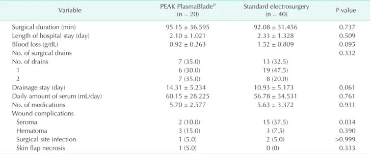
No significant difference was observed between treatment and control groups in these outcomes (Table 2): surgical duration (95.15 ± 36.595 minutes vs. 92.08 ± 31.456 minutes, P = 0.737), length of hospital stay (2.10 ± 1.021 days vs. 2.33 ± 1.328 days, P = 0.509), and number of medications (5.70 ± 2.577 vs. 5.63 ± 3.372, P = 0.931).
Mean drainage duration was 14.31 ± 5.234 days in the PEAK group and 10.93 ± 5.173 days in the control group, but the difference was not significant (P = 0.061). It appears useful to report that 2 patients accidentally removed the drain, before the daily amount of drainage was less than 50 mL. Both patients were in the standard electrosurgery group and both removed it after 9 days. Also, the daily amount of drainage was similar between the 2 groups (60.15 ± 28.225 mL/day vs. 56.78 ± 34.531 mL/day, P = 0.761). In order to evaluate blood loss we calculated the difference in the hemoglobin count between the preoperative and the postoperative examination. Mean blood
Table 1. Patient characteristics
Variable PEAK PlasmaBladea)
(n = 20) Standard electrosurgery
(n = 40) P-value
Age (yr) 62.80 ± 9.908 59.43 ± 11.648 0.272
Body mass index (kg/m2) 24.94 ± 6.786 24.78 ± 4.209 0.910
Patients with diabetes mellitus 1 (5.0) 3 (7.5) >0.999
Patients with hypertension 8 (40.0) 16 (40) >0.999
Breast surgery >0.999
Mastectomies 8 (40.0) 16 (40.0)
Quadrantectomies 12 (60.0) 24 (60.0)
Axillary surgery >0.999
None 1 (5.0) 2 (5.0)
Only sentinel lymph node biopsy 12 (60.0) 24 (60.0)
Axillary lymph node dissection 7 (35.0) 14 (35.0)
No. of axillary lymph nodes removed 7.00 ± 7.766 6.98 ± 7.724 0.991
No. of lymph nodes involved 2.25 ± 4.940 0.88 ± 1.727 0.118
Breast reconstruction after mastectomy 3 (15.0) 6 (15.0) >0.999
Tumor size (cm) 2.43 ± 2.499 1.95 ± 1.442 0.344
Suture technique for skin closure 0.390
Only subcuticular continuous sutures 7 (35.0) 16 (40.0)
Subcuticular continuous + simple interrupted sutures 4 (20.0) 14 (35.0)
Only staples 7 (35.0) 9 (22.5)
Subcuticular continuous sutures + staples 1 (5.0) 0 (0)
Staples + simple interrupted sutures 1 (5.0) 1 (2.5)
Permanence of the sutures (day) 13.60 ± 3.52 14.00 ± 2.82 0.686
Antibiotic prophylaxis 20 (100) 40 (100) >0.999
Postoperative antibiotic therapy 19 (95.0) 34 (85.0) 0.407
Values are presented as mean ± standard deviation or number (%).
Significance level was set at 5%.
a)PEAK PlasmaBlade (Medtronic, Minneapolis, MN, USA).
Table 2. Results
Variable PEAK PlasmaBladea)
(n = 20) Standard electrosurgery
(n = 40) P-value
Surgical duration (min) 95.15 ± 36.595 92.08 ± 31.456 0.737
Length of hospital stay (day) 2.10 ± 1.021 2.33 ± 1.328 0.509
Blood loss (g/dL) 0.92 ± 0.263 1.52 ± 0.809 0.095
No. of surgical drains 0.332
No. of drains 7 (35.0) 13 (32.5)
1 6 (30.0) 19 (47.5)
2 7 (35.0) 8 (20.0)
Drainage stay (day) 14.31 ± 5.234 10.93 ± 5.173 0.061
Daily amount of serum (mL/day) 60.15 ± 28.225 56.78 ± 34.531 0.761
No. of medications 5.70 ± 2.577 5.63 ± 3.372 0.931
Wound complications
Seroma 2 (10.0) 15 (37.5) 0.034
Hematoma 3 (15.0) 3 (7.5) 0.390
Surgical site infection 1 (5.0) 2 (5.0) >0.999
Skin flap necrosis 1 (5.0) 0 (0) 0.333
Values are presented as mean ± standard deviation or number (%).
Significance level was set at 5%.
a)PEAK PlasmaBlade (Medtronic, Minneapolis, MN, USA).
loss was 0.92 ± 0.263 g/dL in the PEAK group and 1.52 ± 0.809 g/dL in the control group. This difference between the 2 groups was not significant (P = 0.095).
A significant difference between the 2 groups was detected in incidence of seroma: only 2 patients out of 20 (10%) in the PEAK group versus 15 out of 40 (37.5%) in the conventional electrosurgery group developed this complication (P = 0.034).
Other wound complications occurred with no significant difference between the 2 groups. Hematoma occurred in 3 patients in each group; therefore, a difference in proportion was noticed, but this difference was not significant (15% vs. 7.5%, P
= 0.390). Surgical site infection was observed with the same frequency between the 2 groups (5%, P > 0.999). We observed only one case of skin flap necrosis and it was in the PEAK group (5% vs. 0%, P = 0.333).
DISCUSSION
The purpose of this study was to evaluate if PEAK Plasma- Blade, due to its properties, could improve the wound-healing process in breast surgery (mainly in quadrantectomy and mastectomy).
Our Senology Research Centre (University of Insubria in Varese - ASST Settelaghi) is composed of a single surgical team that provides standard medical and surgical treatments. These features enabled proper case and control selection, significantly reducing the possibility of hidden variables.
The major limitation of the study was the enrollment of few subjects (we used PEAK PlasmaBlade for only 20 subjects).
This is a possible explanation of why many of the results did not reach a statistical difference, such as the difference in hemoglobin count between preoperative and postoperative examination and drainage duration. Hemoglobin count was used as an indirect measure of blood loss. Mean blood loss was 0.92 ± 0.263 g/dL in the PEAK group and 1.52 ± 0.809 g/
dL in the control group (P = 0.095). The preoperative value was usually taken a couple weeks before operation. In addition, many factors, besides blood loss during operation, could have influenced the difference in hemoglobin value. For these reasons, we cannot definitively state that PEAK PlasmaBlade reduced blood loss. Drain duration was the other variable that resulted close to significance (14.31 ± 5.234 days in the PEAK group vs. 10.93 ± 5.173 days in the control group, P = 0.061). Clarification is required for these data: 2 patients in the standard electrosurgery group prematurely removed the drain by accident.
However, despite these considerations, this study showed a significant result, which is in support of the PEAK PlasmaBlade:
a lower incidence of seroma. The reduction of seroma was very clear (only 2 patients out of 20 that were treated with the PEAK PlasmaBlade versus 15 out of 40 that were treated with standard
electrosurgery, P = 0.034). This represents a very interesting result, considering the fact that seroma is one of the most common wound complications in breast surgery. According to Srivastava et al. [10] in a survey about seroma formation after breast surgery, it is so frequent that it is now believed to be a side effect of surgery rather than a complication. The etiology of seroma is multifactorial. Several factors have been accepted:
increased BMI, early shoulder movements in the postoperative period and surgical factors including technique, extent of dissection and the surgical devices used for dissection, such as electrocautery [10,11]. A detailed analysis showed that the use of single or multiple drains, early or late removal, and drains with or without suction do not influence the incidence of seroma [10].
Only very early removal, within 24 hours, seems to increase formation of seroma.
Many hypotheses have been postulated in the literature to explain the association between electrocautery and seroma [12-14]. Some authors emphasized the role of thrombosis of subdermal vessels caused by cautery [15], others stressed the effect on subcutaneous fat [12], others further state an association between tissue devitalization and seroma [16].
These fluid collections need to be removed with fine-needle aspiration, because they can lead to wound dehiscence, increase the risk of infections, and may delay adjuvant therapy.
Therefore, seroma represents a further inconvenience for patients that have already undergone breast surgery. For this reason, the research of new instruments and surgical techniques that might reduce its incidence is desirable.
If we compare new and old surgical devices, we have to consider also the cost effectiveness. Price represents perhaps the greatest issue limiting the use of PEAK PlasmaBlade, as the device costs significantly more than a conventional electrocautery tip (cost of which is approximately €1,50). Further studies, based on a larger sample size, should ascertain if the routine use of this new technology, reducing complications and, eventually, the number of medications and hospitalization, could lead to an overall cost saving.
PEAK PlasmaBlade may therefore be a valid option for surgical device in breast cancer surgery. However, in order to validate or deny the results of this study, it is necessary to enroll more subjects and to consider the impact of this instrument on axillary lymph node dissection. If a lower incidence of seroma is confirmed in a larger sample size, the use of the PEAK PlasmaBlade in breast surgery should be taken into serious consideration.
CONFLICTS OF INTEREST
No potential conflict of interest relevant to this article was reported.
ACKNOWLEDGMENTS
In order to conduct this study, Medtronic (Medtronic, Min-
neap olis, MN, USA) provided us 1 PULSAR II generator and 20 PEAK PlasmaBlade devices.
REFERENCES
1. Aird LN, Brown CJ. Systematic review and meta-analysis of electrocautery versus scalpel for surgical skin incisions. Am J Surg 2012;204:216-21.
2. Miller E, Paull DE, Morrissey K, Cortese A, Nowak E. Scalpel versus electrocautery in modified radical mastectomy. Am Surg 1988;54:284-6.
3. O'Connor JL, Bloom DA. William T. Bovie and electrosurgery. Surgery 1996;119:390- 6.
4. Cho MJ, Yang JH, Yu YB, Park KS, Chung HW, So Y, et al. Validity of breast-specific gamma imaging for Breast Imaging Re- port ing and Data System 4 lesions on mammography and/or ultrasound. Ann Surg Treat Res 2016;90:194-200.
5. Loh SA, Carlson GA, Chang EI, Huang E, Palanker D, Gurtner GC. Comparative heal ing of surgical incisions created by the PEAK PlasmaBlade, conventional elec- trosurgery, and a scalpel. Plast Reconstr Surg 2009;124:1849-59.
6. Ruidiaz ME, Messmer D, Atmodjo DY, Vose JG, Huang EJ, Kummel AC, et al.
Com pa rative healing of human cutaneous surgical incisions created by the PEAK PlasmaBlade, conventional electrosurgery,
and a standard scalpel. Plast Reconstr Surg 2011;128:104-11.
7. Chang EI, Carlson GA, Vose JG, Huang EJ, Yang GP. Comparative healing of rat fascia following incision with three surgical instruments. J Surg Res 2011;167:e47-54.
8. Isik F. Discussion: comparative healing of human cutaneous surgical incisions created by the PEAK PlasmaBlade, con- ventional electrosurgery, and a stand ard scalpel. Plast Reconstr Surg 2011;128:112- 3.
9. Dogan L, Gulcelik MA, Yuksel M, Uyar O, Erdogan O, Reis E. The effect of plas- makinetic cautery on wound healing and com plications in mastectomy. J Breast Cancer 2013;16:198-201.
10. Srivastava V, Basu S, Shukla VK. Seroma formation after breast cancer surgery:
what we have learned in the last two decades. J Breast Cancer 2012;15:373-80.
11. Hashemi E, Kaviani A, Najafi M, Ebrahimi M, Hooshmand H, Montazeri A. Seroma formation after surgery for breast cancer.
World J Surg Oncol 2004;2:44.
12. Porter KA, O'Connor S, Rimm E, Lopez M. Electrocautery as a factor in seroma formation following mastectomy. Am J
Surg 1998;176:8-11.
13. Yilmaz KB, Dogan L, Nalbant H, Akinci M, Karaman N, Ozaslan C, et al. Compar- ing scalpel, electrocautery and ultrasonic dissector effects: the impact on wound complications and pro-inflammatory cytokine levels in wound fluid from mastectomy patients. J Breast Cancer 2011;14:58-63.
14. Keogh GW, Doughty JC, McArdle CS, Cooke TG. Seroma formation related to electrocautery in breast surgery: a pro- spective randomized trial. Breast 1998;
7:39-41.
15. Hoefer RA Jr, DuBois JJ, Ostrow LB, Silver LF. Wound complications following mo- dified radical mastectomy: an analysis of perioperative factors. J Am Osteopath Assoc 1990;90:47-53.
16. Madden JE, Edlich RF, Custer JR, Panek PH, Thul J, Wangensteen OH. Studies in the management of the contaminated wound. IV. Resistance to infection of surgical wounds made by knife, elec- trosurgery, and laser. Am J Surg 1970;
119:222-4.