Received March 2, 2015, Revised June 25, 2015, Accepted for publication June 29, 2015
Corresponding author: Weon Ju Lee, Department of Dermatology, Kyungpook National University School of Medicine, 130 Dongdeok-ro, Jung-gu, Daegu 41944, Korea. Tel: 82-53-420-5838, Fax: 82-53-426-0770, E-mail: [email protected]
This is an Open Access article distributed under the terms of the Creative Commons Attribution Non-Commercial License (http://creativecommons.
org/licenses/by-nc/4.0) which permits unrestricted non-commercial use, distribution, and reproduction in any medium, provided the original work is properly cited.
Copyright © The Korean Dermatological Association and The Korean Society for Investigative Dermatology
Ann Dermatol Vol. 28, No. 2, 2016 http://dx.doi.org/10.5021/ad.2016.28.2.186
ORIGINAL ARTICLE
Efficacy of Red or Infrared Light-Emitting Diodes in a Mouse Model of Propionibacterium acnes-Induced
Inflammation
Weon Ju Lee, Kyou Chae Lee, Min Ji Kim, Yong Hyun Jang, Seok-Jong Lee, Do Won Kim
Department of Dermatology, Kyungpook National University School of Medicine, Daegu, Korea
Background: Laser/light-based devices may provide an alter- native to conventional acne therapeutics in some patients with nonresponsive acne. Objective: We investigated the ef- ficacy of red or infrared light-emitting diode (LED) devices in a mouse model of Propionibacterium acnes-induced in- flammation through clinical examination and histopatho- logical and immunohistochemical studies. Methods: A hu- man-derived Propionibacterium acnes suspension (109 col- ony-forming units /μl) was injected into the back of an HR-1 mouse. Then, a 28.9 J/cm2 650-nm red LED or 9.3 J/cm2 830-nm infrared LED was applied to the mouse with P. acnes-induced inflammation once daily for 2 weeks. Two weeks after treatment, histological findings with hematox- ylin and eosin staining and expression levels of inflammatory biomarkers (integrin α6, neutrophils, interleukin [IL]-1β, and matrix metalloproteinase [MMP]-2/9) were evaluated in tissue specimens using immunohistochemical staining.
Results: Mice treated with red and infrared LED showed clin- ical improvement in inflammatory nodules compared to mice in the control group. Red LED was much more effective than infrared LED. Epidermal hyperplasia, comedone-like cysts, and integrin α6 expression improved to a similar ex- tent in the red and infrared LED treatment groups and control
group. Neutrophil, IL-1β, MMP-2, and MMP-9 expression after treatment with red and infrared LED decreased consid- erably compared to expression in the control group.
Conclusion: In a mouse model of P. acnes-induced in- flammatory nodules, red and infrared LED devices may be an alternative to conventional acne therapies. In addition, a mouse model of P. acnes-induced inflammatory nodules is helpful for laboratory research of acne. (Ann Dermatol 28(2) 186∼191, 2016)
-Keywords-
Acne, Light, Mice, Propionibacterium acnes
INTRODUCTION
Acne is a very common skin disorder occurring in the fol- licular infundibulum1,2. Propionibacterium acnes colo- nization in the follicular infundibulum is a major cause of acne and plays an important role in inducing an in- flammatory event3. In addition, P. acnes can cause abnor- mal differentiation and proliferation in epidermal keratino- cytes1. Therefore, acne animal models can be established using P. acnes. The establishment of animal models for acne is helpful to expand research fields and to develop new therapeutic modalities. Although various animal models for acne such as the Mexican hairless dog and the Rhino mouse exist, an elucidative model is still needed4. Our animal model of inflammatory events was established with the injection of P. acnes into the back of an HR-1 mouse.
Laser/light-based devices may provide an alternative to conventional acne therapeutics in some patients. Many types of laser/light devices have been introduced to im- prove or cure inflammatory acne. Light-emitting diode
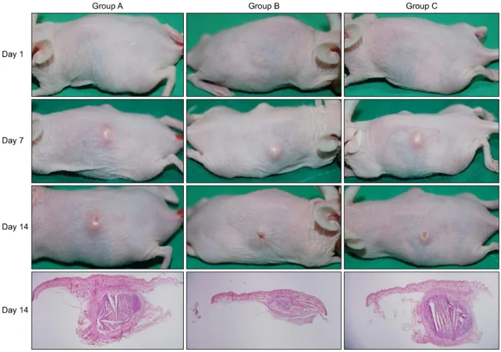
Fig. 1. Clinical inflammatory lesions in group B and group C improved greatly compared to those in group A.
(LED) devices are one of the most popular light-based de- vices for the treatment of acne. LED devices, especially those emitting blue and red light, directly or indirectly tar- get P. acnes5.
In this study, we investigated the efficacy of red or infrared LED devices in a mouse model of P. acnes-induced in- flammation through clinical examination, pathology, and immunohistochemical studies.
MATERIALS AND METHODS
Preparation of the mouse model with injection of Propionibacterium acnes
Six-week-old female Hos:HR-1 mice (HR-1; SLC Inc., Hamamatsu, Japan) were kept under conventional labo- ratory conditions (at 20oC∼24oC in a humidified atmos- phere of 40%∼60%) after 1 week of acclimation. P. acnes (ATCC 11828) were isolated from the pustular lesions of Korean patients with moderate inflammatory acne. P. acnes from post-log phase cultures were grown on brain heart infusion agar and harvested, heat killed (95oC, 5
min), and lyophilized prior to injection. A P. acnes sus- pension was prepared with a concentration of 109 col- ony-forming units/μl. With a 30-gauge needle, the P. acnes suspension was injected intradermally in 20-μl ali- quots on both sides of the mouse back. Four mice in group A were injected with P. acnes and were not treated with red or infrared LED. Three mice in group B were treated with red LED after P. acnes injection, and three mice in group C were treated with infrared LED after P. acnes injection. The animal study protocol was approved by the Ethics Committee for animal studies at Kyoungpook National University, Korea (KNU 2014-0135).
Light-emitting diode irradiation of the Propionibacterium acnes-injected mice
After P. acnes injection, the mice were irradiated using a 28.9 J/cm2 650-nm red LED in group B and a 9.3 J/cm2 830-nm infrared LED in group C once daily for 2 weeks.
Evaluation of clinical changes
The degree of clinical inflammatory changes was eval-
Fig. 2. Improvement in epidermal hyperplasia and microcomedone-like cysts in group B and group C was similar to that in group A.
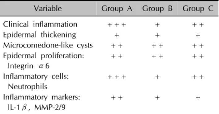
Table 1. Summary of the histological analysis and immuno- histochemical profile results
Variable Group A Group B Group C
Clinical inflammation +++ + ++
Epidermal thickening + + +
Microcomedone-like cysts ++ ++ ++
Epidermal proliferation:
Integrin α6
++ ++ ++
Inflammatory cells:
Neutrophils
+++ + ++
Inflammatory markers:
IL-1β, MMP-2/9
++ + +
Group A: P. acnes-treated group, Group B: P. acnes and red LED-treated group, Group C: P. acnes and infrared LED-treated group. IL: interleukin, MMP: matrix metalloproteinase, +: mild, ++: moderate, +++: prominent or dense.
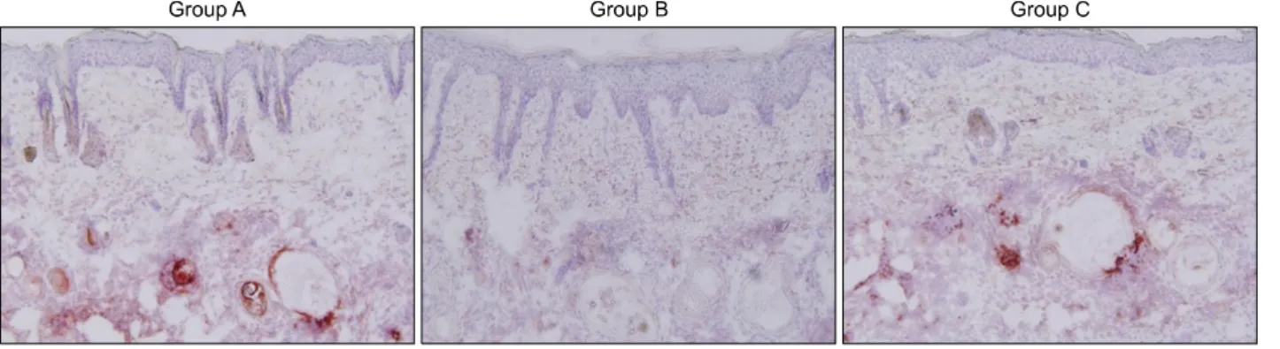
Fig. 3. Integrin α6 expression in group B and group C was similar to that in group A.
uated using digital photography at baseline and at weeks 1 and 2 after P. acnes injection.
Histological examination and immunohistochemical stains
Tissue samples of mice were taken 2 weeks after P. acnes injection. Eight tissue samples were collected from group A, 6 tissue samples from group B, and 6 tissue samples from group C. Paraffin-embedded tissue sections of 3-μm thickness were processed for light microscopy. Hematoxylin and eosin (H&E) and immunohistochemical staining was performed using standard techniques. The primary anti- bodies were as follows: integrin α6 (diluted 1:150; Santa Cruz Biotechnology Inc., Dallas, TX, USA), neutrophils (diluted 1:80; Abcam, Cambridge, UK), interleukin (IL)-1 β (diluted 1:150; Abcam), matrix metalloproteinase (MMP)-2 (diluted 1:300; Abcam), and MMP-9 (diluted 1:250; Abcam). The histological changes in each group were evaluated semiquantitatively with a 3-point scale:
mild (+), moderate (++), and prominent (+++), with an emphasis on changes in inflammation, epidermal/fol- licular wall thickness, and formation of cystic structures containing keratinized plugs (microcomedone-like cystic structures) in the dermis. The degree of immunohisto- chemical staining in each group was also evaluated semi-
quantitatively with a 3-point scale: mild (+), moderate (++), and dense (+++).
RESULTS
Clinical changes
Improvement in the inflammatory nodules of group B and group C was compared to that observed in group A (Fig. 1,
Fig. 5. Interleukin-1β expression decreased in group B and group C compared to that in group A.
Fig. 4. Neutrophil infiltration decreased in group B and group C compared to that in group A.
Table 1). Group B showed more improvement in in- flammatory nodules than group C, and group C showed more improvement in inflammatory nodules than group A.
Histopathological findings
Tissue samples obtained from the three groups (A, B, and C) were stained with H&E. Stains from the treatment groups were compared to those of the control group with- out P. acnes injection. Improvement in epidermal hyper- plasia and thickening in group B and group C was similar to that observed in group A (Fig. 2, Table 1). The number and size of the microcomedone-like cysts in the upper dermis above the focus of inflammation in group B and group C decreased to an extent similar to that in group A (Fig. 2, Table 1).
Immunohistochemical stains
1) Epidermal proliferation marker: integrin α6
Improvement in the expression of integrin α6, the marker for epidermal proliferation, in the basal epidermis and mi- crocomedone-like cyst wall in group B and group C was similar to that in group A (Fig. 3, Table 1).
2) Inflammatory cells: neutrophils
The infiltration of neutrophils into the dermis was de- creased in group B and group C compared to that ob- served in group A (Fig. 4, Table 1). The infiltration of neu- trophils in group B decreased to a greater extent than that in group C.
3) Inflammatory markers: interleukin-1β
IL-1β expression in the basal epidermis after treatment decreased in group B and group C compared to that in group A (Fig. 5, Table 1). IL-1β expression decreased to a greater extent in group B than in group C.
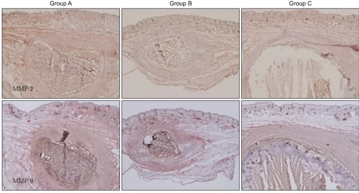
4) Tissue remodeling markers: matrix metalloproteinase-2 and matrix metalloproteinase-9
MMP-2 expression in the dermis decreased to a greater ex- tent in group B and group C than in group A (Fig. 6, Table 1). MMP-9 expression in the dermis decreased to a greater extent in group B and group C than in group A (Fig. 6, Table 1).
Fig. 6. Matrix metalloproteinase (MMP)-2 and MMP-9 expression decreased greatly in group B and group C compared to that in group A.
DISCUSSION
Currently, several animal models can be used for acne research. Among them, rabbit ears and Rhino mice have been used frequently to determine the compound come- dogenicity of acne lesions6,7. However, the rabbit-ear and Rhino mice models have some limitations, including a lack of bacterial colonization and inflammation4,7. Therefore, these models are not helpful for inflammatory acne research. In addition, the use of rabbits may be incon- venient for large-scale drug screening and vaccinations. As proven by our previous study, P. acnes injection into the HR-1 mouse induces acneiform inflammatory nodules with overlying epidermal hyperplasia and superficial sec- ondary microcomedones. Therefore, the HR-1 mouse is suitable for inflammatory acne research.
In this study, HR-1 mice were used to determine the effi- cacy of red or infrared LED treatment of inflammatory acne. Laser/light-based devices are an alternative to con- ventional acne therapeutics in some patients who experi- ence side effects or recurrence with conventional treatments. Treatment with ultraviolet (UV) A and UVB was found to have a marginal beneficial effect for acne8. Blue light is most effective in the photoactivation of the endogenous porphyrin component of P. acnes, because the 407∼420-nm wavelength light has the strongest por- phyrin photoexcitation coefficient. However, blue light
cannot access the deeper dermis9. On the contrary, red light penetrates deeper to the sebaceous glands and has anti-inflammatory properties, as apparent from its influ- ence on cytokine release from macrophages10,11. According to an action spectrum for the inactivation of P. acnes, the sensitivity of P. acnes is the highest with shorter wave- lengths and decreases with increasing wavelength12. A combination of blue and red LED application appears to have excellent potential in the treatment of mild-to-severe acne13. The infrared laser light, at such strengths as 1,450 nm and 1,540 nm, selectively produces an injury zone in the dermis, where the sebaceous glands are located, suffi- cient enough to inhibit high sebum production, leading to acne improvement9.
In this study, both red and infrared LED had anti-in- flammatory effects on HR-1 mice with inflammatory nod- ules composed mainly of neutrophils and histiocytes. Red LED was much more effective than infrared LED. A de- crease in the expression of inflammatory markers, includ- ing neutrophils, IL-1β, MMP-2, and MMP-9, was shown in HR-1 mice after treatment with red or infrared LED.
Their expressions after treatment with red LED decreased to a greater extent than those after treatment with infrared LED. However, the epidermal hyperplasia of the HR-1 mice after red or infrared LED treatment was similar to that after no LED treatment. The size and number of micro- comedone-like cysts after red or infrared LED treatment
were similar to those after no LED treatment. In addition, the expression of integrin α6 after red or infrared LED treatment was similar to that after no LED treatment.
Unfortunately, a limitation to this study is that these ani- mal models do not fully represent inflammatory acne.
In conclusion, red or infrared LED has the potential to re- lieve inflammatory acne and decrease the expression of inflammatory biomarkers; red LED is much more effective than infrared LED. HR-1 mice with P. acnes-induced in- flammation represent a good model for the investigation of inflammatory acne.
ACKNOWLEDGMENT
This research was supported by the Basic Science Research Program through the National Research Foundation of Korea (NRF) funded by the Ministry of Education, Science and Technology (2012R1A1A2007017); Daegugyeong Institute for Regional Program Evaluation.
REFERENCES
1. Shaheen B, Gonzalez M. Acne sans P. acnes. J Eur Acad Dermatol Venereol 2013;27:1-10.
2. Huh SY, Na JI, Huh CH, Park KC. The effect of photodynamic therapy using indole-3-acetic Acid and green light on acne vulgaris. Ann Dermatol 2012;24:56-60.
3. Jeremy AH, Holland DB, Roberts SG, Thomson KF, Cunliffe WJ. Inflammatory events are involved in acne lesion
initiation. J Invest Dermatol 2003;121:20-27.
4. Mirshahpanah P, Maibach HI. Models in acnegenesis.
Cutan Ocul Toxicol 2007;26:195-202.
5. Mariwalla K, Rohrer TE. Use of lasers and light-based therapies for treatment of acne vulgaris. Lasers Surg Med 2005;37:333-342.
6. Nakano K, Kiyokane K, Benvenuto-Andrade C, González S.
Real-timereflectance confocal microscopy, a noninvasive tool for in vivo quantitative evaluation of comedolysis in the rhino mouse model. Skin Pharmacol Physiol 2007;
20:29-36.
7. Nakatsuji T, Shi Y, Zhu W, Huang CP, Chen YR, Lee DY, et al. Bioengineering a humanized acne microenvironment model: proteomics analysis of host responses to Propio- nibacterium acnes infection in vivo. Proteomics 2008;8:
3406-3415.
8. Charakida A, Seaton ED, Charakida M, Mouser P, Avgerinos A, Chu AC. Phototherapy in the treatment of acne vulgaris:
what is its role? Am J Clin Dermatol 2004;5:211-216.
9. Elman M, Lebzelter J. Light therapy in the treatment of acne vulgaris. Dermatol Surg 2004;30:139-146.
10. Ross EV. Optical treatments for acne. Dermatol Ther 2005;
18:253-266.
11. Young S, Bolton P, Dyson M, Harvey W, Diamantopoulos C. Macrophage responsiveness to light therapy. Lasers Surg Med 1989;9:497-505.
12. Kjeldstad B. Different photoinactivation mechanisms in Propionibacterium acnes for near-ultraviolet and visible light. Photochem Photobiol 1987;46:363-366.
13. Goldberg DJ, Russell BA. Combination blue (415 nm) and red (633 nm) LED phototherapy in the treatment of mild to severe acne vulgaris. J Cosmet Laser Ther 2006;8:71-75.