Development of New Vitrification Method for Preimplantation Mouse Embryo
A-Na Ha
1, Md. Fakruzzaman
1, Kyeong-Lim Lee
1, Erdan Wang
1, Jae-Ik Lee
1, Chan-Sik Min
3and Il-Keun Kong
1,2,*1
Division of Applied Life Science (BK21), Graduate School of Gyeongsang National University, Jinju 660-701, Republic of Korea
2
Institute of Agriculture and Life Science, Jinju 660-701, Republic of Korea
3
Gyeongsangnamdo Agriculture Research & Extension Services, Jinju 660-985, Republic of Korea
ABSTRACT
The purpose of this study was attempted to new methods in mammalian embryos vitrification. This method was affected to increase of the embryo vitrification efficiency and it would be applied to the field of embryo transfer to recipient by modified loading method of embryo into 0.25 ml plastic straw. The frozen mouse embryos were carried out warmed from two different cell stages (8-cell and blastocyst, respectively) by attachment of an embryo in the vitrification straw (aV) method. All groups were cultured in M-16 medium to determine the development and survivability for 24 h, respectively. Results shown that, the survivability of two different groups were significantly different (94.8% vs. 70.9%). Total cell number was not significantly different the non-frozen blastocyst (99.7 ± 12.4) compared to the post-thaw blastocyst (94.8 ± 15.1). From the 8-cell embryo, total cell number of frozen blastocysts were significantly lower than others groups (74.7 ± 14.6, p<0.05). In the case of cell death analysis, the blastocysts from non-frozen and frozen-thawed 8-cell group were not different (0.0 ± 0.0 vs. 1.9 ± 3.1, p>0.05). However, the apoptotic nuclei of blastocyst were significantly observed the frozen-thawed group (5.4 ± 4.4) compared to non-frozen group (p<0.05). Therefore, this new method of embryos using in-straw dilution and direct transfer into other species would be more simple procedure of embryo transfer rather than step-wise dilution method and cryopreservation ves- sels, so we can be applied in animal as well as human embryo cryopreservation in further.
(Key words : vitrification, mouse embryos, step-wise, direct transfer, cell number)
†
This work was partly supported by Next-Generation BioGreen 21 Program (Grant No. PJ009587022013), IPET (Grant No. 110020-5 and 112020-3) and a scholarship from the BK21 program. A-Na Ha and Kyeong-Lim Lee were supported by BK21 fellowship in Graduate School of Gyeongsang National University, Republic of Korea.
*
Correspondence : E-mail : ikong7900@gmail.com
INTRODUCTION
In various stages, many studies were developing for cryo- preservation of mammalian. The cryopreservation of mouse embryos is meaningful as well as useful models for the cryo- preservation of livestock and human embryos (Quinn and Kerin, 1986). The first successful frozen of mammalian emb- ryos was achieved with 8-cell mouse embryos (Rall and Fahy, 1985). Also, many various techniques of cryopreservation were developed such as conventional freezing method compared with slow freezing, rapid freezing, ultra-rapid freezing and vitri- fication. The vitrification is the solidification of the solution brought about not by crystallization but by elevation in vis- cosity during cooling (Kong et al., 2000). Vitrification may freeze very fast and easily using the small volume. However,
a major disadvantage of vitrification is the requirement for a
high concentration of cryoprotectant and its accompanying emb-
ryos toxicity. To improve the efficiency of the vitrification
method, many different cryoprotectants and vitrification using
the various tools have been studies. The most commonly used
extracellular cryoprotectants for mammalian embryo vitrifica-
tion including of DMSO, glycerol, ethylene glycol, propylene
glycol (Kasai et al., 1990; Yang et al., 2007b). Frozen of intra
cellular part used non-permeability cryoprotectants as sucrose,
trehalose and propylene-pyrrolidone. Moreover, the vitification
was used to various modified straws which is droplets me-
thods, OPS (Open pulled straw) (Vajta et al., 1998), CPS
(close pulled straw) (Chen et al., 2000), GPS (Glass pulled
straw) (Kong et al., 2000), and Hemi-straw (Vanderzwalmen
et al., 2003).
During the cell frozen, the water of extent inner cell may be formed ice crystal. In cryopreservation process, damage of embryos can be induced by several factors, including of extra- cellular and intracellular ice formation, solution effects, osmo- tic activity, or physical damage by growing ice crystals (Van Den Abbeel and Van Steirteghem, 2000). During cryopre- servation, the addition and removal of penetrating cryoprotec- tive agents may create an osmotic imbalance across the cell membrane. This imbalance may alter the function, morphology and cytoskeletal organization (Hotamisligil et al., 1996). Al- though excessive volumetric changes can be reduced by adding and removing cryoprotectants in a step wise fashion, prolonged exposure of cells to cryoprotectants at non-freezing temperature may induce toxic effects (Rall and Fahy, 1985).
When cryopreserving embryos, cryoprotective component in the freezing medium appears to be indispensable. Cryopro- tectants of low molecular weight such as glycerol, ethylene glycol permeate the cell membrane and exert intracellular cryoprotective action (El-Gayar et al., 2008).
Freezing and thawing during vitrification, reduce the expo- sure time of cells to cryoprotectants at non-freezing tempera- ture and may reduce toxic stress to oocytes and embryos.
Moreover, vitrification and thawing also minimize chilling in- jury, as the cells are exposed to critical temperature zones for comparatively short interval (Dhali et al., 2009; Vajta et al., 1998). Leibo et al., (1984) demonstrated that on a one-step dilution method which was direct thawing with sucrose in straw after frozen with glycerol in bovine. Also, frozen emb- ryos were transferred directly into recipients after thawing without to dilution the cryoprotectants in bovine (Dochi et al., 1998).
In many studies demonstrated, the pregnancies of many animals have developed after step-wise removal of the cryo- protectants thought in straw dilution methods (Kuwayama et al., 1992). For prevention to pathogenic infection of cross-con- tamination by LN
2,embryo vitrification used to ‘straw in straw’
system was adopted in human embryos (Isachenko et al., 2005).
Direct transfer following in-straw dilution of cryoprotectants has become a widely adopted practice for the transfer of bo- vine embryos frozen by conventional slow rate (Dochi et al., 1998) and has the advantage that manipulation of embryos, through dilution media, at the time of transfer is not necessary (Pugh et al., 2000). These simplified regimens of thawing and in-straw diluting of vitrified embryo offers the possibility to
conduct a direct transfer of embryos without the necessity of a microscope and other laboratory equipment.
Therefore, this study is carried out to establish the straw loading vitrification method for improve the post-thaw via- bility efficiency of cryopreserved embryo and apply in assisted reproductive technology (ART) of human and domestic animal species.
MATERIALS AND METHODS
All chemicals and media were purchased from Sigma Che- mical Co. (St. Louis, MO, USA) unless otherwise stated.
1. Animal Care and Embryos Collection
ICR mice were housed and bred in the animal installation of Gyeongsang National University. The mice were kept in light and temperature controlled condition as follows light and dark for 12 h at 22 ± 2 ℃ temperature. The age of female and male mice used 4 to 8 and 10 to 14 weeks, respectively. Do- nor mice were induced superovulation by intraperitoneal injec- tion of 5 IU of PMSG (Daesung Microbiological Labs. Co. Ltd.
Republic of Korea) and 5 IU of hCG (Daesung Microbiolo- gical Labs. Co. Ltd. Republic of Korea) 48 h apart of PMSG.
Synchronization of recipient was done injected with hCG after1-day delay of donor treatment. After 3 ~4 days of super- ovulation, mice were killed by cervical dislocation and ovi- ducts were removed. The 8-cell embryos and blastocysts were collected from oviduct and cervix by flushing.
2. Vitrification and Thawing Procedure
The vitrification procedure was done according to (Yang et al., 2007a) with minor modification. Briefly, the embryos were washed in D-PBS added with 0.3% BSA (DB) media several times for equilibratrion. Embryos were putted in DB added 10% ethylene glycol (EG) for 30 sec and then putted in 3%
Ficoll and 0.5 M sucrose (FS solution) in DB added 40%
ethylene glycol for 30 sec. Finally, embryos were loaded in
0.25 ml straw (FHK, Japan) by aV method. All of thawing
procedure was described by Inabaet al., (2011) with minor
modification. The frozen straw was thawed at the 20 ℃ water
for 5 sec and shakes up-down side slowly to dilution, and then
cut out the top sealing part with straw cutter. After pour emb-
ryos in the dish, the post-thawed embryos were washed in 0.5
M sucrose solution for 2.5 min and 0.25 M sucrose for 2.5
min. Finally, the post-thawed blastocysts and 8-cell embryos
were transferred into 20 μl droplets of M-16 medium, and then cultured at 37 ℃ in a humidified atmosphere of 5% CO
2for 24 h, respectively.
3. Terminal Deoxynucleotidyl Transferase dUTP Nick End Labeling (TUNEL) Staining
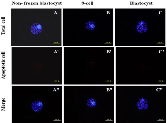
To analysis the total and apoptosis cells, post-thawed 8-cell and blastocysts were washed 2 ~3 times in PBS-PVP and fixed in a well of 4-well dish containing 700 μl of 4% (w/v) para- formaldehyde in PBS-PVP for 1 h at room temperature. After fixation, embryos were stored at 4 ℃ until TUNEL assay was performed. The TUNEL assay was performed according to the manufacturer protocol using the In Situ Cell Death Detection Kit (Fluorescein, Roche Diagnostics Corp.; USA; Cat. 1684795).
Briefly, the fixed embryos were washed twice with PBS-PVP before being incubation in permeabilization solution [0.5%
(v/v) Triton X-100, 0.1% (w/v) sodium citrate] for 30 min at room temperature. After permeabilization, the embryos were washed twice in PBS-PVP. The embryos were then incubated in fluorescence-conjugated dUTP and terminal deoxynucleotide transferase (TUNEL reagent, Roche) for 1 hrs at room tempe- rature in the dark. The TUNEL stained embryos were then washed in PBS-PVP and counterstained with 10 μg/ml Hoe- chst 33342 in PBS-PVP for 10 min at room temperature in the dark to label all nuclei. The blastocysts were then washed twice in PBS-PVP to remove excess Hoechst 33342 and mounted on glass slides under coverslips to evaluate the nuclear configu- ration. Cell numbers of each blastocyst was counted using an epifluorescence microscope (Olympus IX71, Japan) equipped with a mercury lamp. The positive cell was indicated by a bright red fluorescence indicating apoptotic cells and the total cell number was determined by green/blue fluorescence.
4. Statistical Analysis
Embryo survivability and the rates of re-expansion were expressed as percentages (%). The number of total and apop- totic cells was expressed as mean ± S.D. Statistical differences were analyzed by one-way ANOVA (SPSS Inc., Chicago, IL, USA). Significant differences between groups was performed by Duncan’s multiple range test. Statistical significance was taken at p<0.05.
RESULTS
1. Developmental Competence of Post-thawed Embryo Vitrified by
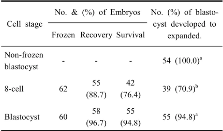
Table 1. Effect of cell stage on the development competence of vitrified embryo by different cell stage
Cell stage
No. & (%) of Embryos No. (%) of blasto- cyst developed to
expanded.
Frozen Recovery Survival Non-frozen
blastocyst - - - 54 (100.0)
a8-cell 62 55
(88.7) 42
(76.4) 39 (70.9)
bBlastocyst 60 58
(96.7) 55
(94.8) 55 (94.8)
a* a,b
Values with different superscripts in same column were significantly different (p<0.05).
*
Non-frozen blastocyst group was cultured in M-16 medium for 24 h.
Two Different Cell Stages
Total 6 biological replications were used for developmental competence of post-thawed embryos. The development rate of post-thawed vitrified blastocyst was highest in 8-cell and blas- tocyst groups (Table 1). The post-thaw re-expansion rate in control was not significantly higher than blastocyst group, but significantly higher than in 8-cell groups (non-frozen blas- tocyst, 100% vs. blastocyst, 94.8% vs. 8-cell, 70.9 %, p<0.05).
2. Assessment of Embryo Quality among Two Different Cell Stages In Table 2, the total cell numbers of post-thaw blastocyst was reduced regardless of the embryo stage used (control, 99.7
± 12.4 vs. blastocyst, 94.8 ± 15.1 and 8-cell, 74.7 ± 14.6, p<0.05). Even total cell numbers in blastocyst group was the
Table 2. Comparison of total and apoptosis cell number of vitrified 8-cell and blastocyst with non-frozen blastocyst
Treatments No. of emb- ryos stained
Total cell no.
(Mean ± S.D.)
Apoptotic cell no.
(Mean ± S.D.) Non-frozen
blastocyst 15 99.7 ± 12.4
a0.0 ± 0.0
cPost-thaw
8-cell 15 74.7 ± 14.6
b1.9 ± 3.1
cdPost-thaw
blastocyst 15 94.8 ± 15.1
a5.4 ± 4.4
d* a~d