Evaluation of Sperm Sex-Sorting Method using Flow Cytometry in Hanwoo (Korean Native Cattle)
Han-Jun Yoo
1, Kyung-Jin Lee
2, Yong-Seung Lee
2, Chang-Woo Lee
4, Joung-Jun Park
1, Hee-Tae Cheong
3, Boo-Keun Yang
2and Choon-Keun Park
2,*1
Developmental Biotechnology Laboratory, Myung-poomHanwoo Consulting, Hoengseong 225-807, Korea
2
College of Animal Life Sciences, Kangwon National University, Chuncheon 200-701, Korea
3
School of Veterinary Medicine, Kangwon National University, Chuncheon 200-701, Korea
4
Gangwon Province Livestock Research Center, Hoengseong 225-807, Korea
ABSTRACT
This study evaluated a method of sorting X and Y chromosomes based on size using the forward angle light scatter related refractive index (FSC) of a flow cytometer. Hanwoo bulls sperm were separated to X and Y chromosomes by the parameters of FSC or Hoechst 33342 intensity. As a result, using monitor program linked flow cytometry during sorting processing, the purities were 97 ± 0.57 or 96 ± 0.67% for the X-fraction and 96 ± 0.33 or 97 ± 1.33% for the Y-fraction in the two sperm sorting methods. There were no differences in the X and Y ratios (X and Y %) bet- ween the sperm sorting methods based on FSC or DNA content. The proportions of female and male embryos used for in vitro fertilization and development were 66.03 ± 3.31 or 69.37 ± 1.41%, and 70.56 ± 2.42 or 56.11 ± 3.09% when sperm were processed using the sex sorting method by FSC or Hoechst 33342. In conclusion, further study is needed to determine the optimum procedure and improve the nozzle to enhancing sorting accuracy or efficiency. Also, the fin- dings of this study do not negate the possibility that the difference method of sperm sorting cannot use a UV laser beam.
(Key words : flow cytometry, sperm sorting, FSC, sheath fluid buffer, Hanwoo)
†
This work was supported by Grant No. 109007-3 from Agricultural R&D Promotion Center (ARPC), Republic of Korea.
*
Correspondence : E-mail : parkck@kangwon.ac.kr
INTRODUCTION
Predetermination of sex in livestock offspring is in great de- mand and is of critical importance for efficient animal farming (Shim et al., 2001). Sex selection is especially useful in dairy cattle due to their economic importance and the need for heifer calves for herd replacement and milk production (Maxwell et al., 2004). To date, the only scientifically proven method based on the difference in DNA content between the X and Y chro- mosomes in mammals has been fully validated using flow cytometric sperm sorting (Johnson, 1995; Johnson and Welch 1999). However, in these studies, the preparation procedures negatively affected the viability of DNA. Sex selection via flow cytometric sorting involves staining of sperm with Hoechst 33342, which penetrates the living sperm membrane and binds to the DNA of the sperm nucleus (Choi et al., 2009), in combination with ultraviolet (UV) laser treatment.
Johnson et al. (1989) postulated that fluorochrome dyes re- duce embryonic viability by mid-gestation. This coincides with the findings of Spinaci et al. (2005) in boar sperm. Also,
weaker laser intensity diminishes the resolution and, indirectly, the sorting rate. There are few published reports of attempts to sort bull sperm from frozen-thawed semen (Lu et al., 1999;
Hollinshead et al., 2004b), and the limiting factor appears to be the low resolution of the X and Y populations due to the egg yolk. Another limiting factor to the use of frozen-thawed bull sperm for sorting is poor post-thaw viability compared to that of non-sorted frozen-thawed sperm (Hollinshead et al., 2004b). Also, another paper reported that the process must be carried out one cell at a time, resulting in an inherently slow system because millions or billions of sperm are needed for conventional artificial insemination (AI) (Johnson et al., 1999).
The current technology requires that each sperm be separately
interrogated for DNA content, thus limiting the number of
sorted X- or Y- sperm in cattle, sheep, swine and horses to ap-
proximately 12 ~20 million sperm per hour (Johnson and
Welch, 1999; de Graaf et al., 2007). Therefore, intensive re-
search is required to further improve the existing technology
or to identify alternatives and/or combinations with other bio-
techniques. On the one hand, in Korea, there are no commer-
cialization sperm sorting system because of the limitations on sorting speed, sorting accuracy, sperm ability and number of sorted sperm. Thus, the progress in this field has been slow.
Therefore, the objective of this study was to evaluate Han- woo (Korean native cattle) sperm sorted into X- and Y-chro- mosomes by using two evaluation methods from a flow cy- tometry. Sperm were prepared on the basis of differences in DNA mass and FSC for flow sorting, and sexed sperm were measured for the parameters of sex ratio using PCR.
MATERIALS AND METHODS
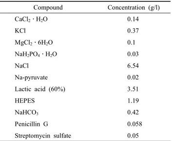
1. Preparation of Sheath Fluid Buffer
Sheath fluid is an isotonic buffer saline solution that is pumped through the flow chamber, causing single cells to flow through the middle of the stream. Preparation of sheath fluid buffer, known as HEPES sheath flow, used in stallions (Buss, 2005) was performed from each of the regents shown in Table 1 in a mixture with distilled water. Briefly, metal ions such as CaCl
2․H
2O, MgCl
2․6H
2O (Sigma-Aldrich, St. Louis, MO, USA), which were involved in chelation, dissolved in 800 ml distilled water. After completely dissolved, remaining powder reagents were added, and then the remaining liquid compo- nents were dissolved to incomplete compounds of sheath flow.
The sheath flow buffer is adjusted to 1 l with distilled water and pH 7.2. The filtration was performed through 0.2 μm pore size filters. Generally, this solution was prepared as a five-fold stock solution and was stored at 4 ℃ before use. The shelf-life
Table 1. Composition of HEPES sheath fluid buffer for sex- sorting of bull sperm using flow cytometry
Compound Concentration (g/l)
CaCl
2․H
2O 0.14
KCl 0.37
MgCl
2․6H
2O 0.1
NaH
2PO
4․H
2O 0.03
NaCl 6.54
Na-pyruvate 0.02
Lactic acid (60%) 3.51
HEPES 1.19
NaHCO
30.42
Penicillin G 0.058
Streptomycin sulfate 0.05
of the solution was considered to be only one week because instability of the stream during flow cytometric sperm sorting is observed after longer storage times.
2. Cryopreservation and Thawing of Sperm
For all experiments herein, Korean native cattle (Hanwoo) semen was collected using an artificial vagina and a teaser at the Hoengseong Livestock Cooperative Farm. Immediately after collection, motility and concentration of each bull sperm sample was assured using a phase-contrast microscope and a hemocy- tometer, respectively. Collected semen (volume: 5 ~15 ml;
density: 2 ~10 × 10
8ml; live ratio >75%) was diluted 1:1 (v/v) with Triladyl containing 20% egg yolk, and the semen tube was placed in a 500 ml, 35 ℃ water jacket before being cooled to 4 ℃ over 6 h in a refrigerator. The second dilution to a concentration of 10 × 10
6sperm per ml was then performed with an aliquot of semen diluted with the same freezing exten- der as was used in the first dilution. At this point, sperm were loaded into 0.5 ml straws, frozen and submerged in liquid nitrogen for storage. For use, semen straws were thawed for 45 s in a 37 ℃ water bath and dried. Semen was placed on a continuous density gradient tube to remove non- viable sperm and cryodiluent
3. Sperm Pretreatment
Percoll, a sterile colloidal silica suspension and isotonic salt gradient solution, was diluted to a 65% fraction with 10x HEPES buffer. The 65% Percoll (Nidacon, Göteborg, Sweden) was prepared by placing 1 ml in a 1.5 ml micro tube. The semen (0.5 ml) was placed over the gradient and centrifuged at 1,700 rpm (Micro 12, Hanil Science Industrial, Korea) for 15 min. The sperm pellets were assessed for concentration and resuspended in 1 ml of HEPES sheath flow buffer. The sperm was then subjected to one more washing step in order to im- prove the resolution of flow cytometry for sexing.
4. Hoechst33342 Staining
Sperm preparation and staining were based on the method
described by Johnson et al. (1989) in order to maintain via-
bility through sorting and fertilization. Briefly, pretreated ali-
quots of fresh or frozen-thawed semen of 1 ~5 × 10
7ml were
stained with 40 μM Hoechst 33342 (Sigma-Aldrich, St. Louis,
MO, USA) and were incubated for 30 min at 38 ℃ to foster
fluorochrome penetration. Finally, extra fluoro-dye, which was
not bound to the adenine-thymine regions of the sperm DNA,
was eliminated via centrifugation (1,500 rpm, 5 min). Also, an unstained control samples was prepared as a reference com- parison of intensity of Hoechst 33342 on flow cytometry du- ring fluorescence compensation and sex-sort processing using FSC. Generally, samples were prepared for separation in se- veral loading tubes because stained sperm aggregate during sperm sorting after 18 ~25 min.
5. Sex-sorting
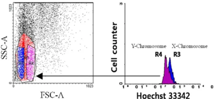
We based our sorting method on a histogram of X- and Y- chromosomes that had undergone Hoechst 33342 staining to produce a dot plot of sperm cell population using reverse order.
Fig. 1 shows blue or purple coloring between the R3 and R4 regions, which matched the dot plot and histogram created using FACSDiva software. This result suggests that each re- gion may independently perform the important role in separate X- and Y-chromosome as criterion of sperm sex-sorting. Intact sperm were sorted using a flow cytometer/cell sorter (BD FACs Aria Ⅱ; BD, USA). Intact, viable Hanwoo sperm were flow-cytometrically separated into X and Y populations on the basis of relative DNA contents and FSC (Fig. 2). Twenty to forty percent of living intact sperm were oriented with this process, and a sorting rate of approximately 50 to 200 sperm/
sec was achieved for each population. The sperm cells were measured and sorted using a 100 μm nozzle and a HEPES sheath fluid pressure of 20 psi. Sperm were sorted in single- cell mode into each 5 ml round tube that had been treated with 0.4% BSA solution to prevent the sperm from sticking to the wall of the tube, resulting in a final concentration of at least 1 ~5 × 10
6ml per tube. The tubes were then stored at room temperature (25 ℃; RT) until analysis at RT. After a to-
Fig. 1. R3 and R4 peaks of the histogram matched the gated region of the dot plot created by BD FACSDiva software.
Independently-gated R3 and R4 regions on the dot plot coincide with the intensity peak of Hoechst 33342 on the histogram.
Fig. 2. Visualized histogram of X- and Y-chromosomes after Hoe- chst 33342 staining and a dot plot of sperm cell population.
Hanwoo sperm were flow-cytometrically separated into X and Y populations based on differences in DNA content and size according to forward angle light scatter related refractive index. Twenty to forty percent of living intact sperm were oriented with this process, and sorting rates of approximately 50 to 200 sperm/sec were achieved for each population. With regard to FSC, the red and green regions indicate the X- and Y-enriched population, respec- tively.
tal of 1 ~5 × 10
6sperm cells were collected, the sperm were concentrated via centrifugation at 1,500 rpm for 7 min. The resultant pellet was resuspended with HEPES buffer and 2%
egg yolk and was processed for immediate use.
6. In vitro Embryo Production
Ovaries were collected from cows at a local slaughterhouse and transported to the laboratory. Oocytes were aspirated from antral follicles with an 18-gauge needle fixed to a 10 ml disposable syringe. The cumulus-oocyte complexes (COCs) were selected and then cultured in vitro maturation media at 38.5 ℃, 5% CO
2. Then, COCs were transferred into each 100 μl of TCM 199 containing EGF and 10% FBS that had been previously coated with mineral oil in a polystyrene culture dish (35 × 10 mm, Nunc, Roskilde, Denmark) and were equi- librated in 5% CO
2in air for approximately 4 h. After 20 ~22 h of incubation in maturation culture, oocytes were removed from cumulus cells by treatment with 300 IU/ml of hyalu- ronidase and repeated pipetting. After washing with Tyrode's albumin lactate pyruvate (TALP-Fer), oocytes were transferred into a 50 μl drop of TALP-Fer covered with mineral oil con- taining 0.4% (w/v) BSA (Fraction V). The dishes were stored in 38.5 ℃, 5% CO
2until the sperm were used for insemination.
For IVF, sex-sorted sperm were washed in TALP-SP for
remove egg yolk and resuspended in 200 ~500 μl TALP-SP.
A maximum of 50 μl of sperm suspension was added to 50 μl of the fertilization drops containing 10 oocytes. After 16 h of insemination, oocytes were removed from the fertilization medium and cultured in 100 μl of CR1aa containing 10%
FBS at 38.5 ℃, 5% CO
2.
7. Sex Determination Using the Polymerase Chain Reaction (PCR) Assay
To detect the S4 gene regions of 16 ~32 cell stage embryos produced via IVF with sex-sorted Hanwoo sperm using the above two sex-sorting methods, embryos were analyzed using genomic PCR. In brief, the zonapellucida of the embryos was removed by repetitive pipetting, and the cytoplasms of the embryos were washed three times with PBS and transferred into DNA extract tubes (AccuPower
ⓇGenomic DNA Extrac- tion Kit; Bioneer, Daejeon, Korea) with 200 μl of deionized water. Also, DNA of male and female positive controls were extract from each hair root. DNA extraction was performed in the following steps: DNA binding with binding buffer and proteinase K for 10 min, washing with washing buffer 1, 2 at 8,000 rpm, 1 min and extract DNA by elution buffer. Extracted genomic DNA was transferred into a PCR mixture tube (Accu- Power
ⓇPCR PreMix; Bioneer, Daejeon, Korea), then 10 pmol of each primer were added to the PCR tube, in addition to 1 U of Taq DNA polymerase, each deoxynucleosidetriphosphate at a concentration of 250 μM, 50 mMTris-HCl (pH 8.3), 40 mMKCl, 1.5 mM MgCl
2and gel loading dye. The final volume was adjusted to 20 μl with distilled water. PCR amplification was performed according to the standard protocol using the forward primer 5'- CAA GTG CTG CAG AGG ATG TGG AG -3' and the reverse primer 5'- GAG TGA GAT TTC TGG
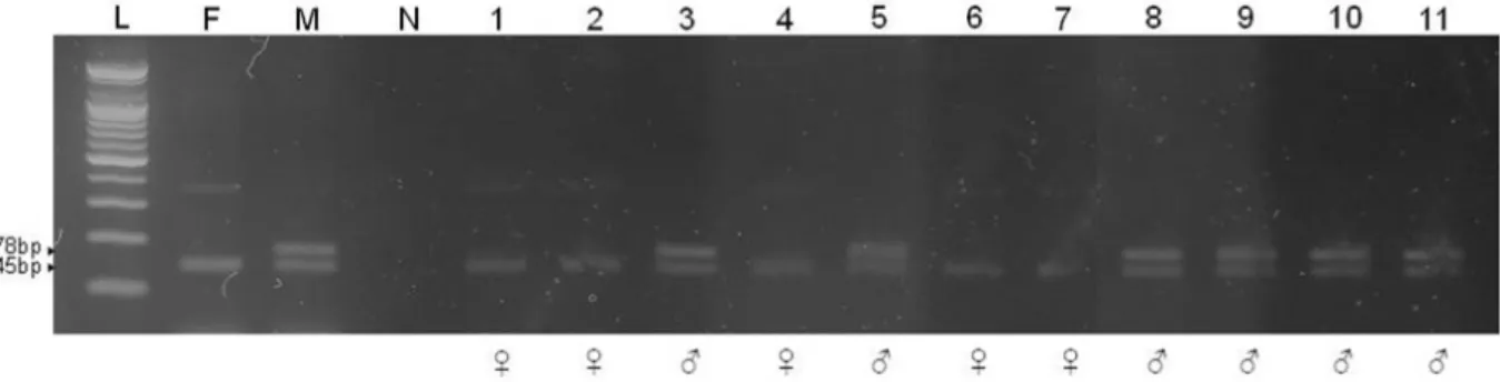
Fig. 3. Embryo sexing after PCR with the S4BF/R primer set to determine the efficiency of the sex-sorting method based on FSC and DNA content. The primers were designed to amplify a male-specific product (178 bp) in addition to a product common to both sexes (145 bp) from the repeated sequence, named S4 (DDBJ/GenBank database accession number D16357). L: 100 bp DNA ladder, F: positive control of female oocyte, M: positive control of male sperm, N: negative control, 1~11: bovine embryos.
ATC ATA TGG CTA CT -3'. The primers were designed to amplify a male-specific product (178 bp) from the repeated S4 bovine sequence (DDBJ/GenBank database accession number D16357), in addition to a product common to both sexes (145 bp) (Kageyama et al., 2004). The PCR reaction conditions consisted of denaturation at 95 ℃ for 5 min, followed by 35 amplification cycles of denaturation at 95 ℃ for 30 sec; annea- ling at 54 ℃ for 45 sec; extension at 72℃ for 45 sec. An addi- tional extension was performed at 72 ℃ for 5 min. After am- plification, a 10 μl aliquot of the PCR reaction mixture was subjected to electrophoresis in a 2% agarose gel and 1x TAE buffer (40 mMTris-acetate, pH 8.0, 1 mM EDTA). The gels were stained with 0.5 μg/ml ethidium bromide, and amplified DNA bands were visualized under ultraviolet illumination.
8. Statistical Analysis
Statistical analysis was performed with analysis of variance (ANOVA) using SAS (version 9.1, SAS Institute Inc., Cary, NC, USA). Differences among sperm analysis mean values from the sperm sorting method results were processed using Duncan's multiple range tests. Significance was defined at a level of p<0.05.
RESULTS
To demonstrate sorting accuracy, we identified embryospro- duced through in vitro fertilization using sexed semen (Fig. 3).
As shown in Table 2, there was a correlation between sorting
accuracy and sex ratio. Using a monitoring program linked
flow cytometry during sorting processing, purities were found
to be 97 ± 0.57 or 96 ± 0.67% for the X-fraction and 96 ± 0.33
or 97 ± 1.33% for the Y-fraction by two sperm sorting me- thods using parameters FSC or Hoechst 33342. Repetition of the experiment using sperm obtained from different bulls re- sulted in similar histograms and purities for both X- and Y-fractions. No differences in the × or Y ratio (X or Y%) were found between sperm sorting method based on differences in DNA content or FSC (Table 2). The X and Y ratios of the embryos by PCR were 66.03 ± 3.31% and 70.56 ± 2.42%, res- pectively, when sorted according to FSC using flow cytometry.
In the other sorting method using Hoechst 33342, the results were demonstrated 69.37 ± 1.41% for the X-ratio and 56.11 ±
3.09% for the Y-ratio. On the other hand, all sorted sperm showed very low fertility (Table 2).
DISCUSSTION
It has been reported that the sorting processes accelerate the maturation of sorted frozen-thawed ram sperm, reducing their fertilizing lifespan (Hollinshead et al., 2003; Hollinshead et al., 2004a). Also, several groups (Cran et al., 1993; Lu et al., 1999) have reported similar cleavage rates but reduced blas- tocyst development following fertilization in vitro with sorted bovine sperm. Additionally, lower pregnancy rates after ferti- lization with sex-sorted compared to non-sorted sperm have been reported in a number of studies (Johnson, 1995; Seidel et al., 1999; Hollinshead et al., 2002: Seidel and Garner, 2002).
Capacitation-like changes have been observed after the sorting of both ram and boar sperm (Catt et al., 1997; Maxwell et al., 1998; Maxwell and Johnson, 1999), similar to those observed
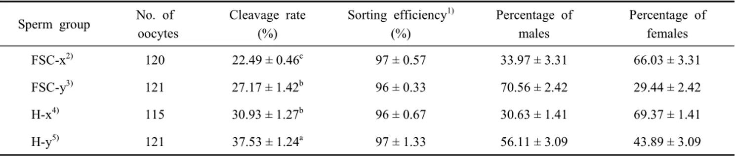
Table 2. The proportions of male and female embryos rates at 48 h after IVF with sex-sorted sperm in Hanwoo cattle
Sperm group No. of oocytes
Cleavage rate (%)
Sorting efficiency
1)(%)
Percentage of males
Percentage of females
FSC-x
2)120 22.49 ± 0.46
c97 ± 0.57 33.97 ± 3.31 66.03 ± 3.31
FSC-y
3)121 27.17 ± 1.42
b96 ± 0.33 70.56 ± 2.42 29.44 ± 2.42
H-x
4)115 30.93 ± 1.27
b96 ± 0.67 30.63 ± 1.41 69.37 ± 1.41
H-y
5)121 37.53 ± 1.24
a97 ± 1.33 56.11 ± 3.09 43.89 ± 3.09
(Mean ± SEM, p<0.05)
1)
This parameter indicates correctly separated ratio of sperm in flow cytometry.
2)
FSC-x: Sorted X sperm without near UV laser beam in flow cytometry.
3)
FSC-y: Sorted Y sperm without near UV laser beam in flow cytometry.
4)
H-x: Sorted X sperm using differences in DNA content by Hoechst 33342.
5)