The Regulation Mechanisms of Kinesin Motor Proteins
Sang Jun Park1†, Joung-Su Seog2†, Il Soo Moon3 and Dae-Hyun Seog4*
1Department of Pharmaceutical Engineering, Inje University, Gimhae 50834, Korea
2Department of Nursing, Suseong College, Daegu 42078, Korea
3Departments of Anatomy, College of Medicine, Dongguk University, Gyeongju 38066, Korea
4Department of Biochemistry, Inje University College of Medicine, Busan 614-735, Korea Received April 17, 2017 /Revised April 25, 2017 /Accepted April 27, 2017
Proper intracellular transport is essential for normal cell function. Intracellular transport is mediated by microtubule-dependent molecular motor proteins, as well as kinesin and cytoplasmic dynein, which move their cargo along long, microtubule tracks in cells. Kinesins are ATP-dependent plus-end-directed motor proteins in the intracellular transport of organelles, vesicles, RNA complexes, and protein complexes. The mislocalization of these different types of cargo has been linked to cell dysfunction and degeneration. The cargo transport of kinesins can be described by the following steps:
binding to the appropriate cargo and/or adaptor proteins, activation of the kinesin’s motility and movement along the microtubule, and the release of the cargo at the correct destination. Recently, sev- eral studies have revealed the mechanisms for the regulation of kinesin motor activity, including cargo loading and unloading. Intracellular cargo transport is also modulated by adaptor proteins, which link the kinesins to their cargo. The regulatory proteins, which include protein kinases and phosphatases, regulate kinesin motor activity directly through the phosphorylation or dephosphorylation of kinesins and indirectly through the modification of adaptor proteins, such as c-Jun NH-terminal kinase-inter- acting proteins, or of the microtubule network. These findings lay the groundwork for understanding how kinesins are differentially engaged in intracellular cargo transport. In addition, understanding the regulatory mechanisms of each kinesin is an area of key interest within cell biology and neurophysiology. In this study, we reviewed kinesins’ regulation proteins and discuss how their regu- lation affects cargo recognition and transport.
Key words: Adaptor protein, kinesin, phosphorylation, protein kinase, Rab protein
†Author contributed equally.
*Corresponding author
*Tel : +82-51-890-6974, Fax : +82-51-894-5801
*E-mail : [email protected]
This is an Open-Access article distributed under the terms of the Creative Commons Attribution Non-Commercial License (http://creativecommons.org/licenses/by-nc/3.0) which permits unrestricted non-commercial use, distribution, and reproduction in any medium, provided the original work is properly cited.
Journal of Life Science 2017 Vol. 27. No. 7. 840~848 DOI : https://doi.org/10.5352/JLS.2017.27.7.840
Introduction
The intracellular transport of membrane-bounded vesicles and organelles contributes for morphogenesis and function- ing of the cell. Long-distance intracellular transport is de- pendents almost entirely on microtubule tracks [16]. Micro- tubules are polymerized with two subunits, α- and β-tubule at plus ends, organized in a radial array from the cell center toward the cell periphery [16]. In contrast to other cell types, microtubules are formed in a parallel unipolar array in the neuronal dendrites with plus ends oriented outward the cor- tex [17]. Two ATP-dependent microtubule motor proteins,
kinesins and cytoplasmic dynein have directional motility on polar microtubule tracks in the cells [16]. Kinesins pri- mary drive the transport of cargos along the microtubule tracks to pus-ends directly (Fig. 1). By contrast, cytoplasmic dynein associates an essential accessory proteins complex known as dynactin, which function transport of cargo to mi- crotubule tracks to minus-ends directly [16]. Kinesins make up a large superfamily, with up to 45 members expressed in mammalian cells [34]. A standardized nomenclature groups kinesin genes into 14 subfamilies that share the mo- tor domain similarity and the structural similarity [28].
The intracellular transport of various cargos in cells un- derlies many essential cellular functions, including the pro- tein secretion, cell growth and cell signaling, trafficking of RNA complexes, protein and organelle degradation, and dis- tribution of organelles. Kinesin-1 motors, a major motor for intracellular transport drive a wide range of cargos includ- ing various vesicles, mitochondria, and RNA particles [18].
Kinesin-2 motors drive the fodrin-positive vesicles [53], N-cadherin and β-catenin [54], and N-methyl-D-aspartate - Review -
Fig. 1. Regulation of kinesins-mediated intracellular cargo transport by proteins kinases and Rab proteins. Kinesins drive the micro- tubule-dependent movement of organelles, vesicles, RNA granules, and proteins from the cell body to the cell periphery.
Cargo-bound kinesins are regulated by several protein kinases and Rab proteins, such as glycogen synthase kinase 3 beta (GSK3β), c-Jun N-terminal kinase 3 (JNK3), extracellular signal-regulated kinases-1 and 2 (ERK1/2), protein phosphatase 2C-like serine/threonine phosphatases X2 (POPX2), cyclin-dependent kinase 5 (Cdk5), cyclin-dependent kinase 1 (Cdk1), Calcium/calmodulin-dependent protein kinase II (CaMKIIα), protein kinase A (PKA), protein kinase C (PKC), protein phos- phatase PP2A, and casein kinase 2 (CK2). Plus (+) and minus (-) refer to the polarity of microtubules. ER; endoplasmic reticulum, Nu; nucleus, MTOC; microtubule-organizing center.
(NMDA) receptor containing vesicles in the cells [49].
Kinesin-3 motors drive the synaptic vesicle precursors and dense core vesicles [18]. Dysfunction of these kinesin-medi- ated cargo transports is relevant to abnormal neurogenesis and various neurological disorders such as Huntington's Disease (HD), Alzheimer's Disease (AD), Charcot–Marie–
Tooth Type 2F (CMT2F), and Amyotrophic Lateral Sclerosis (ALS) [15].
The binding proteins of kinesins have been studied in de- tail to the specific cargo level. However, less is well studied about how the motor activities are regulated when bound to cargo and released it. The autoinhibition of kinesin-1 is an example of this regulation (Fig. 1) [4]. Kinesin-mediated cargo transport is also modulated by adaptor proteins. [18].
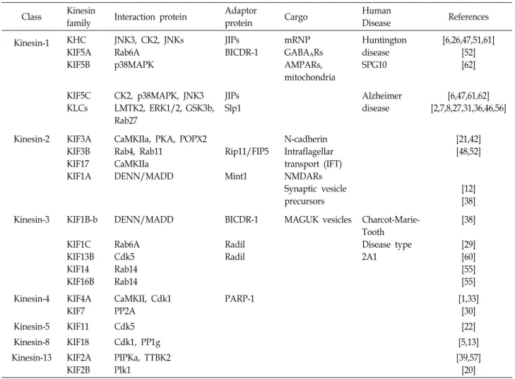
Adaptor proteins can effectively control the directionality of cargo-associated motor [50]. Kinesins and adaptor proteins directly bind to the modification proteins, including protein kinase, phosphatase, and small G-proteins (Table 1) [10].
Theses kinesin regulation mechanisms by autoinhibition or
kinesin modification proteins allow for sustained the cell ho- meostasis [4].
In this review, we discuss the recent progress regarding the following questions. How do kinesins regulate loading and unloading their cargo? How do kinesins regulate the binding to microtubules? What is the biological significance of these regulation mechanisms?
Kinesin-1
Kinesin-1 is a tetramer consisting of two kinesin heavy chains (KHC; KIF5A, KIF5B, and KIF5C) that contain the mo- tor domain and two kinesin light chains (KLCs) [23]. KLCs interact with the carboxyl (C)-terminal tail domain of KHC and regulate the cargo binding and the autoinihibition of the motor [4]. By binding with adaptor proteins or by direct interaction with cargos, kinesin-1 transport many different cargos, including mitochondria, mRNA-protein complex (mRNP), α-amino-3-hydroxy-5-methylisoxazole-4-proprionate
Table 1. Modification proteins interactions with Kinesins
Class Kinesin
family Interaction protein Adaptor
protein Cargo Human
Disease References
Kinesin-1 KHC KIF5A KIF5B
JNK3, CK2, JNKs Rab6A
p38MAPK
JIPs BICDR-1
mRNP GABAARs AMPARs, mitochondria
Huntington disease SPG10
[6,26,47,51,61]
[52]
[62]
KIF5C KLCs
CK2, p38MAPK, JNK3 LMTK2, ERK1/2, GSK3b, Rab27
JIPs Slp1
Alzheimer disease
[6,47,61,62]
[2,7,8,27,31,36,46,56]
Kinesin-2 KIF3A KIF3B KIF17 KIF1A
CaMKIIa, PKA, POPX2 Rab4, Rab11 CaMKIIa DENN/MADD
Rip11/FIP5
Mint1
N-cadherin Intraflagellar transport (IFT) NMDARs Synaptic vesicle precursors
[21,42]
[48,52]
[12]
[38]
Kinesin-3 KIF1B-b KIF1C KIF13B KIF14 KIF16B
DENN/MADD Rab6A Cdk5 Rab14 Rab14
BICDR-1 Radil Radil
MAGUK vesicles Charcot-Marie- Tooth Disease type 2A1
[38]
[29]
[60]
[55]
[55]
Kinesin-4 KIF4A KIF7
CaMKII, Cdk1 PP2A
PARP-1 [1,33]
[30]
Kinesin-5 KIF11 Cdk5 [22]
Kinesin-8 KIF18 Cdk1, PP1g [5,13]
Kinesin-13 KIF2A KIF2B
PIPKa, TTBK2 Plk1
[39,57]
[20]
(AMPA) receptor vesicles, γ-aminobutyric acid (GABA) re- ceptor vesicles, and brain derived neurotrophic factor (BDNF) vesicles [18].
Phosphorylation
Kinesin-1 is phosphoproteins and the phosphorylation of KHCs can regulate the cargo loading and unloading [9].
Protein kinases can regulate the intracellular cargo transport through the direct phosphorylation of KIF5s or adapter pro- teins [11]. c-Jun N-terminal kinases (JNKs) are activated by cellular stress and regulate the intracellular transport by di- rect or indirect phosphorylation of kinesin-1, and adapter proteins [26]. JNKs directly phosphorylate KIF5B and induce its dissociation from microtubules [51]. JNKs also regulate the intracellular transport by phosphorylation of adaptor protein, such as JNK-interacting protein 1 (JIP1). In cells, phosphorylation of JIP1 at Ser 421 stabilizes its binding with kinesin-1 and activates the intracellular transport of amyloid precursor proteins (APP) containing vesicles [59]. These re-
sults show that JNKs-mediated phosphorylation of kinesin-1 and/or adaptor protein can modulate of the intracellular car- go transport in cells. Other JNKs, JNK3 blocks the intra- cellular transport by direct phosphorylation at Ser 176 of KIF5C, reducing microtubules binding affinity [6]. The mo- tor domain of KIF5s contains the consensus JNK3 phospho- tylation site corresponding to Ser 175 [6]. Phosphorylation of this residue has been implicated in HD [11]. Placement of negative charge at Ser 175 of KIF5s leads to a lower stall force and decreased velocity of motor. However, the ATPase activity, and microtubule-binding affinity are unchanged be- tween wild-type motor domain and mutant construct. A sed- imentation velocity experiment showed that a mutant fa- vored the autoinhibited conformation [6]. This result sug- gests that cargo is transported by phosphorylated kinesin-1.
Another protein kinase, casein kinase 2 (CK2) directly phos- phorylates at Thr 338 which is located in the non-motor re- gion of KIF5C [47]. In cultured cells, reducing CK2 ex- pression decreased the lipid droplet transport, consistent
with a deceased number of active kinesin-I [61]. This data indicates that the protein kinase CK2 upregulates kine- sin-based intracellular transport and the activity of car- go-bound kinesin-1.
p38 mitogen-activated protein kinases (p38 MAPKs) sig- naling pathway plays important roles in skeletal myogenesis and is activated in response to pro-inflammatory cytokines [43]. p38 MAPK directly phosphorylates Ser 175/Ser 176 of KIF5C and thereby inhibits the fast intracellular transport of membrane-bound organelle [62]. p38 MAPK also phos- phorylates the tail domains of neurofilaments (NFs) and reg- ulates their attachment between neurofilaments and motor proteins [35]. Two isoforms of KLC have been identified in mammals; the neuronal tissue-specific KLC1 and the ubiq- uitous KLC2 [45]. KLCs were phosphorylated by various protein kinases including extracellular signal-regulated kin- ases-1 and 2 (ERK1/2), glycogen synthase kinase-3 beta (GSK3β), and lemur tyrosine kinase-2 (LMTK2). ERK1/2 is playing a key role in neuronal differentiation and is acti- vated in response to mitogens and growth factors [46].
ERK1/2 directly phosphorylates Ser 460 of KLC1 and pre- vents binding APP-labeled vesicles to kinesin-1, leading to a decrease the APP-contained vesicle transport in neuron [56]. However, ERK1/2-mediated phosphorylation of KLC1 has no effect on the kinesin-1-mediated intracellular trans- port of other cargoes, including collapsin response mediator protein 2 (CRMP2) [56]. GSK3β was identified as a regulator of glycogen metabolism in cells [8]. GSK3β directly phos- phorylate KLC2 and adapter proteins [36]. In mammalian cells, GSK3β acts as a negative regulator of the intracellular transport [7]. In Drosophila, overexpression of GSK3β inhibits the intracellular transport of mitochondria and APP-contain- ing vesicles by kinesin-1 [7]. In squid axoplasm, GSK3β re- leases the cargos from kinesin-1 without influencing the mi- crotubule binding or ATPase activity of KHCs [36]. GSK3β has also been shown to regulate the intracellular cargo trans- port through phosphorylation of adapter proteins. Collapsin response mediator protein-2 (CRMP-2) acts as adaptor pro- tein between kinesin-1 and cargoes, including such as TrkB- containing vesicles [24]. GSK3β-mediated phosphorylation of CRMP-2 inhibits the binding with KLC1 and CRMP-2 and blocks the intracellular transport of kinesin-1 cargoes [2].
LMTK2 plays important roles as a susceptibility gene for prostate cancer [58]. LMTK2 modulates phosphorylation of KLC2 by GSK3β. Using small interfering RNA, loss of LMTK2 was reduced the binding affinity of the kinesin-1
and Smad2 cargo [31]. Thus, phosphorylation of KLCs and adaptor proteins by various protein kinases modulates the cargo loading and unloading to kinesin-1.
Rab GTPase
Rab proteins are a family of monomeric guanine nucleo- tide (G)-binding proteins which have emerged as regulators of the intracellular transport in cells (Fig. 1) [52]. Interestingly, Rab27A and Rab27B associate with the tetratricopeptide re- peat (TPR) of KLCs via adaptor protein, Slps (synaptotag- min-like proteins) [2]. Inhibition of the Rab27a/Slp3/kine- sin-1 complex formation impairs lytic granules secretion to the immune synapse [27]. This data indicate that the binding with Rab27 protein and kinesin-1 or adaptor proteins modu- lates the intracellular cargo transport.
Kinesin-2
Four kinesin-2 subfamilies exist: the heterotrimeric com- plex composed of KIF3A, KIF3B, KIF3C motor subunits and KAP3, the non-motor subunit, which binds the cargo and the homodimeric complex of KIF17 motor subunit [28].
Kinesin-2 drives the various cargoes such as fodrin-positive vesicles [53], N-cadherin and β-catenin [54], NMDA receptor vesicles [49], and associate with Rab7-positive late endosome [3].
Phosphorylation
Using quantitative phosphoanalyses, protein kinase A (PKA) and Calcium/calmodulin-dependent protein kinase II (CaMKIIα) directly phosphorylates Ser 689, 694, and 698 of KIF3A [21]. In cultured cells, protein kinase inhibitors treat- ment and transfection of mutagenesis constructs of KIF3A revealed that cargo transport was enhanced by phosphor- ylation of the KIF3A [21]. Also, phosphorylation of KIF3 was upregulated cargo-loading activity, the association of KIF3 and cargos. Protein phosphatase 2C-like serine/threonine phosphatases X2 (POPX2), a serine-threonine phosphatase, interact with KAP3. POPX2 dephosphorylates Ser 690 of KIF3A and KIF3A-S690A mutant blocks the cargo transport [42]. Thus, POPX2 acts as a negative regulator of the cargo transport. These results suggest that phosphorylation and dephosphorylation pair of KIF3 regulate cargo-loading in the intracellular cargo transport process. Interestingly, CaMKIIα also binds to the tail region of KIF17 and phosphorylates at Ser 1029 of KIF17. Phosphorylation of KIF17 dissociates
Mint1, adaptor protein from the KIF17 and releases the car- gos from KIF17 [12]. This result suggests that phosphor- ylation of KIF17 is an important mechanism for car- go-unloading. Phosphorylation of KIF3 upregulates the car- go-loading activity. However, the phosphorylation of KIF17 upregulates the cargo-unloading activity. These contrasting results may represent differences in the regulation mecha- nism of cargo loading and unloading between kinesin-2 motors.
Rab GTPase
Protein sorting, targeting and recycling of endocytosed membrane proteins are regulated by various Rab GTPases, such as Rab4, Rab8, and Rab11 [52]. Rip11/FIP5 was origi- nally identified as a Rab11-binding protein and act as adap- tor protein that recruit various proteins that regulate mem- brane transport [44]. The siRNA-based protein knockdown experiment showed Rip11/FIP5 regulates the sorting of in- ternalized receptors to a recycling pathway. Interestingly, the tail domain of KIF3B directly binds to Rip11/FIP5 [48].
This interaction between Rip11/FIP5-Rab11 complex and KIF3 suggests that Rab GTPases are involved in the regu- lation of kinesin-2-mediated protein transport.
Kinesin-3
Kinesin-3 comprises five subfamilies, namely the KIF1, KIF13, KIF14, KIF16, and KIF28 [28]. This family is reported to be monomeric [40] or homodimeric structure [18].
Kinesin-3 drives the synaptic vesicle precursors, which con- tain synaptic vesicle proteins such as synaptophysin, syn- aptotagmin, and Rab3A [18, 28].
Phosphorylation
Cyclin-dependent kinase 5 (Cdk5) is regulates several sig- nal processes in the cell function, including cell growth, and cell migration [25]. The inhibition of Cdk5 activity inhibited the intracellular transport of membrane-bound organelles and secretory vesicles [25]. Cdk5 phosphorylates Thr-506, a residue located in the forkhead-associated (FHA) domain of KIF13B [60]. The inhibition of Cdk5 activity modulated the association of KIF13B and transient receptor potential va- nilloid 1 (TRPV1). The overexpression of Cdk5 in cells was promoted TRPV1 intracellular transport by activating the motor-cargo loading [60]. This result indicates that phos- phorylation of KIF13 is an important mechanism for car-
go-loading and upregulates intracellular cargo transport to cell surface.
Rab GTPase
Rab3A is required for the intracellular vesicle transport of in cells [52]. Differentially expressed in normal and neo- plastic cells (DENN)/mitogen-activated protein kinase-acti- vating death domain (MADD) binds with the stalk domain of KIF1A and KIF1β. Rab3A indirectly associates with KIF1A and KIF1β via DENN/MADD (Rab-GEP) and regulates the interaction between KIF1A/KIF1β and DENN/ MADD [38].
GTP-Rab3A transports presynatic precursor vesicles more effectively than GDP-Rab3A [38]. This result suggests that Rab3A regulates the KIF1A and KIF1β-mediated cargo transport. Another Rab protein, Rab6A directly binds the motor domain and the C-terminal region of KIF1C [29]. This association of KIF1C and Rab6A inhibits the interaction be- tween KIF1C and microtubule track and slows intracellular cargo transport to the cell surface [29]. The association of Rab6A also directly regulates the motor activity of KIF1C.
Rab14 directly associates with KIF16B and regulates the Golgi-to-endosome trafficking of the fibroblast growth factor receptor (FGER) containing vesicles during embryonic de- velopment [55]. These results revealed that Rab proteins reg- ulate the microtubule affinity and cargo-loading of kinesin-3.
Kinesin-4
Kinesin-4 can be classified into five subfamilies: KIF4, KIF7, KIF21, KIF27, and NcKIF21 [28]. Kinesin-4 acts in the intracellular cargo transport, the microtubule dynamics, and the cell signaling [18]. For example, KIF4A regulates the sta- bilization of microtubules during cell division [33], and KIF7 regulates Hedgehog (Hh) signaling at cilia [14], and KIF21 regulates microtubule growth and participates in organizing microtubule arrays at the cell edge [37].
Phosphorylation
Genetic study indicates that KIF7 regulates Hh signaling in vertebrates. Hh signaling leads to ciliary accumulation and affects the localization of transcriptional regulators in cells [41]. The protein phosphatase 2A (PP2A) interacts with KIF7 and promotes the dephosphorylation of KIF7. The de- phosphorylation of KIF7 promotes the trafficking of KIF7 and Gli proteins to the tips of cilia and for the transcriptional output of Hh signaling. [30]. KIF4 regulates programmed
cell death by interacting directly with poly ADP-ribose poly- merase-1 (PARP-1), a nuclear enzyme that modifies various nuclear proteins with poly ADP-ribosylation to maintain cell homeostasis. [1]. When cells are stimulated by membrane depolarization via Ca2+ influx into nucleoplasm, CaMKII in- duces the dissociation of KIF4 from PARP-1, resulting in up- regulation of PARP-1 activity. After dissociation from PARP- 1, KIF4 enters into the cytoplasm from the nucleus [33].
Other Kinesins
Phosphorylation
Kinesin-5 is a homotetrameric protein and essential role in the mitotic spindle [28]. Cdk5 is the protein kinase respon- sible for phosphorylating kinesin-5 at Thr 926, which is im- portant for kinesin-5 to associate with microtubules. Cdk5- mediated phosphorylation of Kinesin-5 also associates pref- erentially with microtubules rich in tyrosinated tubulin [22].
This result provides that Cdk5-mediated phosphorylation of kinesin-5 regulates the localization of kinesin-5 on dendritic microtubules, as they are known to be less detyrosinated than axonal microtubules.
KIF2 is a member of the kinesin-13 and regulates micro- tubule dynamics at growth cones [19]. Phosphatidylinositol 4-phosphate 5-kinase alpha (PIPKα) directly binds KIF2A [39]. The microtubule-depolymerizing activity of KIF2A was enhanced in the presence of PIPKα in vitro and in vivo. PIPKα also suppresses the elongation of axon branches in a KIF2A- dependent manner [39]. Tau-tubulin kinase 2 (TTBK2) di- rectly phosphotylates Ser 135 of KIF2A and inactivates mi- crotubule-depolymerizing activity by phosphorylation of KIF2A [57]. TTBK2 depletion reduces microtubule lifetime and impairs the cell migration. Overexpression of non- phosphorylatable type KIF2A also reduces microtubule life- time and slowdowns the cell migration [57]. Using quantita- tive phosphoanalyses by mass spectrometry, KIF2B had identified the multiple phosphorylation sites. Polo-like kin- ase 1 (Plk1) directly phosphorylates KIF2B at Thr 125 and Ser 204, and that these two sites regulate KIF2B function [20]. Phosphorylation of Ser 204 of KIF2B is required for the kinetochore localization, and phosphorylation of Thr 125 of KIF2B is required for KIF2B activity in the kinetochore-mi- crotubule attachments [20]. These results provide that vari- ous protein kinases promote the phosphorylation of KIF2, and regulate the microtubule dynamics.
KIF18A, a member of the kinesin-8, plays critical roles in
various cellular processes, including cell motility, cell divi- sion, microtubule dynamics and the intracellular transport [32]. KIF18A directly interacts with protein phosphatase 1 (PP1) through a conserved RVxF motif [5]. PP1 induces met- aphase plate thinning by dephosphorylating KIF18A. PP1 and Cdk1 antagonistically regulate KIF18A. Chromosome at- tachment induces Cdk1 inactivation and kinetochore recruit- ment of PP1 [13]. This result provides that chromosome movement is regulated by phosphorylation and dephospor- ylation of KIF18A.
Conclusion and outlook
In this review, we have discussed how various protein kinases and many Rab GTPases control the kinesin motor activity, cargos are loaded to their motors, and the cargos are unloaded upon reaching their destinations. Kinesins and adaptor proteins are major targets for the regulation of intra- cellular cargo transport. The proteins regulating kinesins and adaptor proteins have been rapidly identified in recent years. Given the important role, it is not surprising that alter- ations in protein kinase and Rab GTPase activities block the intracellular cargo transport and may lead to several nerv- ous disorders. Thus, to disclose clearly the different regu- latory mechanisms involved in the intracellular cargo trans- port is major challenge for future research.
Acknowledgment
This research was supported by Basic Science Research Program though the National Research Foundation of Korea (NRF) funded by the Ministry of Education, Science and Technology (NRF-2015R1D1A1A01056820).
References
1. Althaus, F. R. and Richter, C. 1987. ADP-ribosylation of proteins. Enzymology and biological significance. Mol. Biol.
Biochem. Biophys. 37, 1-237.
2. Arimura, N., Kimura, T., Nakamuta, S., Taya, S., Funahashi, Y., Hattori, A., Shimada, A., Ménager, C., Kawabata, S., Fujii, K., Iwamatsu, A., Segal, R. A., Fukuda, M. and Kaibuchi, K. 2009. Anterograde transport of TrkB in axons is mediated by direct interaction with Slp1 and Rab27. Dev. Cell 16, 675-686.
3. Castle, M. J., Perlson, E., Holzbaur, E. L. and Wolfe, J. H.
2014. Long-distance axonal transport of AAV9 is driven by dynein and kinesin-2 and is trafficked in a highly motile Rab7-positive compartment. Mol. Ther. 22, 554-566.
4. Caviston, J. P. and Holzbaur, E. L. 2006. Microtubule motors at the intersection of trafficking and transport. Trends Cell Biol. 16, 530-537.
5. De Wever, V., Nasa, I., Chamousset, D., Lloyd, D., Nimick, M., Xu, H., Trinkle-Mulcahy, L. and Moorhead, G. B. 2014.
The human mitotic kinesin KIF18A binds protein phospha- tase 1 (PP1) through a highly conserved docking motif.
Biochem. Biophys. Res. Commun. 453, 432-437.
6. DeBerg, H. A., Blehm, B. H., Sheung, J., Thompson, A. R., Bookwalter, C. S., Torabi, S. F., Schroer, T. A., Berger, C.
L., Lu, Y., Trybus, K. M. and Selvin, P. R. 2013. Motor do- main phosphorylation modulates kinesin-1 transport. J. Biol.
Chem. 288, 32612-32621.
7. Dolma, K., Iacobucci, G. J., Hong Zheng, K., Shandilya, J., Toska, E., White, J. A. 2nd., Spina, E. and Gunawardena, S.
2014. Presenilin influences glycogen synthase kinase-3 β (GSK-3β) for kinesin-1 and dynein function during axonal transport. Hum. Mol. Genet. 23, 1121-1133.
8. Embi, N., Rylatt, D. B. and Cohen, P. 1980. Glycogen syn- thase kinase-3 from rabbit skeletal muscle. Separation from cyclic-AMP-dependent protein kinase and phosphorylase kinase. Eur. J. Biochem. 107, 519-527.
9. Espeut, J., Gaussen, A., Bieling, P., Morin, V., Prieto, S., Fesquet, D., Surrey, T. and Abrieu, A. 2008. Phosphorylation relieves autoinhibition of the kinetochore motor Cenp-E.
Mol. Cell 29, 637-643.
10. Fu, M. M. and Holzbaur, E. L. 2014. Integrated regulation of motor-driven organelle transport by scaffolding proteins.
Trends Cell Biol. 24, 564-574.
11. Gibbs, K. L., Greensmith, L. and Schiavo, G. 2015. Regula- tion of axonal transport by protein kinases. Trends Biochem.
Sci. 40, 597-610.
12. Guillaud, L., Wong, R. and Hirokawa, N. 2008. Disruption of KIF17-Mint1 interaction by CaMKII-dependent phos- phorylation: a molecular model of kinesin-cargo release.
Nat. Cell Biol. 10, 19-29.
13. Häfner, J., Mayr, M. I., Möckel, M. M. and Mayer, T. U.
2014. Pre-anaphase chromosome oscillations are regulated by the antagonistic activities of Cdk1 and PP1 on Kif18A.
Nat. Commun. 5, 4397.
14. He, M., Subramanian, R., Bangs, F., Omelchenko, T., Liem, K. F. Jr., Kapoor, T. M. and Anderson, K. V. 2014. The kine- sin-4 protein Kif7 regulates mammalian Hedgehog signal- ling by organizing the cilium tip compartment. Nat. Cell Biol.
16, 663-672.
15. Hirokawa, N. and Tanaka, Y. 2015. Kinesin superfamily pro- teins (KIFs): Various functions and their relevance for im- portant phenomena in life and diseases. Exp. Cell Res. 334, 16-25.
16. Hirokawa, N. 1998. Kinesin and dynein superfamily pro- teins and the mechanism of organelle transport. Science 279, 519-526.
17. Hirokawa, N. and Takemura, R. 2005. Molecular motors and mechanisms of directional transport in neurons. Nat. Rev.
Neurosci. 6, 201-214.
18. Hirokawa, N., Niwa, S. and Tanaka, Y. 2010. Molecular mo-
tors in neurons: transport mechanisms and roles in brain function, development, and disease. Neuron 68, 610-638.
19. Homma, N., Takei, Y., Tanaka, Y., Nakata, T., Terada, S., Kikkawa, M., Noda, Y. and Hirokawa, N. 2003. Kinesin su- perfamily protein 2A (KIF2A) functions in suppression of collateral branch extension. Cell 114, 229-239.
20. Hood, E. A., Kettenbach, A. N., Gerber, S. A. and Compton, D. A. 2012. Plk1 regulates the kinesin-13 protein Kif2b to promote faithful chromosome segregation. Mol. Biol. Cell 23, 2264-2274.
21. Ichinose, S., Ogawa, T. and Hirokawa, N. 2015. Mechanism of activity-dependent cargo loading via the phosphoryla- tion of KIF3A by PKA and CaMKIIα. Neuron 87, 1022-1035.
22. Kahn, O. I., Sharma, V., González-Billault, C. and Baas, P.
W. 2015. Effects of kinesin-5 inhibition on dendritic archi- tecture and microtubule organization. Mol. Biol. Cell 26, 66-77.
23. Kanai, Y., Okada, Y., Tanaka, Y., Harada, A., Terada, S. and Hirokawa, N. 2000. KIF5C, a novel neuronal kinesin en- riched in motor neurons. J. Neurosci. 20, 6374-6384.
24. Kawano, Y., Yoshimura, T., Tsuboi, D., Kawabata, S., Kaneko-Kawano, T., Shirataki, H., Takenawa, T. and Kaibuchi, K. 2005. CRMP-2 is involved in kinesin-1-depend- ent transport of the Sra-1/WAVE1 complex and axon formation. Mol. Cell Biol. 25, 9920-9935.
25. Kawauchi, T. 2014. Cdk5 regulates multiple cellular events in neural development, function and disease. Dev. Growth Differ. 56, 335-348.
26. Koch, P., Gehringer, M. and Laufer, S. A. 2015. Inhibitors of c-Jun N-terminal kinases: an update. J. Med. Chem. 58, 72-95.
27. Kurowska, M., Goudin, N., Nehme, N. T., Court, M., Garin, J., Fischer, A., de Saint Basile, G. and Ménasché, G. 2012.
Terminal transport of lytic granules to the immune synapse is mediated by the kinesin-1/Slp3/Rab27a complex. Blood 119, 3879-3889.
28. Lawrence, C. J., Dawe, R. K., Christie, K. R., Cleveland, D.
W., Dawson, S. C., Endow, S. A., Goldstein, L. S., Goodson, H. V., Hirokawa, N., Howard, J., Malmberg, R. L., McIntosh, J. R., Miki, H., Mitchison, T. J., Okada, Y., Reddy, A. S., Saxton, W. M., Schliwa, M., Scholey, J. M., Vale, R. D., Walczak, C. E. and Wordeman, L. 2004. A standardized ki- nesin nomenclature. J. Cell Biol. 167, 19-22.
29. Lee, P. L., Ohlson, M. B. and Pfeffer, S. R. 2015. Rab6 regu- lation of the kinesin family KIF1C motor domain contributes to Golgi tethering. Elife 4, eLife. 06029.
30. Liu, Y. C., Couzens, A. L., Deshwar, A. R. B., McBroom- Cerajewski, L. D., Zhang, X., Puviindran, V., Scott, I. C., Gingras, A. C., Hui, C. C. and Angers, S. 2014. The PPFIA1- PP2A protein complex promotes trafficking of Kif7 to the ciliary tip and Hedgehog signaling. Sci. Signal 7, ra117.
31. Manser, C., Guillot, F., Vagnoni, A., Davies, J., Lau, K. F., McLoughlin, D. M., De Vos, K. J. and Miller, C. C. 2012.
Lemur tyrosine kinase-2 signalling regulates kinesin-1 light chain-2 phosphorylation and binding of Smad2 cargo.
Oncogene 31, 2773-2782.
32. Mayr, M. I., Hümmer, S., Bormann, J., Grüner, T., Adio, .S, Woehlke, G. and Mayer, T. U. 2007. The human kinesin
Kif18A is a motile microtubule depolymerase essential for chromosome congression. Curr. Biol. 17, 488-498.
33. Midorikawa, R., Takei, Y. and Hirokawa, N. 2004. KIF4 mo- tor regulates activity-dependent neuronal survival by sup- pressing PARP-1 enzymatic activity. Cell 125, 371-383.
34. Miki, H., Setou, M., Kaneshiro, K. and Hirokawa, N. 2001.
All kinesin superfamily protein, KIF, genes in mouse and human. Proc. Natl. Acad. Sci. USA 98, 7004-7011.
35. Miller, C. C., Ackerley, S., Brownlees, J., Grierson, A. J., Jacobsen, N. J. and Thornhill, P. 2002. Axonal transport of neurofilaments in normal and disease states. Cell Mol. Life Sci. 59, 323-330.
36. Morfini, G., Szebenyi, G., Elluru, R., Ratner, N. and Brady, S. T. 2002. Glycogen synthase kinase 3 phosphorylates kine- sin light chains and negatively regulates kinesin-based motility. EMBO J. 21, 281-293.
37. Niwa, S. 2015. Kinesin superfamily proteins and the regu- lation of microtubule dynamics in morphogenesis. Anat. Sci.
Int. 90, 1-6.
38. Niwa, S., Tanaka, Y. and Hirokawa, N. 2008. KIF1Bβ and KIF1A-mediated axonal transport of presynaptic regulator Rab3 occurs in a GTP-dependent manner through DENN/
MADD. Nat. Cell Biol. 10, 1269-1279.
39. Noda, Y., Niwa, S., Homma, N., Fukuda, H., Imajo-Ohmi, S. and Hirokawa, N. 2012. Phosphatidylinositol 4-phosphate 5-kinase alpha (PIPKα) regulates neuronal microtubule de- polymerase kinesin, KIF2A and suppresses elongation of ax- on branches. Proc. Natl. Acad. Sci. USA 109, 1725-1730.
40. Okada, Y., Yamazaki, H., Sekine-Aizawa, Y. and Hirokawa, N. 1995. The neuron-specific kinesin superfamily protein KIF1A is a unique monomeric motor for anterograde axonal transport of synaptic vesicle precursors. Cell 81, 769-780.
41. Oro, A. E. 2007. The primary cilia, a 'Rab-id' transit system for hedgehog signaling. Curr. Opin. Cell Biol. 19, 691-696.
42. Phang, H. Q., Hoon, J. L., Lai, S. K., Zeng, Y., Chiam, K.
H., Li, H. Y. and Koh, C. G. 2014. POPX2 phosphatase regu- lates the KIF3 kinesin motor complex. J. Cell Sci. 127, 727-739.
43. Plotnikov, A., Zehorai, E., Procaccia, S. and Seger, R. 2011.
The MAPK cascades: signaling components, nuclear roles and mechanisms of nuclear translocation. Biochim. Biophys.
Acta. 1813, 1619-1633.
44. Prekeris, R., Klumperman, J. and Scheller, R. H. 2000. A Rab11/Rip11 protein complex regulates apical membrane trafficking via recycling endosomes. Mol. Cell 6, 1437-1448.
45. Rahman, A., Friedman, D. S. and Goldstein, L. S. 1998. Two kinesin light chain genes in mice. Identification and charac- terization of the encoded proteins. J. Biol. Chem. 273, 15395- 15403.
46. Roskoski, R. Jr. 2012. ERK1/2 MAP kinases: structure, func- tion, and regulation. Pharmacol. Res. 66, 105-143.
47. Schäfer, B., Götz, C. and Montenarh, M. 2008. The kinesin I family member KIF5C is a novel substrate for protein kin- ase CK2. Biochem. Biophys. Res. Commun. 375, 179-183.
48. Schonteich, E., Wilson, G. M., Burden, J., Hopkins, C. R., Anderson, K., Goldenring, J. R. and Prekeris, R. 2008. The
Rip11/Rab11-FIP5 and kinesin II complex regulates endo- cytic protein recycling. J. Cell Sci. 121, 3824-3833.
49. Setou, M., Nakagawa, T., Seog, D. H. and Hirokawa, N.
2000. Kinesin superfamily motor protein KIF17 and mLin-10 in NMDA receptor-containing vesicle transport. Science 288, 1796-1802.
50. Setou, M., Seog, D. H., Tanaka, Y., Kanai, Y., Takei, Y., Kawagishi, M. and Hirokawa, N. 2002. Glutamate-re- ceptor-interacting protein GRIP1 directly steers kinesin to dendrites. Nature 417, 83-87.
51. Stagi, M., Gorlovoy, P., Larionov, S., Takahashi, K. and Neumann, H. 2006. Unloading kinesin transported cargoes from the tubulin track via the inflammatory c-Jun N-termi- nal kinase pathway. FASEB J. 20, 2573-2575.
52. Stenmark, H. 2009. Rab GTPases as coordinators of vesicle traffic. Nat. Rev. Mol. Cell Biol. 10, 513-525.
53. Takeda, S., Yamazaki, H., Seog, D. H., Kanai, Y., Terada, S. and Hirokawa, N. 2000. Kinesin superfamily protein 3 (KIF3) motor transports fodrin-associating vesicles impor- tant for neurite building. J. Cell Biol. 148, 1255-1265.
54. Teng, J., Rai, T., Tanaka, Y., Takei, Y., Nakata, T., Hirasawa, M., Kulkarni, A. B. and Hirokawa, N. 2005. The KIF3 motor transports N-cadherin and organizes the developing neuro- epithelium. Nat. Cell Biol. 7, 474-482.
55. Ueno, H., Huang, X., Tanaka, Y. and Hirokawa, N. 2011.
KIF16B/Rab14 molecular motor complex is critical for early embryonic development by transporting FGF receptor. Dev.
Cell 20, 60-71.
56. Vagnoni, A., Rodriguez, L., Manser, C., De Vos, K. J. and Miller, C. C. 2011. Phosphorylation of kinesin light chain 1 at serine 460 modulates binding and trafficking of calsyn- tenin-1. J. Cell Sci. 124, 1032-1042.
57. Watanabe, T., Kakeno, M., Matsui, T., Sugiyama, I., Arimura, N., Matsuzawa, K., Shirahige, A., Ishidate, F., Nishioka, T., Taya, S., Hoshino, M. and Kaibuchi, K. 2015. TTBK2 with EB1/3 regulates microtubule dynamics in migrating cells through KIF2A phosphorylation. J. Cell Biol. 210, 737-751.
58. Waters, K. M., Le Marchand, L., Kolonel, L. N., Monroe, K. R., Stram, D. O., Henderson, B. E. and Haiman, C. A.
2009. Generalizability of associations from prostate cancer genome-wide association studies in multiple populations.
Cancer Epidemiol. Biomarkers Prev. 18, 1285-1289.
59. Whitmarsh, A. J. 2006. The JIP family of MAPK scaffold proteins. Biochem. Soc. Trans. 34, 828-832.
60. Xing, B. M., Yang, Y. R., Du, J. X., Chen, H. J., Qi, C., Huang, Z. H., Zhang, Y. and Wang, Y. 2012. Cyclin-dependent kin- ase 5 controls TRPV1 membrane trafficking and the heat sensitivity of nociceptors through KIF13B. J. Neurosci. 32, 14709-14721.
61. Xu, J., Reddy, B. J., Anand, P., Shu, Z., Cermelli, S., Mattson, M. K., Tripathy, S. K., Hoss, M. T., James, N. S., King, S.
J., Huang, L., Bardwell, L. and Gross, S. P. 2012. Casein kin- ase 2 reverses tail-independent inactivation of kinesin-1.
Nat. Commun. 3, 754.
62. Yi, P., Chew, L. L., Zhang, Z., Ren, H., Wang, F., Cong, X., Zheng, L., Luo, Y., Ouyang, H., Low, B. C. and Zhou,
초록:Kinesin 모터 단백질의 조절 기전
박상준1†․석정수2†․문일수3․석대현4*
(1인제대학교 제약공학과, 2수성대학교 간호학과, 3동국대학교 의과대학 해부학교실, 4인제대학교 의과대학 생화학
교실)
세포내 수송 기구는 세포의 작용과 생존에 필수적이다. 이러한 세포내 수송은 긴 미세소관을 따라서 운반체를 운반하는 미세소관 의존 분자 모터 단백질인 kinesin과 cytoplasmic dynein에 의하여 이루어진다. Kinesin은 ATP 의존적으로 미세소관의 plus-end방향으로 이동하는 모터 단백질로 세포내 소기관, 분비소포, RNA 복합체, 단백 질 복합체들을 수송한다. Kinesins에 의한 다양한 운반체의 수송의 이상은 세포의 기능 이상과 연관된다. Kinesins 에 의한 운반체 수송의 기본 단계는: 운반체 혹은 adaptor 단백질과의 결합, kinesin 기능 활성화와 미세소관을 따라서 이동, 그리고 올바른 위치에서 운반체와의 분리 단계로 나뉘어 진다. 최근의 연구결과들에서 kinesin 모터 기능 활성화, 운반체와의 결합, 운반체와의 해리 기전이 확인되고 있으며 세포내 운반체 수송은 kinesin과 운반체 를 연결하는 adaptor 단백질에 의하여서도 조절된다. 단백질 인산화 효소, 탈 인산화 효소를 포함하는 kinesin 모 터 활성 조절 단백질들은 kinesin의 인산화 혹은 탈 인산화를 통하여 직접적으로 세포내 수송을 조절하거나, c-Jun NH-terminal kinase-interacting proteins (JIPs)와 같은 adaptor 단백질들과 미세소관의 간접적 수식을 통하여 세 포내 수송을 조절하기도 한다. 이러한 연구결과들은 세포의 기능과 형태 유지에 관여하는 kinesin에 의한 다양한 세포내 수송 조절 기전을 이해하는데 기초적인 토대가 된다. 또한 각각의 kinesin에 대한 조절 기전을 밝히는 것은 세포생물학과 신경생리학을 이해하는데 중요하므로 본 종설에서는 kinesin에 의한 세포내 수송을 조절하는 단백 질과 kinesin과 수송체와의 결합이 어떻게 조절되는지를 고찰하고자 한다.
Y. T. 2015. KIF5B transports BNIP-2 to regulate p38 mitogen-activated protein kinase activation and myoblast
differentiation. Mol. Biol. Cell 26, 29-42.