Kinesin Superfamily-associated Protein 3 (KAP3) Mediates the Interaction between Kinesin-II Motor Subunits and HS-1-associated Protein X-1 (HAX-1) through Direct Binding
Won Hee Jang and Dae-Hyun Seog*
Departments of Biochemistry, College of Medicine and UHRC, Inje University, Busan 614-735, Korea
Received August 1, 2013 /Revised August 16, 2013 /Accepted August 16, 2013Kinesin-II, a molecular motor, consists of two different motor subunits, KIF3A and KIF3B, and one large kinesin superfamily-associated protein 3 (KAP3), forming a heterotrimeric complex. KAP3 is as- sociated with the tail domains of motor subunits. However, its exact role remains unclear. Here, we demonstrated KAP3 binding to the carboxyl (C)-terminal tail region of HS-associated protein X-1 (HAX-1). HAX-1 bound to the C-terminal region of KAP3, but not to KIFs (KIF3A, KIF3B, and KIF5B) and the kinesin light chain (KLC) in the yeast two-hybrid assays. The interaction was further confirmed in the glutathione S-transferase (GST) pull-down assay and by co-immunoprecipitation. Anti- HAX-1 antibody as well as anti-KIF3A antibody co-immunoprecipitated KIF3B and KAP3 from mouse brain extracts. These results suggest that KAP3 could mediate the interaction between Kinesin-II and HAX-1.
Key words : Microtubule motors, kinesin-II, kinesin superfamily-associated protein 3 (KAP3), HS-associated protein X-1 (HAX-1), adaptor proteins, protein-protein interaction
*Corresponding author
*Tel : +82-51-890-6974, Fax : +82-51-894-5801
*E-mail : [email protected]
This is an Open-Access article distributed under the terms of the Creative Commons Attribution Non-Commercial License (http://creativecommons.org/licenses/by-nc/3.0) which permits unrestricted non-commercial use, distribution, and reproduction in any medium, provided the original work is properly cited.
Journal of Life Science 2013 Vol. 23. No. 8. 978~983 DOI : http://dx.doi.org/10.5352/JLS.2013.23.8.978
Introduction
Kinesins are motor proteins that utilize ATP hydrolysis to drive the transport of cargoes along microtubules [2].
Kinesin-II is the amino (N)-terminal motor domain member of Kinesin superfamily (KIF) [9]. It is a heterotrimeric com- plex of three dissimilar subunits, two motor subunits (KIF3A and KIF3B) and a larger non-motor protein (kinesin super- family-associated protein 3, KAP3) [7]. KIF3A and KIF3B were shown to bind to each other through the coiled-coil stalk domains, while KAP3 was proposed to bind to the tail region of the motor subunits [8]. Kinesin-II is a versatile mo- tor involved in multiple different transports involving a large variety of cargoes [7]. Kinesin-II is ubiquitously ex- pressed, with abundant expression in nerve tissue [8].
Microinjection and immunoprecipitation by anti-KIF3B anti- body revealed that Kinesin-II is thought to play a role in anterograde axonal transport in neuron [22].
In the nerve axons, the formation of cilia and flagella needs the transport of various proteins to the nerve terminal
along microtubules. Chlamydomonas KIF3 homolog FLA10 was localized in flagella and highly concentrated around the flagellar basal bodies [15]. Caenorhabditis elegans KIF3 homo- log Osm3 is essential for the assembly of the cilia of sensory neurons [16]. Kinesin-II in sea urchin has been localized in the connecting cilia. Kinesin-II was shown to be involved in ciliogenesis in the sea urchin embryo. Microinjection of anti-Kinesin-II antibody disrupted the formation of cilia [21].
Thus, Kinesin-II plays important roles in anterograde intra- flagellar transport for the flagella and cilia formation in many species and cell types [8]. Interestingly, knockout mice for Kinesin-II genes showed the randomization of left-right asymmetry [8] which is implicated in certain chronic human disorders such as Bardet-Biedl syndrome [4] and polycystic kidney disease [13].
Previous genetic studies suggest that KAP3 plays an im-
portant role in Kinesin-II functions. Mutation in the FLA3,
a Chlamydomonas KAP3 homolog, was shown to cause loss
of the anterograde intraflagellar transport [12, 15]. In
Drosophila, mutation in the DmKAP3 eliminated the sensory
cilia of neurons [18]. Similarly, mutation in the Caenorhabditis
elegans KAP3 was shown to affect the Kinesin-II mediated
transport into the sensory cilia [16]. However, a study with
recombinant KIF3A/KIF3B and KAP3 suggested that KAP3
has no effect on the motor activity of Kinesin-II [23]. Human
KAP3 was shown to mediate the regulation of the interaction
of small G proteins implicated in vesicle transport with
membranes [19]. Therefore, KAP3 may control the binding of Kinesin-II to vesicular cargoes through the interaction with small G proteins located at membranes. On the basis of these observations, it could be speculated that KAP3 may function as a cargo adapter for Kinesin-II.
Understanding how Kinesin-II binds to its specific cargo is an important question. In this study, we screened for pro- teins that bind to KAP3 and found an interaction with HS-1-associated protein X-1 (HAX-1). HAX-1 was originally identified as a 35 kDa protein that interacts with HS-1, a Src kinase substrate [5]. The KAP3 and HAX-1 interaction suggests that Kinesin-II may transport HAX-1 containing cargo along microtubules in cells.
Materials and Methods Plasmid constructs
Full-length mouse HAX-1 (accession NM_011826.3), and KAP3 (accession NM_010629.2) were amplified by polymer- ase chain reaction (PCR) from Marathon-Ready
TMcDNA li- brary (Clontech, Palo Alto, CA, USA) and cloned into pGEM T-easy vector (Promega Corp, Madison, WI, USA). The C-terminal region of KAP3 was utilized as a template to am- plify the region coding for amino acids 320-772 using the specific primers [23]. The amplified fragment was cloned in- to pGEM T-easy vector (Promega). The resulting recombi- nant plasmid was then cut with EcoRI and XhoI and the in- sert was subcloned into pLexA (Clontech), pJG4-5 (Clontech), and pET41 (Novagen, San Diego, CA, USA).
Screening of KAP3-binding proteins by yeast two- hybrid system
The Matchmaker LexA two-hybrid system was used for screening according to the manufacturer’s manual (Clontech).
In brief, the yeast strain EGY48 carrying the p8op-lacZ re- porter plasmid was transformed with pLexA-KAP3. The yeast cells were subsequently transformed with the mouse brain cDNA library [22] and grown on synthetic dextrose (SD) plates supplemented with glucose but with no histi- dine, tryptophan, or uracil (SD/-His/-Trp/-Ura). The se- lection of positive clones was performed on an SD/-His/
-Trp/-Ura/-Leu plate containing galactose, raffinose, X-gal, and BU salts. Plasmids from positive clones were analyzed by digestion with EcoRI and XhoI. Unique inserts were se- quenced and protein sequence analysis was performed with the BLAST algorithm at the National Center for Biotechnol-
ogy Information (NCBI). Sequence-verified clones were test- ed again for interactions of with the bait in yeast by retransformation.
β -Galactosidase activity in liquid cultures of yeast The β-galactosidase activity of yeast was assayed as de- scribed previously [11, 22]. Mid-log phase yeast cells were collected and permeabilized with 0.1% sodium dodecyl sul- phate (SDS) and chloroform. An excess amount of o-nitro- phenyl-β-D-galactoside (ONPG) was added to yeast lysate, and the mixture was incubated at 30℃, and then the reaction was stopped by increasing pH to 11 by the addition of 1 M Na
2CO
3. The formation of the reaction product, o-nitro- phenol, was determined by measuring absorbance at 420 nm on a spectrophotometer and normalizing for the reaction time. The units of enzyme activity were calculated by the following equation: units=1000 × [(OD
420- 1.75 × OD
550)] / (reaction time × culture volume × OD
600). All experiments were independently performed at least four times [1].
Co-immunoprecipitation and Immunoblot analysis Mouse brains were homogenized in ice-cold homoge- nization buffer (0.32 M sucrose, 4 mM HEPES, pH 7.3) sup- plemented with protease inhibitor cocktail (Sigma-Aldrich, St. Louis, MO, USA). For immunoprecipitation, mouse brain lysate was diluted in the same volume of 2X binding buffer (50 mM HEPES, 200 mM KCl, 0.2% Triton X-100, pH 7.0) and incubated with anti-HAX-1 antibody (Santacruz Biotech- nology, Santa Cruz, CA, USA), anti-KIF3A antibody [22], or control IgG overnight at 4ºC, followed by precipitation with protein-A Sepharose (Amersham Pharmacia, Piscataway, NJ, USA). The beads were collected by brief centrifugation and washed three times with TBS-T (20 mM Tris-HCl, pH 7.5, 0.2 M NaCl, 0.1% Tween 20). The washed beads were re- suspended with Laemmli
’s loading buffer and the proteins were eluted and denatured by boiling for 2 min and then separated by SDS-PAGE. The proteins were transferred from the gel to a nitrocellulose membrane and incubated with an- ti-KAP3 [23], anti-KIF3B [23], and anti-HAX-1 antibodies.
Glutathione S-transferase (GST) pull-down assays
cDNA encoding the C-terminal region of HAX-1 was
cloned into pET41a. The recombinant GST-HAX-1 fusion
protein was expressed in bacterial strain BL21 GOLD
(Stratagene, La Jolla CA, USA) after induction with 0.5 mM
isopropyl thio-β-D-galactopyranoside (IPTG) for 3 hr. The
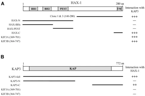
Fig. 1. Identification of the proteins interacting with KAP3 by yeast two-hybrid screening. (A) The domain structure of HAX-1 illustrating that clones 1 and 3 corresponds to the C-terminal fragment of HAX-1. The putative BH, PEST, and TM domains are indicated in gray. To determine minimal KAP3 binding region and binding specificity of HAX-1, several truncated forms of HAX-1, KIF3A, and KIF3B were constructed by PCR and tested in the yeast two-hybrid assay for interaction with KAP3.
+++, interaction with KAP3; -, no interaction with KAP3. aa, the amino acid residue number. (B) The C-terminal region of KAP3 mediates interaction with HAX-1. KAP3 has a KAP domain, depicted as the gray box. Several truncated forms of KAP3, KIF3A, and KIF3B were constructed by PCR and tested in the yeast two-hybrid assay for interaction with HAX-1.
++ or +++, interaction with HAX-1; -, no interaction with HAX-1. aa, the amino acid residue number.
fusion proteins were purified using glutathione-agarose beads (Sigma-Aldrich) according to the manufacturer’s protocol. The mouse brain S2 fraction was incubated over- night at 4℃ with the GST fusion protein-coupled glutathione beads. The beads were pelleted by centrifugation, washed three times with the extraction buffer (1% Triton X-100 in PBS containing 10 μg/ml each aprotinin, leupeptin, and pep- statin and 1 μM phenylmethanesulfonyl fluoride), and once with PBS. The bound proteins were eluted from the gluta- thione beads with 100 μl of Laemmli
’s loading buffer. The pulled-down proteins were analyzed by immunoblotting with anti-KIF3A, anti-KIF3B, anti-KIF5B, and anti-KAP3 an- tibodies [10, 23].
Results
Identification of KAP3 interacting proteins by yeast two-hybrid system
To identify KAP3 interacting proteins, we screened a mouse brain cDNA library by yeast two-hybrid system using the C-terminal region (aa 320-772) of KAP3 as bait. From 5×10
6colonies screened, we obtained 4 positive clones. Two clones (clone 1 and 3) of the 4 clones were identical and
possessed cDNA fragments corresponding to the C-terminal region of HAX-1 (Fig. 1A). HAX-1 was isolated as HS-1 inter- acting protein and suggested to be involved in B cell signal transduction [20]. HAX-1 contains several protein-protein in- teraction domains, the putative two Bcl-2 homology (BH) domains and one proline, glutamic acid, serine, threonine (PEST) domain. The C-terminus of HAX-1 has the putative transmembrane domain (TMD) [5]. To determine the mini- mal binding domain of HAX-1 that is required for the inter- action with KAP3, we constructed several deletion mutants of HAX-1. Yeast two-hybrid assays showed that the minimal domain required for binding was located in the C-terminal region of HAX-1 (Fig. 1A). Unexpectedly, KAP3 did not in- teract with the putative protein-protein interaction domains within the N-terminal region of HAX-1. Next, we determined the minimal binding domain of KAP3. We constructed sev- eral deletion mutants of KAP3 and tested the interaction with HAX-1 by yeast two-hybrid assays. The interaction with HAX-1 was dependent on the C-terminal region of KAP3 (Fig. 1B). Together, these results show that the inter- action between KAP3 and HAX-1 is mediated through their C-terminal regions.
Next we investigated whether HAX-1 interacts with
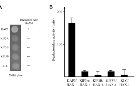
Fig. 2. Interaction between KAP3 and HAX-1. (A) The C-terminal regions of each KIF, KAP3, and KLC were fused to the pLexA DNA binding domain. HAX-1 specifically interacted with KAP3 but not with KIFs and KLC. +, interaction; -, no interaction.
(B) The strength of interactions between KIFs, KAP3, or KLC and HAX-1 were examined quantitatively using β-galactosidase activity in yeast two-hybrid reporter assay.
Fig. 3. Association of Kinesin-II with HAX-1 in the GST pull- down assay and co-immunoprecipitation. (A) Proteins in the mouse brain lysate were allowed to bind to GST alone or GST-HAX-1 fusion proteins. The elution frac- tions were resolved by SDS-PAGE and analyzed by im- muoblotting using anti-KIF3A, KIF3B, KAP3, or KIF5B antibodies. (B) Mouse brain lysates were im- munoprecipitated with anti-KIF3A, HAX-1 antibodies, or preimmune serum, and then the precipitates were immunoblotted with anti-KAP3, or KIF3B antibodies.
KIF3A and KIF3B subunits of Kinesin-II and KIF5B and kine- sin light chain 1 (KLC1) subunits of Kinesin-I. As shown in Fig. 1B, 2A, and 2B, there was no detectable binding of HAX-1 with KIF3A, KIF3B, KIF5B, and KLC1. A quantitative β -galactosidase assay showed that HAX-1 directly bound to KAP3 but not to KIFs (Fig. 2B). These data indicate that the interaction of KAP3 with HAX-1 is specific.
HAX-1 is associated with Kinesin-II
Kinesin-II is composed of a KIF3A/KIF3B heterodimer and KAP3, forming a heterotrimeric complex [23]. Therefore, we next determined whether HAX-1 interacts specifically with KAP3 and whether the interaction includes Kinesin-II at the protein level using GST pull-down experiments.
Recombinant GST-HAX-1 fusion protein was expressed in E. coli. The purified GST fusion protein was allowed to inter- act with mouse brain lysates. Immunoblotting analyses re- vealed that KAP3 interacted with GST-HAX-1, but not with GST. In addition, KIF3A and KIF3B, which bind to the KAP3, were efficiently precipitated with GST-HAX-1 (Fig. 3A). This indicates that HAX-1 interacts with Kinesin-II.
To further confirm whether the binding of KAP3 to HAX-1 mediates the interaction between Kinesin-II and HAX-1, we performed co-immunoprecipitation analyses.
Lysates from mouse brain were incubated with anti-HAX-1 antibody or anti-KIF3A antibody. Protein A-Separose beads precipitated the immuno-complexes, which were then sub- sequently separated by SDS-PAGE and immunoblotted with
anti-KAP3 and anti-KIF3B antibodies. As shown in Fig. 3B, both anti-HAX-1 and anti-KIF3A antibodies efficiently pre- cipitated the Kinesin-II complex, KIF3A/KIF3B heterodimer and KAP3. These results suggest that the interaction of HAX-1 with Kinesin-II is mediated by KAP3.
Discussion
In the search for proteins interacting with KAP3 we have
shown that HAX-1, a multifunctional protein can associate
with KAP3. Using the C-terminal region of KAP3 as bait, we identified HAX-1 in yeast two-hybrid assay of a mouse brain cDNA library. The C-terminal region of HAX-1 inter- acted with KAP3. Furthermore, using a combination of GST pull-down assay and co-immunoprecipitation, we confirmed that KAP3 interacted with HAX-1 at the protein level.
Moreover, we showed that Kinesin-II complex can be co-pre- cipitated with HAX-1. Although we did not identified the HAX-1 containing cargoes, this result suggest that Kinesin-II transports HAX-1 containing cargo through the interaction between KAP3 and HAX-1.
HAX-1 was first identified as a partial cDNA for mRNAs induced upon exposure of macrophages to silica. HAX-1 is ubiquitously expressed and a multifunctional protein [20].
Recent studies have shown that HAX-1 interacts with a num- ber of cellular and viral proteins [5]. HAX-1 interacts with HtrA2 (high temperature requirement protein A2) and Parl (presenilin-associated, rhomboid-like) within mitochondria.
This interaction was shown to be required to suppress apop- tosis in lymphocytes and neurons [3]. In other study, it was suggested that HAX-1 is involved in the formation of cell-matrix contacts through its binding to the polycystic kid- ney disease protein PKD2 as well as to cortactin, an F-actin -associated protein [6]. In additional study, HAX-1 was shown to bind the integrin β
6subunit and regulate the in- ternalization of the integrin α
vβ
6[17]. Reduction of HAX-1 expression level by siRNA showed that integrin dependent cell migration was reduced through down regulation of en- docytosis via a clathrin [17]. A recent study revealed that HAX-1 interacts with the HIV-1 Rev protein and facilitates the export of viral mRNAs. In HAX-1 and Rev co-expressing cells, Rev was transported from the nucleus to the cyto- plasm, where it co-localized with HAX-1 in the cytoplasm [14].
As described in this study, KAP3 directly interacted with HAX-1. These data suggested that KAP3 may link Kinesin-II and HAX-1. We propose one possibility to illustrate how Kinesin-II can mediate the transport of many cargoes.
Different cargoes may use different adaptor proteins that mediate the attachment of Kinesin-II to proteins or mem- brane-bound cargoes. Based on the ability of KAP3 to bind both KIF3A/KIF3B and HAX-1, associated with various cel- lular and viral proteins, we favor the model that HAX-1 may function as an adaptor and mediate the attachment of many different cargoes to Kinesin-II. In this report, we describe that the C-terminal of HAX-1 mediates interaction with
KAP3. This interaction may link Kinesin-II to many different cargoes.
Acknowledgement
This research was supported by Basic Science Research Program though the National Research Foundation of Korea (NRF) funded by the Ministry of Education, Science and Technology (2010-0021296).
References
1. Ausubel, F. M., Brent, R., Kingston, R. E., Moore, D. D., Seidman, J. G., Smith, J. A. and Struhl, K. 1998.
Current Protocols in Molecular Biology
. John Wiley & Sons.2. Brady, S. T. 1985. A novel brain ATPase with properties expected for the fast axonal transport motor.
Nature
317, 73-75.3. Chao, J. R., Parganas, E., Boyd, K., Hong, C. Y., Opferman, J. T. and Ihle, J. N. 2008. HAX1-mediated processing of HtrA2 by Parl allows survival of lymphocytes and neurons.
Nature
452, 98-102.4. Davenport, J. R., Watts, A. J., Roper, V. C., Croyle, M. J., van Groen, T., Wyss, J. M., Nagy, T. R., Kesterson, R. A.
and Yoder, B. K. 2007. Disruption of intraflagellar transport in adult mice leads to obesity and slow-onset cystic kidney disease.
Curr Biol
17, 1586-1594.5. Fadeel, B. and Grzybowska, E. 2009. HAX-1: a multifunc- tional protein with emerging roles in human disease.
Biochim Biophys Acta
1790, 1139-1148.6. Gallagher, A. R., Cedzich, A., Gretz, N., Somlo, S. and Witzgall, R. 2000. The polycystic kidney disease protein PKD2 interacts with HAX-1, a protein associated with the actin cytoskeleton.
Proc Natl Acad Sci USA
97, 4017-4022.7. Hirokawa, N., Niwa, S. and Tanaka, Y. 2010. Molecular mo- tors in neurons: transport mechanisms and roles in brain function, development, and disease.
Neuron
68, 610-638.8. Hirokawa, N., Tanaka, Y. and Okada, Y. 2012. Cilia, KIF3 molecular motor and nodal flow.
Curr Opin Cell Biol
24, 31-39.9. Hirokawa, N. 1998. Kinesin and dynein superfamily pro- teins and the mechanism of organelle transport.
Science
279, 519-526.10. Kanai, Y., Okada, Y., Tanaka, Y., Harada, A., Terada, S. and Hirokawa, N. 2000. KIF5C, a novel neuronal kinesin en- riched in motor neurons.
J Neurosci
20, 6374-6384.11. Kim, S. J., Lee, C. H., Park, H. Y., Yea, S. S., Jang, W. H., Lee, S. K., Park, Y. H., Cha, O. S., Moon, I. S. and Seog, D. H. 2007. JSAP1 interacts with kinesin light chain 1 through conserved binding segments.
J Life Sci
17, 889-895.12. Kozminski, K. G., Beech, P. L. and Rosenbaum, J. L. 1995.
The Chlamydomonas kinesin-like protein FLA10 is involved in motility associated with the flagellar membrane.
J Cell
Biol
131, 1517-1527.초록:Kinesin superfamily-associated protein 3 (KAP3)를 통한 HS-1-associated protein X-1 (HAX-1)과 Kinesin-II의 결합
장원희․석대현*
(인제대학교 의과대학 생화학교실)
Kinesin-II는 다양한 운반체들을 미세소관을 따라 운반하는 motor 단백질의 하나이다. Kinesin-II는 두 개의 motor 단백질 KIF3A와 KIF3B, 그리고 motor 단백질의 말단에 결합하는 kinesin superfamily-associated protein 3 (KAP3)로 구성되어 있다. KAP3는 Kinesin-II의 기능에 중요한 역할을 하는 것으로 알려져 있으나 명확한 기능은 아직 밝혀지지 않았다. 본 연구에서 KAP3와 결합하는 단백질을 분리하기 위하여 효모 two-hybrid system을 사용하여 탐색한 결과 HS-1-associated protein X-1 (HAX-1)을 분리하였다. KAP3은 HAX-1의 C-말단 부위와 결합하며, HAX-1은 KAP3의 C- 말단부위와 결합함을 효모 two-hybrid assay로 확인하였다. 그러나, HAX-1는 KIF3A, KIF3B, KIF5B, 그리고 kinesin light chain (KLC)과는 결합하지 않았다. KAP3와 HAX-1의 단백질 결합은 glutathione
S
-transferase (GST) pull-down assay와 공동면역침강으로 추가 확인하였다. 생쥐의 뇌 파쇄액을 HAX-1 항체와 KIF3A 항체로 면역침강을 행한 결과 Kinesin-II의 구성단백질인 KIF3B와 KAP3가 같이 침강하였다. 이러한 결과들은 KAP3가 Kinesin-II와 HAX-1의 결합을 매개한다는 것을 시사한다.13. Lin, F., Hiesberger, T., Cordes, K., Sinclair, A. M., Goldstein, L. S., Somlo, S. and Igarashi, P. 2003. Kidney-specific in- activation of the KIF3A subunit of kinesin-II inhibits renal ciliogenesis and produces polycystic kidney disease.
Proc Natl Acad Sci USA
100, 5286-5291.14. Modem, S. and Reddy, T. R. 2008. An anti-apoptotic protein, HAX-1, inhibits the HIV-1 rev function by altering its sub-cellular localization.
J Cell Physiol
214, 14-19.15. Mueller, J., Perrone, C. A., Bower, R., Cole, D. G. and Porter, M. E. 2005. The FLA3 KAP subunit is required for local- ization of kinesin-2 to the site of flagellar assembly and processive anterograde intraflagellar transport.
Mol Biol Cell
16, 1341-1354.16. Ou, G., Koga, M., Blacque, O. E., Murayama, T., Ohshima, Y., Schafer, J. C., Li, C., Yoder, B. K., Leroux, M. R. and Scholey, J. M. 2007. Sensory ciliogenesis in Caenorhabditis elegans: assignment of IFT components into distinct mod- ules based on transport and phenotypic profiles.
Mol Biol Cell
18, 1554-1569.17. Ramsay, A. G., Keppler, M. D., Jazayeri, M., Thomas, G.
J., Parsons, M., Violette, S., Weinreb, P., Hart, I. R. and Marshall, J. F. 2007. HS1-associated protein X-1 regulates carcinoma cell migration and invasion via clathrin-mediated endocytosis of integrin alphavbeta6.
Cancer Res
67, 5275- 5284.18. Sarpal, R., Todi, S. V., Sivan-Loukianova, E., Shirolikar, S.,
Subramanian, N., Raff, E. C., Erickson, J. W., Ray, K. and Eberl, D. F. 2003. Drosophila KAP interacts with the kinesin II motor subunit KLP64D to assemble chordotonal sensory cilia, but not sperm tails.
Curr Biol
13, 1687-1696.19. Shimizu, K., Kawabe, H., Minami, S., Honda, T., Takaishi, K., Shirataki, H. and Takai, Y. 1996. SMAP, an Smg GDS-as- sociating protein having arm repeats and phosphorylated by Src tyrosine kinase.
J Biol Chem
271, 27013-27017.20. Suzuki, Y., Demoliere, C., Kitamura, D., Takeshita, H., Deuschle, U. and Watanabe, T. 1997. HAX-1, a novel intra- cellular protein, localized on mitochondria, directly asso- ciates with HS1, a substrate of Src family tyrosine kinases.
J Immunol
158, 2736-2744.21. Tabish, M., Siddiqui, Z. K., Nishikawa, K. and Siddiqui, S.
S. 1995. Exclusive expression of C. elegans osm-3 kinesin gene in chemosensory neurons open to the external environment.
J Mol Biol
247, 377-389.22. Takeda, S., Yamazaki, H., Seog, D. H., Kanai, Y., Terada, S. and Hirokawa, N. 2000. Kinesin superfamily protein 3 (KIF3) motor transports fodrin-associating vesicles im- portant for neurite building.
J Cell Biol
148, 1255-1265.23. Yamazaki, H., Nakata, T., Okada, Y. and Hirokawa, N. 1996.
Cloning and characterization of KAP3: a novel kinesin su- perfamily-associated protein of KIF3A/3B.