다양한 중합방법에 따른 악안면 보철용 폴리우레탄과 자가중합 레진 간의
결합력에 관한 연구
단국대학교 치과대학 치과보철학교실 김 두 열 조 인 호․
A Study on the Adhesiveness between Polyurethane Sheet for M axillofacial Prostheses and Autopolym erizing
Acrylic Resin in Various Polym erization M ethods
Doo-Yeol Kim, In-Ho Cho
Department of Prosthodontics Graduate School Dankook University
The field of maxillofacial prosthetics is concerned with the prosthetic reconstruction of missing head and neck tissue.
Currently, facial prostheses are usually applied in cases of defects caused by the surgical removal of tumors or congenital defects. While silicone has been most widely used for the reconstruction of missing maxillofacial defects, it does not have ideal physical properties. Therefore, bonding a thin polyurethane sheet to silicone prostheses was recommended. In this case skin adhesives were used for the retention of maxillofacial prostheses. But retention of devices has always been problematic. The contributions of implants can be made to solve these problems. Implants have reduced the need for adhesive use, simplifying cleaning procedures and thus extending the life of the prostheses. For implant-retained prostheses, retentive matrix is necessary to hold attachments and/or magnets. The retentive matrix is usually fabricated with autopolymerizing acrylic resin or visible light- polymerized resin.
The purpose of this study was to compare the adhesion-in-peel force of silicone adhesive to autopolymerizing acrylic resin and polyurethane sheet with two different surface textures : pumice polish only or retention groove, and three surface primers : Dow corning 1205 primer or Dow corning S-2260 primer or Factor Ⅱ A-304 primer, and two polymerization methods : room temperature or dry heat oven. The t-peel bond strength of specimens was determined as described in ASTM Standard D1876-72. The results were statistically analyzed using the ANOVA test, multiple range test and t-test
The results were as follows.
1. The t-peel bond strength of A-304 primer was the highest and statistically higher than that of S-2260(p<0.05).
2. The t-peel bond strength of specimens with retention groove was statistically higher than that of specimens polished with pumice(p<0.05).
3. The t-peel bond strength of specimens polymerized in dry heat oven was statistically higher than that of specimens
in room temperature(p<0.01).
Ⅰ 서. 론
악안면 보철물이 가져야 할 가장 중요한 요소는 인체조직과 같은 성질의 물성을 갖는 재료를 이용 하여 견고히 부착되고 오랫동안 변하지 않는 내구 성을 갖는 것이다
8)
.초기 안면부 보철물의 재료로는 나무 상아 밀, , 납 금속 등이었고 근래 가장 널리 사용되는 재료는, 폴리실록산 폴리우레탄 폴리비닐 클로라이드 폴, , , 리메틸 메타애크릴레이트 라텍스 실리콘 등이다, ,
1,17)
. 이중 실리콘은 1960년 Barnhart3)
에 의해 처음사용되었으며 화학적 안정성 강도 내구성 용이한, , , 조작성 때문에 오늘날까지 최상의 재료로 여겨져 왔다
26)
. 그러나 약한 찢김 저항성 약한 접착성 비, , 젖음성 비연마성 세균의 성장 피부오일의 흡수, , , 등이 실리콘의 사용을 제한하였다1-3,28,38)
. 1987년 Udagama38)
는 폴리우레탄 판과 실리콘의 접착을 제 안하였다 폴리우레탄은 높은 접착성을 가진 부드. 럽고 활성있는 재료이며 투명한 폴리우레탄 판이 실리콘을 이장함으로써 변연강도와 심미성이 증가, 하며 접착제와 친화성이 있고 피부오일에 의한 악 안면 보철물의 훼손을 막아준다고 하였다1,20,31,34,37,38)
.
이러한 악안면 보철물의 유지수단으로 과거에는 피부접착제 등에 의존하였지만 최근에는 악안면 임 플랜트가 이용되고 있다 임플랜트 유지 악안면 보. - 철물은 어태치먼트 또는 자석을 수용하기 위한 유 지부(retentive matrix)가 필요하며 이것은 주로 애크
릴릭 레진으로 제작된다.
임플랜트 유지 악안면 보철물은 실리콘 상부 보- 철물과 폴리우레탄 판 그리고 레진 하부구조물이 층을 이루고 있으며 이들의 결합을 위해, primer와 와 같은 의료용 접착 silastic medical adhesive type A
제를 사용한다 이처럼 여러 가지 구성요소가 하나. 의 보철물을 이루게 되므로 각 구성요소 간의 결합 력과 보철물의 유지가 악안면 보철에서 큰 비중 을 차지하고 있다 그리고 이들의 결합력을 증진시. 키기 위하여 primer의 종류 작용시간 중합방법을, , 달리하는 등 지금까지 많은 연구가 있어왔다. 1988 년 Singer 등
35)
은 폴리우레탄 판에 Dow corning 를 도포함으로써 실리콘과의 강한 결 S-2260 primer합력을 보여주었고, 1994년 Wang 등
44)
은 Dow가 보다 높은 결합
corning 1205 primer S-2260 primer 강도를 보인다고 보고하였다.
임플랜트 유지 악안면 보철물에 있어서 결합력의- 저하는 실리콘 상부 보철물과 폴리우레탄 사이 폴, 리우레탄과 레진 하부구조 사이 그리고 임플랜트와 유지장치 및 자석 사이에서 일어나게 된다 이에 본. 연구는 폴리우레탄과 레진 하부구조 사이의 결합력 에 대하여 알아보고자 하였으며 레진 판(resin plate) 을 제작한 후 그 표면에 유지구의 형성 유무 그리고, , 현재 가장 널리 사용되는S-2260, 1205, A-304 세 가 지의 primer를 이용하여 실온법과 건조열 오븐법의 두 가지 중합방법을 이용하여 온성할 경우 각각의, 조건이 폴리우레탄 판과 애크릴릭 레진 판 사이의 결합강도에 미치는 영향을 비교 연구하였다
t-peel .
다양한 중합방법에 따른 악안면 보철용 폴리우레탄과 자가중합 레진 간의
결합력에 관한 연구
단국대학교 치과대학 치과보철학교실 김 두 열 조 인 호․
1. 실험재료
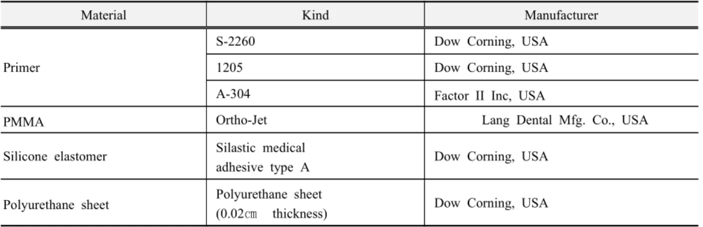
본 실험에서는 Table 1에 정리한 바와 같이 0.02 두께의 폴리우레탄 판(polyurethane sheet; Factor
㎝
과
II Inc., USA) PMMA(Ortho-Jet, Lang Dental Mfg.
그리고 세 종류의
Co., USA) primer; S-2260(Dow 및 Corning, USA), 1205(Dow Corning, USA)
를 사용하였다 A-304(Factor II Inc., USA) .
2. 시편제작
시편은 Table 2에 열거한 바와 같이 제작하였는 데 레진 판의 표면형태에 따라 크게 군으로 나누, 2 고 다시 이것을 중합방법 실온법 건조열 오븐법 에( , ) 따라 군으로 나누며 사용된4 , primer(S-2260, 1205, 의 종류에 따라 군으로 나누어 각 군당 개
A-304) 12 4
씩 총 48개의 시편을 다음과 같이 제작하였다(Fig.
1).
Fig. 1. Schematic drawing of specimen for t-peel test
2 cm
0.2 cm
0.02 cm
Resin plate Primer Adhesive Primer Polyurethane
3 cm 7 cm
Table 1. Summary of materials used in this study
Material Kind Manufacturer
Primer
S-2260 Dow Corning, USA
1205 Dow Corning, USA
A-304 Factor II Inc, USA
PMMA Ortho-Jet Lang Dental Mfg. Co., USA
Silicone elastomer Silastic medical
adhesive type A Dow Corning, USA
Polyurethane sheet Polyurethane sheet
(0.02 ㎝ thickness) Dow Corning, USA
위해 one-piece stainless steel 음형 주형과 two-piece 를 다음과 같이 제작하였다 좌측에 aluminum plates . 는smooth surface를 우측에는 주형에, 1㎜간격으로
깊이의 유지구를 형성하여 를
0.5㎜ groove surface 재현하도록 제작하였다(Fig. 2).
레진 판의 제작 2)
제조회사의 지시에 따라28 cmHg vacuum하에서 진공 혼합된 자가중합 PMMA를 금속음형 주형에 주입하고 주형을 나사로 제 위치시킨 후 표준 의치, 압력기(Tokuyama, Japan)로 50㎏ ㎠/ 의 압력을 가한 상태에서 중합하였다(Fig. 3).
의 도포 3) Primer
를 도포하기 전에 불순물을 제거하기 위해 Primer
거즈에 아세톤을 묻혀 폴리우레탄 판을 2×2 inch
닦은 후 레진 판과 폴리우레탄 판에 각각의, primer;
를 도포하였다 와 S-2260, 1205, A-304 (Fig. 4 5).
레진 판과 폴리우레탄 판의 부착 4)
처리된 폴리우레탄 판과 레진 판을
Primer silastic
를 튜브 medical adhesive type A(Dow Corning, USA) 로 직접 고르게 도포한 후 다시 주형을 나사로 제, 위치시키고 표준 의치 압력기, (Tokuyama, Japan)로
의 압력을 가한 상태에서 중합하였다
50㎏ ㎠/ (Fig.
6).
Fig. 2. Stainless steel mold and aluminum plates
Fig. 3. Fabrication of resin plate
Fig. 4. Application of primer on polyurethane sheet
Fig. 5. Application of primer on resin plate Table 2. Experimental groups and numbers of specimens
Resin Surface Curing Primer Group No. Total
PMMA
Pumice only
Bench curing
A-304 1 4
48
S-2260 2 4
1205 3 4
Dry heat oven
A-304 4 4
S-2260 5 4
1205 6 4
Retention groove
Bench curing
A-304 7 4
S-2260 8 4
1205 9 4
Dry heat oven
A-304 10 4
S-2260 11 4
1205 12 4
Fig. 6. Attachment of resin plate and polyureth- ane sheet with medical adhesive type A
Fig. 7. Polymerization in room temperature
실리콘의 중합 5)
실온법(Room temperature method)
⑴
상온에서 24시간 동안 중합(Fig. 7).
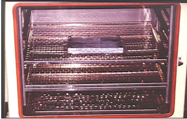
건조열 오븐법(Dry heat oven method)
⑵
상온에서24시간 동안 중합한 후 다시 건조열 오븐, 을 이용하여 에 (Dry-heat oven; Yamato, Japan) 75℃
서 시간 동안 추가중합3 (Fig. 8).
결합강도 측정 6) T-peel
폴리우레탄 판과 레진 판 사이의 t-peel 결합강도 측정은Z020(Zwick, Germany)을 이용하여 미국재료
Fig. 8. Polymerization in dry heat oven
Fig. 9. Measurement of t-peel bond strength
시험협회 즉, ASTM(American Society for Testing 에 근거하여 평균 결합강 Materials) D1876-72 t-peel 도를 측정하였다(Fig. 9).
3. 통계처리
폴리우레탄 판과 레진 판 사이의 t-peel 결합강도 에 레진의 표면형태, primer의 종류 및 중합방법이 미치는 영향을 측정하기 위해 SPSS V 7.0 for
을 사용하였다 Win(SPSS Inc., USA) .
각 군들이 정규분포(normal distribution)를 이루는 지 검사하기 위해 K-S test(Kolmogorov - Smirnov
를 시행하였고 레진의 표면형 Goodness of Fit test) ,
태, primer의 종류 및 중합방법이 결과에 미치는 영 향을 알아보기 위해 ANOVA test를 시행하였으며,
에 대하여 와
primer one- way ANOVA test multiple 를 시행하였고 레진의 표면처리방법과 중 range test ,
합방법에 대하여independent t-test를 시행하여 변수 간의 유의성을 분석하였다.
Ⅲ 실험결과.
각 군에 대한t-peel 결합강도의 평균과 표준편차 를 도표로 정리하면 다음과 같다(Table 3).
에 대한 평균과 표준편차는 와 같으
Primer Table 4
며 이들 간의 multiple range test 결과 A-304의 결합 력이 가장 크게 나타났고, S-2260과는 유의한 차이 가 있었다(Table 5).
Table 4. Mean and standard deviation of t-peel bond strength for primers (unit : g/ ) ㎝
Mean Standard deviation
S-2260 1922.59 530.99
1205 2446.76 583.90
A-304 2631.56 736.57
Table 5. Result of multiple range test for primers
S-2260 1205 A-304
S-2260 1205
A-304 *
* : Statistically significant (P<0.05)
Table 6. Result of independent t-test according to surface treatment (unit : g/㎝ )
Mean Standard deviation P-values Groove 2552.131 719.333
0.025 Pumice 2115.141 575.477
Table 7. Result of independent t-test according to curing method (unit : g/ ) ㎝
Mean Standard deviation P-values Bench curing 1822.777 402.710
0.000 Dry heat oven 2844.496 491.347
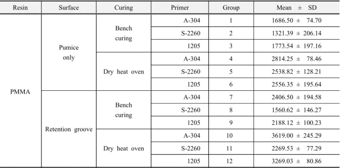
Table 3. Result of measurement of t-peel bond strength (unit : g/ ) ㎝
Resin Surface Curing Primer Group Mean ± SD
PMMA
Pumice only
Bench curing
A-304 1 1686.50 ± 74.70
S-2260 2 1321.39 ± 206.14
1205 3 1773.54 ± 197.16
Dry heat oven
A-304 4 2814.25 ± 78.46
S-2260 5 2538.82 ± 128.21
1205 6 2556.35 ± 195.64
Retention groove
Bench curing
A-304 7 2406.50 ± 194.58
S-2260 8 1560.62 ± 146.27
1205 9 2188.12 ± 100.23
Dry heat oven
A-304 10 3619.00 ± 245.29
S-2260 11 2269.53 ± 77.29
1205 12 3269.03 ± 80.86
표면처리방법에 대한 independent t-test의 결과 유 지구를 형성한 군이 형성하지 않은 군에 비해 유의 성 있게 결합력이 크게 나타났다(P<0.05) (Table 6).
중합방법에 대한independent t-test의 결과 건조열 오븐법이 실온법에 비해 유의성 있게 결합력이 크 게 나타났다(P<0.01)(Table 7).
각 군에 대한 비교에서 유지구를 형성한 후, 를 도포하고 건조열 오븐법으로 중합한 군
A-304 10
이 가장 높은t-peel 결합강도를 나타냈으며 유지구, 를 형성하지 않고 S-2260을 도포한 후 실온법으로, 중합한 군이 가장 낮은2 t-peel 결합강도를 나타냈 다(Table 8).
Ⅳ 총괄 및 고안.
악안면 보철은 착탈식 또는 고정성 인공 대체물 로써 안면부 구조를 대체 혹은 수복하는 보철학의 한 분야이다
13)
. 종양의 수술 사고 또는 선천적 기형, 으로 인한 안면 결손부를 가진 환자들은 외과적으 로 기능과 심미를 회복하는 것이 이상적이지만 많 은 경우 보철적 수복을 필요로 한다37)
. 이런 악안면 보철물의 중요한 목적은 결손부의 형태와 기능을 회복시켜 개인의 정신적 손상을 덜어주고 사회복귀 를 쉽게 할 수 있도록 하는 것이다6,15,37)
. 악안면 보 철에 있어서 성공의 범주는 심미적이고 기능적이며 생체적합성이 좋고 유지력이 우수하여야 한다는 것이다 악안면 보철의 성공은 또한 결손부의 상태. , 보철의사의 숙련도 그리고 사용되어지는 재료에, 의해서 일차적으로 결정된다
4,15,19,41)
.
악안면 보철용 재료에서 요구되는 이상적 성질에 대하여Lewis 등
18)
은 다음과 같이 제시하였다 첫째. 는 낮은 점도 긴 작업시간 색상 조절의 가능 낮은, , , 중합온도 주형 제작의 용이성 등이고 둘째는 높은, 인장강도 높은 찢김강도 체적 안정성 화학물질과, , , 자외선으로부터의 저항성 등이며 셋째는 무독성, 비알러지성 비발암성 용이한 세척성 경량성 접착, , , , 제와의 적합성 등이다.그러나 지난 20여년 동안 새롭고 향상된 재료의 개발을 위해 노력하였으나 아직까지 인체조직을 대 체하는 이상적인 단일재료는 없다
10,21,22,31,34,36,37)
그 . 러므로 악안면 보철물은 여러 가지 재료를 함께 사 용하여 악안면 보철물이 가져야 할 요구조건을 만 족시켜야한다.
년 치과생체재료협회
1973 (Dental Biomaterials 의 심포지움
Research Priorities)
19,27)
에서 악안면 보철 물에 대한 더 좋은 재료의 필요성을 확인하였고 오 늘날까지 재료의 개발과 함께 악안면 보철물의 각 구성요소 간의 결합력을 증진시키기 위한 많은 연 구가 이루어져왔다. Barnhart3)
에 의하여 실리콘이 악안면 보철에 사용된 이래 Gonzalez 등11)
과 Goldberg 등9)
은 폴리우레탄의 물리적 기계적 특성, 에 대하여 연구하였고, Udagama와 Drane37)
은Group 2 1321.39
Group 8 1560.62
Group 1 1686.50
Group 3 1773.54
Group 9 2188.12
Group 11 2269.53
Group 7 2406.50 2406.50
Group 5 2538.82
Group 6 2556.35
Group 4 2814.25
Group 12 3269.03
Group 10 3619.00
methyl triacetoxy silane cross-linked silicone제재인 를 소개하였으며 silastic medical adhesive type A , Udagama
38)
는 또한silastic medical adhesive type A와 의 두 가지 를 사용하여 폴리우 S-2260, 1205 primer레탄 판과 실리콘의 결합력에 대하여 비교 연구하 였다 그리고. Farah 등
7)
은 MDX4-4210과 silastic 를 여러 비율로 혼합하여 좀 medical adhesive type A더 부드럽고 유연한 재료를 소개하기 위해 노력하 였고, Singer 등
31)
은 실리콘과 폴리우레탄과의 결“ 합력에 있어서 primer의 효과 에 관한 논문에서”의 종류 그리고 와 의
primer , adhesive A MDX4-4210 혼합비에 따라서 비교 연구하였으며, Wang 등
40)
은 실리콘과 폴리우레탄 사이의 결합력을primer의 종 류 그리고, primer의 작용시간과 중합방법에 따라서 비교 연구하였다 그러나 이러한 노력에도 불구하. 고Chen 등5)
의 연구에 의하면 악안면 보철물의 평 균 수명은 약10개월 정도이며 그 원인으로는 자외 선에 대한 노출 공기오염 알콜이나 휘발유와 같은, , 세척제에 의한 보철물의 급속한 변성이라고 하였 다.한편 Jebreil
16)
의 연구에 의하면 환자들이 보철물 의 착용을 꺼리는 이유 중 하나로 유지력의 저하를 들었는데 보철물의 구성요소 간의 결합력과 함께, 안면부에서 보철물이 탈락되지 않도록 하는 유지력 또한 중요하다 적절한 유지를 제공하는 것은 많은. 보철물에서 흔히 부딪히는 일이지만 특히 악안면 보철물의 유지는 보철물 제작에 결정적으로 중요한요소이다
8,41)
. 악안면 보철물의 유지방법에는 해부학적 언더컷 피부접착제의 사용 언더컷과 접착제, , 의 병용 그리고 최근의 골유착성 임플랜트에 의한, 것들이 있다 초기 보철물의 유지는 철사나 안경테. , 스프링 머리띠 등에 의한 단지 기계적인 것에 의존, 하였는데 이러한 방법들은 예를 들어 안경을 벗으, 면 결손부가 노출되는 등 비심미적이고 조잡함 때 문에 부적절한 방법이었다
12,13,23-25,29,32,35,39)
. 양면 테 이프 방향족 시멘트 수용성 또는 실리콘계 접착제, , 는 과거에 흔히 사용된 대 주요 접착제인데 이들3 은 적절한 유지를 얻기 위하여 광범위한 부위를 피 개하여야 했으며 눈이나 입 주위 머리카락이 있는, 부위 안면근육의 운동이 큰 부위까지는 연장이 불, 가능하고 피부자극이나 알러지와 같은 부작용이 나 타나며 땀에 의해 접착력이 저하된다 또한 테이프.는 실리콘에 잘 붙지 않고 방향족 시멘트는 얇은 변 연부를 말아 올리거나 색소침착 등에 의해 심미성 이 저하되며 실리콘 접착제는 세척을 위한 용해제 에 의해 보철물의 훼손을 초래하게 된다
12,13,23,32,35,39)
.
이러한 종래의 유지수단과 관련된 많은 단점을 극복할 수 있게 한 두개 안면부 임플랜트에 의한 악 안면 보철은1979년에 처음으로 임상에 적용되었으 며 임플랜트의 사용은 피부접착제의 필요성을 줄, 여주고 세척과정을 단순화함으로써 보철물의 수명 을 연장시켰다 또한 일상생활에 있어서 보철물의. 탈락에 대한 두려움을 줄여주었다 악안면 보철물. 이 이러한 임플랜트에 연결되기 위해서는 어태치먼 트나 자석을 부착하기 위한 유지부가 필요한데 이 것은 일반적으로 자가중합 레진이나 광중합 레진으 로 만들어지며 보철물의 무게를 감소시키고 부가적 인 언더컷을 이용할 수 있게 한다
12,30,34)
.Taft 등
34)
은 레진 판을 pumice 처리한 것과 를 준 것에 과 의 두 가지 retention bead S-2260 1205를 도포하여 그 유지력에 대한 비교 실험 결 primer
과 1205가 레진 판의 표면처리방법에 관계없이 에 비해 결합력이 더 좋다고 하였다 본 실험
S-2260 .
에서는 레진 하부구조에 retention bead 와 유사한 역할을 할 것으로 사료되는groove를 형성한 후 폴, 리우레탄과의 결합력에 대하여 알아보고자 하였으 며 그 결과, groove를 형성한 것이 더 높은 결합력을 보였다.
폴리우레탄 판을 실리콘에 이장할 때와 마찬가지 로 보철물의 레진 유지부와 폴리우레탄 판 사이에 도primer를 도포하는데 이는 폴리우레탄 판과 레진 의 접착강도를 증진시키기 위함이며 primer에 의한 접착력의 증가는 이것이 라디칼을 형성하여 표면결 합력을 변화시키기 때문이다
34)
. 본 실험에서는 세 가지의 를 비교하였는 A-304, S-2260, 1205 primer데A-304가 가장 높은 결합력을 보였으며, S-2260과 는 유의한 차이를 보였다 이러한 결합력의 차이에. 대해 Wang 등
40)
은 primer내의 silane coupling agent 의 서로 다른 활성 유기기능 군(reactive organo- 을 가지는 피막 성분 때문이라고 하 functional group)였다 본 실험에서는 또한. silastic medical adhesive 와 의 중합방법에 따른 결합력의 차이 type A pimer
에 대해서도 알아보았는데 건조열 오븐에서 추가,
시 건조열 오븐이나 마이크로 웨이브의 사용은 중 합을 가속화시켜 보철물의 제작시간은 단축시키지 만 결합력은 증가시키지 않는다고 하였으나 제조, 사에 따르면 완전한 중합에는“ 24시간 정도가 소요 되며 수 일에 걸쳐 결합력은 증가한다”
14)
고 한 것으 로 미루어 보아 본 실험에서의 건조열 오븐 내의 추 가중합은 결합력을 증진시킨 것으로 사료된다.표 은 각 군의 결합력의 순서를 나타내며 이에8 따르면 보철물 제작시 레진 유지부에 유지구를 형 성하고A-304 primer를 도포한 후 건조열 오븐에서, 추가중합하는 것이 권장되며 악안면 보철물에 사용 되는 이상적인 단일 재료가 개발되기까지 보철물의 각 구성요소들을 결합시키는 재료의 개발과 그것의 적용방법에 대한 다양한 연구가 계속되어야 하겠 다.
Ⅴ 결. 론
최근 안면 결손부의 수복시 주로 임플랜트에 의 해 유지를 얻는다 이를 위해서 보철물내에 레진 하. 부구조가 필요한데 본 연구는 레진 판의 표면을, 만 처리한 것과 유지구를 형성한 것으로 나 pumice
누고 흔히 사용되는A-304, S-2260, 1205 세 가지의 를 사용하여 실온에서 중합한 것과 건조열 오 primer
븐에서 추가중합한 것의 t-peel 결합강도를 미국재 료협회의 규격에 따라 측정 비교 분석한 결과 다음, 과 같은 결론을 얻었다.
에 대한 비교에서 가 가장 큰 결합력 1. Primer A-304
을 나타내었고S-2260과는 유의한 차이가 있었다 (p<0.05).
레진 판의 표면형태에 대한 비교에서 유지구를 2.
형성한 군이 하지 않은 군에 비해t-peel 결합강도 에 있어서 유의한 차이가 있었다(p 0.05).<
중합방법에 대한 비교에서 건조열 오븐법이 실 3.
온법에 비하여 t-peel 결합강도에 있어서 유의한 차이가 있었다(p 0.01).<