(1865), most of which discovered in various marine habi- tats (Gerlach and Riemann 1974). After that, 11 species of the genus Phanoderma were transferred to the genus Meta- phanoderma by Platonova (1984). Consequently, 22 valid species are currently recognized in the genus Phanoderma (Wieser 1953; Gerlach and Riemann 1974; Lorenzen 1981; Platonova 1984; Zograf et al. 2015). It can be dis- tinguished from other genera of this family in having the attenuated head, well-developed cephalic capsule and pha- ryngeal capsule, and a tubular precloacal supplement (Smol et al. 2013; Zograf et al. 2015).
During a survey of the free-living marine nematodes of Korea, a new species of the genus Phanoderma was ob- tained from washings of sediments from rocky intertidal seagrass bed of the East Sea, Korea. In the present study, we provide taxonomic descriptions and illustrations of the new marine nematode using differential interference con- trast (DIC) microscope. This is the first taxonomic report
https://doi.org/10.11626/KJEB.2019.37.3.396
INTRODUCTION
The family Phanodermatid ae Filipjev, 1927 consists of two subfamilies and nine genera, as the apparent cosmo- politan distribution of free-living marine nematodes (Smol et al. 2013). The family Phanodermat idae is characterized by the presence of a head capsule, small buccal cavity, and cellular structure in the pharyngeal region (Inglis 1964;
Lorenzen 1981; Platonova 1984; Smol et al. 2013). The family Phanoderma tidae is classified into two subfamilies according to the structure of the complex pharyngeal-ce- phalic capsule (Platonova 1984). The subfamily Phanoder- mat inae Filipjev, 1927 is characterized by having a well-de- veloped cephalic capsule and pharyngeal capsule with one small dorsal and two large subventral outgrowths that point forward, and is composed of two genera, Phanoder- ma Bastian, 1865 and Metaphanoderma Platonova, 1984.
The genus Phanoderma was first erected by Bastian
Original article
A new free-living marine nematode species of the genus Phanoderma Bastian, 1865 (Enoplida: Phanodermatidae) from the East Sea, Korea
Hyo Jin Lee and Hyun Soo Rho*
East Sea Environment Research Center, Korea Institute of Ocean Science & Technology, Uljin 36315, Republic of Korea
Korean J. Environ. Biol.
37(3) : 396-405(2019) ISSN 1226-9999(print) ISSN 2287-7851(online)
* Corresponding author Hyun Soo Rho Tel. 054-780-5345 E-mail. [email protected]
Received: 26 July 2019 Revised: 18 September 2019
Revision accepted: 18 September 2019
Abstract: A new species of free-living marine nematode is described from intertidal sediments of the East Sea, Korea. Phanoderma koreense sp. nov. is characterized by the presence of well-developed pharyngeal and cephalic capsule, six inner labial sensilla present as minute papillae around with circular groove, long and slender spicules with 4-5 serrated distal end, located at the base of the precloacal supplement, a series of eight to nine stout and short setae on the ventral cloacal region and conico-cylindrical tail with two pairs of blunt setae. In this study, we provide taxonomic descriptions and illustrations of a new species by differential interference contrast microscope and a pictorial key to the valid species of Phanoderma Bastian, 1865. This is the first record of the genus Phanoderma in the East Sea, Korea.
Keywords: taxonomy, marine nematodes, Phanoderma, East Sea, Korea
on the genus Phanoderma in the East Sea, Korea.
MATERIALS AND METHODS
Sampling of taxa. The marine nematodes were obtained from washings of intertidal sediments, which were collect- ed from rocky intertidal seagrass bed on the eastern coast of Korea. Samples were filtered through a sieve with 67 μm mesh in the field after freshwater rinsing for less than a minute for osmotic shock, and then fixed in 5% formalin (Kristensen 1989).
Sample processing and preparation of specimens. The nematodes were sorted from the mixed meiobenthos un- der LEICA 205 C stereomicroscope. The nematodes were transferred to glycerol and mounted between two cover slips on a double sided slide for morphological observa- tions (Shirayama et al. 1993).
Light microscopy. Specimens were measured, examined and drawn using Nomarski differential interference con- trast (DIC) with an Olympus BX53 microscope equipped with a drawing tube and a Olympus DP26 digital camera with Olympus CellSens corresponding imaging software.
Terminology and abbreviations. Measurements are in μm. Abbreviations used in the text are as follows: a, body length divided by maximum body diameter; abd, anal body diameter; b, body length divided by pharynx length;
bcl, length of buccal cavity; bcw, width of buccal cavity;
bd, body diameter at the base of pharynx; c, body length divided by tail length; cs, cephalic setae length; dps, dis- tance of the precloacal supplement from cloacal opening;
ep, distance from anterior to anterior edge of excretory pore; gub, gubernaculum length; hd, diameter at the level of cephalic setae; L, total body length; M, maximum body diameter; N (%), nerve ring distance from the anterior end as percentage of pharynx length; nr, distance from anterior end to nerve ring; ol, distance from anterior end to anteri- or edge of ocelli; ph, pharynx length; spic, spicules length;
sup, precloacal supplement length; t, tail length; v, distance from anterior end to vulva; V (%), vulva distance from an- terior end as percentage of total body length.
SYSTEMATIC ACCOUNTS
Class Enoplea Inglis, 1983 Order Enoplida Filipjev, 1929
Family Phanodermatidae Filipjev, 1927 Genus Phanoderma Bastian, 1865
Type species
Phanoderma cocksi Bastian, 1865 Other valid species
Phanoderma albidum Bastian, 1865 Phanoderma annulocaudatum Allgén, 1939 Phanoderma brachyductum Wieser, 1953 Phanoderma coecum Allgén, 1947 Phanoderma hawaiiense Allgén, 1951 Phanoderma islandicum Ditlevsen, 1926 Phanoderma laticolle (Marion, 1870) Phanoderma longisetum Allgén, 1939 Phanoderma macrolaimum (Linstow, 1908)
Phanoderma nasutum Schuurmans Stekhoven, 1950 Phanoderma necta Gerlach, 1957
Phanoderma ocellatum (Cobb, 1920) Phanoderma paracampbelli Allgén, 1958
Phanoderma rigidum Schuurmans Stekhoven, 1946 Phanoderma robustum Allgén, 1957
Phanoderma segmentum Murphy, 1963 Phanoderma serratum Ditlevsen, 1930 Phanoderma setigerum (Marion, 1870) Phanoderma steineri Ditlevsen, 1918 Phanoderma tenuicaudatum Allgén, 1951 Phanoderma unicum Inglis, 1964
Phanoderma koreense sp. nov. (Figs. 1 - 3; Table 1)
Type material. Holotype. Male, slide (KIOST NEM-1- 2486) deposited in the nematode collection at the spec- imen conservation room of the Bio-Resources Bank of Marine Nematodes (BRBMN), Korea Institute of Ocean Science & Technology.
Paratypes. Seven paratypes, four males (KIOST NEM-1- 2487-KIOST NEM-1-2490) and three females (KIOST NEM-1-2491-KIOST NEM-1-2493) deposited in the nematode collection at the specimen conservation room of the Bio-Resources Bank of Marine Nematodes, Korea In- stitute of Ocean Science & Technology. All the specimens collected on 29 June 2017 from the type locality by HJ Lee. All are mounted in anhydrous glycerin between two coverslips on double sided slide, sealed with nail polish.
Etymology. The specific name is taken from the Korea, the type locality.
Type locality and habitat. Jumun-ri, Jumunjin-eup, Gang-
won-do, Korea (37°54ʹ23.76ʺN, 128°49ʹ39.40ʺE). The
nematodes were obtained from the sediments of rocky in-
tertidal seagrass bed on the eastern coast of Korea collected
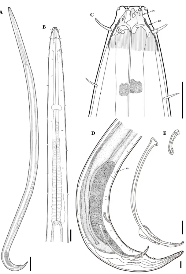
Fig. 1. Phanoderma koreense sp. nov., holotype male in lateral view: A, habitus; B, anterior region; C, head region; D, spicule and tail region;
E, spicule, gubernaculum and precloacal supplement. cc, cephalic capsule; pc, pharyngeal capsule; vs, ventral cloacal setae(Scale bars:
A=200μm; B, E=50μm; C, D=20μm).
A
B
C
D E
Fig. 2. Phanoderma koreense sp. nov., paratype females in lateral view: A, habitus; B, anterior region; C, vulva region; D, head region; E, tail region(Scale bars: A=200μm; B, E=50μm; C=100μm; D=20μm).
A D
E C
B
at a depth of 1 m by hands with scoop sampler. Sediments were composed of tiny shell gravels and coarse detritus.
Diagnosis. Head attenuated. Six inner labial sensilla present as minute papillae around with circular groove.
Well-developed trifurcate pharyngeal capsule, and cephal- ic capsule with posteriorly bearing longitudinal striations.
Two very small tooth-like subventral projection part pres- ent. Long and slender spicules with 4-5 serrated distal end, beyond precloacal supplement. Gubernaculum short and thick. A series of eight to nine stout and short setae present on ventral cloacal region. Conico-cylindrical tail with two pairs of blunt setae in both sexes.
Measurements. See Table 1 for detailed measurements and morphometric ratios.
Description. Males: Body long (3,746-4,233 μm), typi-
cally cylindrical appearance (Fig. 1A). Anterior end gradu- ally narrow just in front of nerve ring, cervical region 4-5 times thinner than rest of body region. Maximum body diameter at mid body level 104-122 μm. Cuticle smooth and thick with a few scattered short somatic setae through- out body. Head diameter 21-27 μm, distinctly narrowed.
Six inner labial sensilla present as minute papillae around with circular groove (Figs. 1C, 3B). Six outer labial sen- silla and four cephalic setae in one circle; cephalic setae 11-12 μm long, 45-52% of head diameter. Buccal cavity small but surrounded by cuticular capsules without teeth.
Well-developed trifurcate pharyngeal capsule with big and wide extremely sclerotized outgrowths (Figs. 1C, 3A). Two tooth-like subventral projection part present, very small and cuticularized. Cephalic capsule with solid structure di-
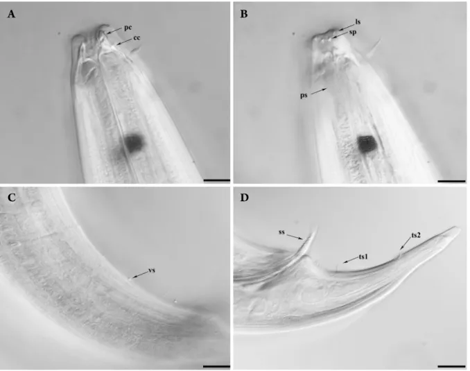
Fig. 3. Phanoderma koreense sp. nov., DIC photomicrographs, holotype male in lateral view: A, head region; B, head region showing labi- al sensilla and subventral projection part; C, ventral cloacal seta; D, tail region showing serrated spicule distal end and two tail setae. cc, cephalic capsule; ls, labial sensilla; pc, pharyngeal capsule; ps, posterior striation; sp, subventral projection; ss, serrated distal end; ts1, tail setae 1; ts2, tail setae 2; vs, ventral cloacal setae (Scale bars: A, B=40μm; B, C=20μm).
A B
C D
vided into two parts: anterior section with thick walls and without grooves, and posterior section with relatively thin walls and longitudinal striations (Figs. 1C, 3B). Amphids inconspicuous, ocelli 7 μm wide, located at 38-45 μm from anterior end (Fig. 1C). Pharynx muscular, 831-915 μm long, cylindrical anteriorly and enlarged at posterior end with characteristic crenelated outline. Excretory pore con- spicuous, situated at 86-95 μm from anterior end. Nerve ring encircling pharynx, situated at 36-40% of pharynx length from anterior end (Fig. 1B). Tail 135-142 μm long, consists of proximal conical and distal cylindrical parts with two pairs of blunt setae (Figs. 1D, 3D). Spicules elon- gated, strongly curved, 191-219 μm long, extend beyond precloacal supplement, distally sharpened with 4-5 serrat- ed distal end (Fig. 1E). Gubernaculum 38-42 μm, relative- ly short and thick. Precloacal supplement tubular shaped, 39-43 μm long, situated at 114-137 μm in front of cloacal opening, about 53-65% of spicules length. Conspicuous 8-9 stout, short setae located on ventral cloacal region in a row (Figs. 1D, 3C).
Females: Similar to male in general appearance (Fig. 2B, D). Body length 4,263-4,367 μm with cylindrical body,
distinctly tapering anterior end. Maximum body diameter at mid body level 120-130 μm (Fig. 2A). Reproductive sys- tem didelphic, ovary symmetrical, reflexed. Vulva 2,396- 2,484 μm from anterior end, situated at 55-58% of total body length (Fig. 2C). Tail length 177-188 μm long, about 2.6-2.7 times of anal body diameter. Conico-cylindrical tail with two pairs of blunt setae (Fig. 2E).
Differential diagnosis and relationships. Phanoderma koreense sp. nov. is characterized by the combination of the following features: (1) the presence of well-developed trifurcate pharyngeal capsule and cephalic capsule with posteriorly bearing longitudinal striations; (2) six inner labial sensilla present as minute papillae around with circu- lar groove (3) long and slender spicules with 4-5 serrated distal end, beyond the precloacal supplement; (4) the pres- ence of relatively short, thick gubernaculum; (5) a series of eight to nine stout and short setae located on the ventral cloacal region; and (6) conico-cylindrical tail with two pairs of blunt setae in both sexes. Phanoderma koreense sp.
nov. is closely related P. cocksi Bastian, 1865 and P. serratum Ditlevsen, 1930 based on very long and slender spicules with serrated distal end and cephalic capsule with posteri-
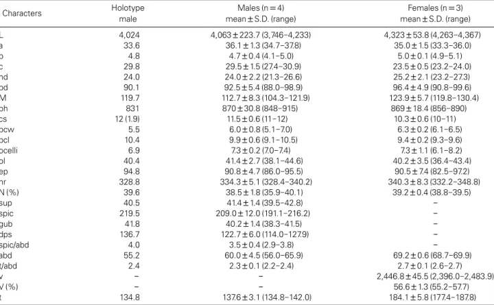
Table 1. Morphometrics of Phanoderma koreense sp. nov.(in µm)
Characters Holotype
male
Males(n=4) mean±S.D.(range)
Females(n=3) mean±S.D.(range)
L 4,024 4,063±223.7(3,746-4,233) 4,323±53.8(4,263-4,367)
a 33.6 36.1±1.3(34.7-37.8) 35.0±1.5(33.3-36.0)
b 4.8 4.7±0.4(4.1-5.0) 5.0±0.1(4.9-5.1)
c 29.8 29.5±1.5(27.4-30.9) 23.5±0.5(23.2-24.0)
hd 24.0 24.0±2.2(21.3-26.6) 25.2±2.1(23.2-27.3)
bd 90.1 92.5±5.4(88.0-98.9) 96.4±4.9(90.8-99.6)
M 119.7 112.7±8.3(104.3-121.9) 123.9±5.7(119.8-130.4)
ph 831 870±30.8(848-915) 869±18.4(856-890)
cs 12(1.9) 11.5±0.6(11-12) 10.3±0.6(10-11)
bcw 5.5 6.0±0.8(5.1-7.0) 6.3±0.2(6.1-6.5)
bcl 10.4 9.9±0.6(9.1-10.5) 9.4±0.2(9.3-9.6)
ocelli 6.9 7.3±0.2(7.0-7.4) 7.3±1.1(6.1-8.2)
ol 40.4 41.4±2.7(38.1-44.6) 40.2±3.5(36.4-43.4)
ep 94.8 90.8±4.7(86.0-95.5) 90.5±7.4(82.5-97.2)
nr 328.8 334.3±5.1(328.4-340.2) 340.3±8.3(332.2-348.8)
N(%) 39.6 38.5±1.8(35.9-40.1) 39.2±0.4(38.8-39.5)
sup 40.5 41.4±1.4(39.5-42.8) -
spic 219.5 209.0±12.0(191.1-216.2) -
gub 41.8 40.2±1.4(38.3-41.5) -
dps 136.7 122.7±6.0(114.0-127.9) -
spic/abd 4.0 3.5±0.4(2.9-3.8) -
abd 55.2 60.0±4.5(56.0-65.9) 69.2±0.6(68.7-69.9)
t/abd 2.4 2.3±0.1(2.2-2.4) 2.7±0.1(2.6-2.7)
v - - 2,446.8±45.5(2,396.0-2,483.9)
V(%) - - 56.6±1.3(55.2-57.7)
t 134.8 137.6±3.1(134.8-142.0) 184.1±5.8(177.4-187.8)
orly longitudinal striations. However, Phanoderma koreense
sp. nov. differs from P. cocksi by the length (c
=27-31 vs. c
=35 in Gerlach 1962) and shape (conico-cylindrical vs.
conical) of tail in males, and present of eight to nine stout
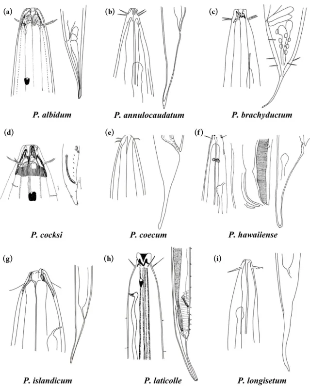
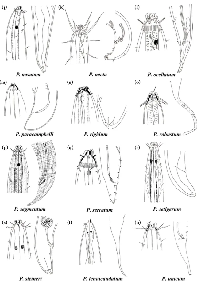
Fig. 4. Pictorial key to the valid species of the genus Phanoderma except for P. macrolaimum. Source of figures: (a) Platt & Warwick(1983);
(b) Allgén(1939); (c) Schuurmans Stekhoven(1950); (d) Platt & Warwick(1983); (e) Allgén(1947); (f) Allgén(1951); (g) Ditlevsen(1926); (h) Marion(1870); (i) Allgén(1939); (j) Schuurmans Stekhoven(1950); (k) Gerlach(1957); (l) Gerlach(1962); (m) Allgén(1958); (n) Schuurmans Stekhoven(1946); (o) Allgén(1957); (p) Murphy(1963); (q) Inglis(1971); (r) Marion(1870); (s) Ditlevsen(1918); (t) Allgén (1951); (u) Inglis (1964).
(a) (b) (c)
(d) (e) (f)
(g) (h) (i)
Fig. 4. Continued.
(j) (k) (l)
(m) (n) (o)
(p) (q) (r)
(s) (t) (u)
and short setae located on the ventral cloacal region (absent in P. cocksi). Phanoderma koreense sp. nov. is distinguished from P. serratum by the relatively longer spicules (191- 219 μm vs. 154 μm), shorter tail in males (c
=27-31 vs.
c
=35.7), and the presence of conspicuous gubernaculum.
Phanoderma koreense sp. nov. also resembles P. ocellatum (Cobb, 1920) and P. segmentum Murphy, 1963, mainly by possessing the conico-cylindrical tail and long spicules stretched over the precloacal supplement. However, Phan- oderma koreense sp. nov. is distinguished from P. ocellatum by having the longer body size (3,746-4,233 μm vs. 2,600 μm in male), spicules with serrated distal end, and a series of eight to nine stout and short setae on the ventral cloacal region. Phanoderma koreense sp. nov. differs from P. segmen- tum by relatively shorter body length in males (3,746-4,233 μm vs. 4,750 μm in Murphy 1963), more slender body shape in males (a
=34-38 vs. a
=44 in Murphy 1963), and the presence of spicules with serrated distal end (absent in the original description of P. segmentum). Also, Phanoder- ma segmentum have five distinct segments of the spicules, a feature not shared by Phanoderma koreense sp. nov.
Taxonomic remarks. The genus Phanoderma Bastian, 1865 differs from the other genera of this family in having the attenuated head, well-developed pharyngeal capsule and cephalic capsule, and a tubular precloacal supplement (Smol et al. 2013; Zograf et al. 2015). The checklist of the genus Phanoderma has been recorrected in detail by Ger- lach and Riemann (1974) including their synonymy and repeated findings. Phanodermopsis necta Gerlach, 1957 was relocated to Phanoderma on the basis of the presence of a precloacal supplement (Lorenzen 1981; Zograf et al.
2015). Platonova (1984) rearranged 11 species of the ge- nus Phanoderma into the genus Metaphanoderma on the basis of the morphological differences, such as the shape of pharyngeal and cephalic capsule. However, the descrip- tions of several species are insufficient, depicting very few characteristics and providing poor illustrations of the im- portant characteristics of cephalic and spicule structures, and many species descriptions were based on only females or juveniles (Platonova 1984; Zograf et al. 2015). Conse- quently, 22 valid species are recorded in the genus Phano- derma (Wieser 1953; Gerlach and Riemann 1974; Loren- zen 1981; Zograf et al. 2015). We herein provide a pictorial identification key to species of the genus Phanoderma (Fig.
4). The illustration of Phanoderma macrolaimum (Linstow, 1908) could not be found in the original description, how- ever, we analyzed and differentiated the taxonomic key characters based on the original description of the species.
ACKNOWLEDGEMENT
This research was supported by the Marine Biotech- nology Program (No. 20170431) of the Korea Institute of Marine Science and Technology Promotion (KIMST) funded by the Ministry of Oceans and Fisheries (MOF) (PM61440).
REFERENCES
Allgén CA. 1939. Über einige im reinen Schalensand der West- küste Norwegens freilebende Nematoden. Festschr. Embrik Strand (Riga) 5:404-425.
Allgén CA. 1947. West American marine nematodes (Papers from Dr. Th. Mortensen’s Pacific Expedition 1914-16. LXXV).
Vidensk. Meddr. Dansk. Naturh. Foren. 110:65-219.
Allgén CA. 1951. Pacific freeliving marine nematodes (Papers from Dr. Th. Mortensen’s Pacific Expedition 1914-16. LXXVI).
Vidensk. Meddr. Dansk. Naturh. Foren. 113:263-411.
Allgén CA. 1957. Zur Kenntnis norwegischer Nematoden XXIV.
Über einige für Norwegen neue freilebende marine Nema- toden. Det Kongelige Norske Videnskabers Selskabs 30:22- 28.
Allgén CA. 1958. Über einige freilebende marine Nematoden von der Ostküste Südamerikas (Uruguay, Nordküste Argenti- nas). Zool. Anz. 160:205-217.
Bastian HC. 1865. Monograph of the Anguillulidae, or free nema- toids, marine, land, and freshwater, with descriptions of 100 new species. Trans. Linn. Soc. Lond. 25:73-184.
Cobb NA. 1920. One hundred new nemas (type species of 100 new genera). Contributions to a Science of Nematology 9:217-343.
De Coninck LA and JH Schuurmans stekhoven. 1933. The freeliving marine nemas of the Belgian Coast. II With general remarks on the structure and the system of nemas. Mém.
Mus. R. his. Nat. Belg. 58:3-163.
Ditlevsen H. 1918. Marine freeliving nematodes from Danish waters. Vidensk. Meddr. Dansk. Naturh. Foren. 7:147-214.
Ditlevsen H. 1926. Free-living nematodes. Dan. Ingolf-Exp. 6:1- 42.
Ditlevsen H. 1930. Marine free-living nematodes from New Zea- land. Vidensk. Meddr. Dansk. Naturh. Foren. 87:202-242.
Filipjev IN. 1918. Free-living marine nematodes of the Sevasto- pol area. Transactions of the Zoological Laboratory and the Sevastopol Biological Station of the Russian Academy of Sciences 4:1-203.
Filipjev IN. 1927. Les nvématodes libres des mers septentrio-
nales appartenant á la famille des Enoplidae. Arch. Natur- gesch. 91:1-216.
Gerlach SA. 1957. Die Nematodenfauna des sandstrandes an der kuste von Mittelbrasilien (Brasilianische Meeres-Nematoden IV). Mitt. Zool. Mus. Berlin 33:411-459.
Gerlach SA and F Riemann. 1974. The Bremerhaven checklist of aquatic nematodes. Veröff. Inst. Meeresforsch. Bremerhaven 4:1-736.
Inglis WG. 1964. The marine Enoplida (Nematoda): a comparative study of the head. Bull. Br. Mus. Nat. Hist. Zool. 11:165-376.
Kristensen RM. 1989. Marine Tardigrada from the southeastern United States coastal waters I. Paradoxipus orzeliscoides n. gen., n. sp. (Arthrotardigrada: Halechiniscidae). Trans. Am.
Math. Soc. 108:262-282.
Linstow OV. 1908. Zoologische und Anthropologische Ergebisse einer Forschungsreise im Westlichen und Zentralen Südafrika ausgefürhrt in den Jahren 1903-1905. II. Helminthes. Nem- atoden und Acanthocephalen. Denkschr. Medicine Naturwis Gschichte. Jenia. 13:19-28.
Lorenzen S. 1981. Entwurf eines phylogenetischen systems der freilebenden nematoden. Veröff. Inst. Meeresforsch. Bremer- haven 7:1-472.
Marion AF. 1870. Recherches zoologiques et anatomiques sur des Nématoides non parasites, marins. Annls. Sci. Nat.
13:1-100.
Marion AF. 1875. Révision des Nématoides du Golf de Marseille.
C. R. Hebd. Séances Acad. Sci. 80:499-501.
Mawson PM. 1956. Free -living nematodes. Section I: Eno- ploidea from Antarctic stations. Report of the British, Austra-
lian and New Zealand Antarctic Research Expedition 6:37-74.
Murphy DG. 1963. Three new species of marine nematodes from the Pacific near Depot bay, Oregon. Proc. Helminthol.
Soc. Wash. 30:249-256.
Platonova TA. 1984. Materials for revision of the family Phanoder- matidae (Enoplida, Nematoda). Zoolgicheskii Zhurnal 63:507- 516.
Platt HM and RM Warwick. 1983. Freeliving marine nematodes part I British Enoplids. Synopses of the British Fauna. Cam- bridge University Press.
Schuurmans Stekhoven JH. 1946. Freilebende marine Nema- toden des Skagerraks und der Umgebung von Stocklholm.
Ark. Zool. 16:1-91.
Schuurmans Stekhoven JH. 1950. The freeliving marine nemas of the Mediterranean: I. The bay of Villefranche. Mém. Mus. R.
His. Nat. Belg. pp. 1-220.
Shirayama Y, T Kaku and R Higgins. 1993. Double-sided micro- scopic observation of meiofauna using an HS-slide. Benth.
Res. 44:41-44.
Smol N, A Muthumbi and J Sharma. 2013. Handbook of zoology.
Gastrotricha, Cycloneuralia and Gnathifera. Vol 2. Nematoda.
7.3 Order Enoplida. Walter De Gruyter. pp. 193-246.
Wieser W. 1953. Free-living marine nematodes. I. Enoploidea.
Lund. Univ. Arsskrift 49:1-155.
Zograf JK, AT Yulia and NP Olga. 2015. Description of new spe- cies of Phanodrmopsis(Enoplida, Phanodermatidae) with key to genera of family Phanodermatidae and pictorial key to Phanodermopsis species. Zootaxa 4032:277-289.