DOI: https://doi.org/10.3339/jkspn.2019.23.2.111 ISSN 2384-0250 (online)
Vitamin D Dependent Rickets Type 1A Caused by CYP27B1 Mutation
Vitamin D dependent rickets type 1A (VDDR1A) is an autosomal recessive disorder caused by mutations in CYP27B1. Clinical findings are growth retardation, hypo- tonia, muscle weakness, hypocalcemic seizures, and radiological features of rickets.
We aimed to present the VDDR1A case with a genetic study of CYP27B1. The 14- month-old boy was admitted to the hospital due to a seizure. Serum calcium, phosphorus, alkaline phosphatase, parathyroid hormone (PTH), 25(OH) vitamin D, and 1,25(OH)2 vitamin D values were 5.1 mg/dL, 3.7 mg/dL, 705 IU/L, 429 pg/mL, 24.9 ng/mL, and 8.8 pg/mL, respectively. Radiological study showed cupping and fraying of the distal ulna and radius. The molecular genetic study revealed that the patient had a compound heterozygous mutation, Phe443Profs*24 and c.589+
1G>A, in CYP27B1. Genetic analysis of the family members presented that the mother was heterozygous for the mutation c.589+1G>A, and that the father was heterozygous for Phe443Profs*24. The patient was treated with calcium lactate and calcitriol. Until now, six Korean patients with VDDR1A have been studied.
Including this case, Korean patients with VDDR1A were found to have only three different mutations in 14 alleles, indicating that the mutation in the CYP27B1 gene is homogeneous in the Korean population.
Key words: Rickets, CYP27B1 gene, Vitamin D dependent rickets type 1A
Na Ry Bak, M.D.
Eun Song Song, M.D.
Eun Mi Yang, M.D.
Chan Jong Kim, M.D.
Department of Pediatrics, Chonnam National University Medical School &
Children’s Hospital, Gwangju, Korea
Corresponding author:
Eun Mi Yang, M.D. and Chan Jong Kim, M.D.
Department of Pediatrics, Chonnam National University Medical School &
Children’s Hospital, 42 Jebong-ro, Dong- Gu, Gwangju 61469, Korea
Tel: +82.62-220-6647 Fax: +82.62-222-6103
E-mail: [email protected] [email protected]
Received: 3 April 2019 Revised: 7 June 2019 Accepted: 22 June 2019
This is an open-access article distributed under the terms of the Creative Commons Attribu tion Non-Commercial License (http://
crea tivecom mons.org/licenses/by-nc/4.0/) which permits unrestricted non-commercial use, distribution, and reproduction in any medium, provided the original work is properly cited.
Copyright © 2019 The Korean Society of Pediatric Nephrology
Introduction
There are two types of vitamin D: ergocalciferol (D2) and cholecalciferol (D3). Two types of vitamin D are biologically inactive prohormones, and it is necessary to undergo two enzymatic conversions in the liver and kidney1,2). CYP2R1 catalyzes the initial hydroxylation step in the liver converting vita
min D to 25(OH) vitamin D (25OHD). Albeit 25OHD is the most plentiful type of vitamin D in circulation, it has minimal ability bind to the vitamin D receptor (VDR)3). The 25OHD 1αhydroxylase gene (CYP27B1) encodes the 1αhydroxylase enzyme, which catalyzes 25OHD to 1,25(OH)2 vitamin D [1,25(OH)2D]. The 1,25(OH)2D is the active type of vitamin D that acts via the VDR to increase the expression of genes. It is stimulated by parathyroid hormone (PTH), hy pocalcemia, and hypophosphatemia4,5).
Vitamin D dependent rickets type 1A (VDDR1A), also known as vitamin D 1αhydroxylase deficiency or pseudovitamin D deficiency rickets, is an auto
somal recessively inherited disorder resulting from defects in the CYP27B1.
The associated clinical findings are growth retardation, hypotonia, muscle
weakness, hypocalcemic seizures, and radiological features of rickets. Biochemical findings show hypocalcemia, ele
vated PTH, and decreased values of 1,25 (OH)2D in spite of nor mal or elevated concentrations of 25OHD. Generally, a good response to medication with alfacalcidol or calcitriol is estimated for patients with VDDR1A6,7). Calcitriol and calcium treatment was modi fied based on the laboratory and clinical features. Treat ment results in the normaliza
tion of laboratory parameters and radiographic abnorma
lities within 3 months.
So far, six Korean patients with VDDR1A have been in
vestigated at a genetic study810). We now report the results of clinical and molecular genetic studies of CYP27B1 mu
tations in one additional patient and review the findings in all reported Korean patients with CYP27B1 defects.
Case report
A 14monthold boy was admitted in our hospital due to a generalized tonic seizure lasting for two minutes. He was born to nonconsanguineous Korean parents as their first baby at 40 weeks of gestation. The boy was healthy at birth and weighed 4,220 g. The parents were healthy and they do not have any hereditary disorders. At the age of 9 months, he had the ability to crawl, and at 13 months, he was able to walk alone. At a physical examination, he was 77.0 cm tall (25–50 percentile) and weighed 9.5 kg (10–25 percentile).
Laboratory profile showed as follows; serum total calcium of 4.8 mg/dL (normal range, 8.4–10.2 mg/dL); ionized calcium of 1.4 mEq/L (normal range, 2.2–2.6 mEq/L);
phos phorus of 3.7 mg/dL (normal range, 3.8–6.5 mg/dL), alkaline phosphatase of 705 U/L (normal range, 145–420 U/L); and PTH of 429 pg/mL (normal range, 10–65 pg/mL).
25OHD level was 24.9 ng/mL (normal range, 30–90 pg/
mL) and 1,25(OH)2D was 8.8 pg/mL (normal range, 20–
55 pg/mL). Radiological study showed cupping and fraying of the distal ulna and radius (Fig. 1). The above laboratory and radiological signs were compatible with a diagnosis of rickets. A decreased 1,25(OH)2D level with relatively normal 25OHD and very high PTH concentrations indi
cated a diagnosis of a genetic type of rickets, precisely 1α
hydroxylase deficiency.
A mutation analysis of the CYP27B1 gene was performed.
Genomic DNA was extracted from leukocytes. All coding exons and intron in flanking regions of CYP27B1 were amplified by polymerase chain reaction and directly se
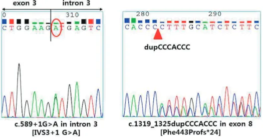
quenced. The primers used for DNA sequencing of patients were as previously described8). Molecular genetic study revealed that the patient had a compound heterozygous mutation for the 7bp duplication 13191325dupCCCACCC (Phe443Profs*24) and c.589+1G>A in the CYP27B1 gene (Fig. 2). The family members revealed that the mother was heterozygous for the mutation c.589+1G>A, and the father was heterozygous for Phe443Profs*24. G to A substitution in the first nucleotide of intron 3 (c.589+1G>A) was reported previously in Japanese and Korean patients811). The Phe443 Profs*24 mutation was reported in multiple ethnic groups including Korean population8).
The boy was treated with calcitriol and calcium lactate.
Compliance with the supplementation was good, resulting in quick improvement of the abnormal clinical, laboratory, and radiographic findings. The patient showed normoc
alcemia without recurrent hypocalcemic seizures. Radio
logic study showed marked improvement after 5 months of treatment (Fig. 1).
Discussion
There are four types of genetic errors in vitamin D me
A B
Fig. 1. Radiographic features of the patient. (A) X-ray finding showed cupping and fraying of the metaphyseal regions of radius and ulnar at presentation. (B) After 5 months of treatment, bone lesions were markedly improved.
tabolism, and it can cause rickets. The first type is the VDDR1A, which is caused by defects in the CYP27B1 gene.
A mutation in the CYP2R1 gene that results in 25hydr
oxylase deficiency causes vitamin D dependent rickets type 1B (VDDR1B). Vitamin D dependent rickets type 2A (VDDR2A), also referred to as vitamin D resistant rickets (VDDR), is due to a defect in the VDR. VDDR2B is a rare type of rickets owing to abnormal expression of a hormone response elementbinding protein that disrupts the normal function of VDR13,14).
The clinical and laboratory findings in patients with VDDR1A are similar to those seen in patients with nutri
tional rickets or hypophosphatemic rickets. However, VDDR1A can be discriminated from hypophosphatemic rickets by increased PTH and decreased 1,25(OH)2D con
centrations and nutritional rickets by a normal 25OHD value. The hypophosphatemia in VDDR1A is an effect of high PTH level and renal excretion of phosphate. PTH pro
motes calcium reabsorption in the renal proximal tubules, and also promotes serum calcium through bone resorption
15). It also increases the alkaline phosphatase level15). Most of the reported patients with VDDR1A showed low or un
detectable 1,25(OH)2D levels. But there have been a few reported cases of patients with VDDR1A with normal 1,25(OH)2D levels6,16,17). Nishikawa et al.18) presented that liver mitochondrial CYP27A1 can catalyse 1αhydroxyla
tion of 25OHD. A little bit increment of 1,25(OH)2D con
centrations has been reported in CYP27B1 knockout mice
after high vitamin D supplementation, suggesting the con
version from 25OHD to 1,25(OH)2D by a nonCYP27B1 enzyme14). Wang et al.17) reported two patients with VDDR1A who had normal 1,25(OH)2D values. Both ex hibited a mild phenotype, and their mutations had partial enzyme func
tion in vitro. So, VDDR1A should be sus pected in cases with a mild or severe phenotype of rickets and normal or elevated values of 25OHD albeit 1,25(OH) 2D concentra
tions are within the normal values.
The CYP27B1 gene is constituted of 9 exons spanning 5 kb19). Until now, more than 70 mutations have been re
ported in the CYP27B1 gene in multiple ethnic groups10,14). Several mutations in the CYP27B1 gene are more common in specific populations8,12). Deletion of guanine 958 (958 delG) was reported on 20 French Canadian allele, and a microsatellite haplotype study revealed that these arose from a single ancestor12). A seven nucleotide duplication in exon 8 (Phe443Profs*24) was reported in 14 affected alleles, and these arose from different populations with diverse microsatellite haplotypes12).
To date, six Korean patients with VDDR1A have been in
vestigated at a genetic study, and three different mutations have been reported810). There are two frequent mutation points; c.589+1G>A in seven alleles and Phe443Profs*24 in four alleles. Our patient was also found to have two mu
tations common in the Korean population: c.589+1G>A and Phe443Profs*24. Thus, Korean patients with VDDR1A have been found to have only three different mutations in
Fig. 2. Mutation analysis of the proband revealed a compound heterozygous mutation for c.589+1G>A and Phe443Profs*24 in the CYP27B1 gene.
Fig. 2. Mutation analysis of the proband revealed a compound heterozygous mutation for c.589+1G>A and Phe443Profs*24 in the CYP27B1 gene.
14 alleles.
Including our case, the clinical characteristics of seven patients with vitamin D dependent rickets type 1A were summarized in Table 1. The most prevalent mutation is the c.589+1G>A splice donor site mutation, affecting 8 out of 14 alleles in Korean patients with VDDR1A. This muta
tion also have been reported in Japanese patients11). G to A change in the first nucleotide of an intron is a relatively frequent mutation and generally results in the skipping of the exon or initiation of potential splice sites17). The c.589+
1G>A mutation introduces a premature stop codon, and it causes disruption of 1αhydroxylase activity.
The mutation of Phe443Profs*24 had been found in 5 of 14 alleles in Korean patients. This seven nucleotide dupli
cation (CCCACCC) causes a premature TGA stop signal.
It eliminates the hemebinding domain of 1αhydroxylase and thus inactivate enzyme activity. Wang et al.12) presented that six families of different ethnic groups had this muta
tion in association with four diverse microsatellite haplo
types. Kitanaka et al.11) reported 10 different CYP27B1 gene mutations in 20 Japanese alleles, indicating that there was no single ancestor effect in Japanese population.
In conclusion, we present a case study of VDDR1A in which the patient was found to have a compound hetero
zygous mutation, Phe443Profs*24 and c.589+1G>A, in the CYP27B1 gene. Korean patients have been found to have only three different mutations in 14 alleles. This suggests that the mutation of the CYP27B1 gene is relatively homo
geneous in the Korean population.
Conflict of interest
No potential conflict of interest relevant to this article was reported.
Patient consent
This study was approved by the institutional review board (IRB), and the consent was waived due to the nature of the retrospective study [IRB number CNUHEXP
2018295].
References
1. Miller WL, Portale AA. Vitamin D 1α-hydroxylase. Trends Endo- crinol Metab 2000;11:315-9.
2. Portale AA, Miller WL. Rickets due to hereditary abnormalities of vitamin D synthesis or action. In: Glorieux FH, Pettifor JM, Jupp- ner H, eds. Pediatric bone. San Diego: Academic Press, 2003;583- 98.
3. Portale AA, Miller WL. Human 25-hydroxyvitamin D 1α-hydroxy- lase: cloning, mutations, and gene expression. Pediatr Nephrol 2000;14:620-5.
4. Breslau NA. Normal and abnormal regulation of 1,25(OH)2D syn- thesis. Am J Med Sci 1988;296:417-25.
5. Kumar R, Harnden D, DeLuca HF. Metabolism of 1,25-dihydroxy vitamin D3: evidence for side-chain oxidation. Biochemistry 1976;15:2420-3.
6. Demir K, Kattan WE, Zou M, Durmaz E, BinEssa H, Nalbantoğlu Ö, et al. Novel CYB27B1 gene mutations in patients with vitamin D-dependent rickets Type 1A. PLoS One 2015;10:e0131376.
Table 1. Clinical Characteristics of Seven Patients with Vitamin D Dependent Rickets Type 1A Patient
no.
Age at diagnosis
(months) Presentation Serum Ca(mg/dL) Serum P
(mg/dL) Serum ALP
(IU/L) Serum PTH
(pg/mL) Serum 25OHD
(ng/mL) Serum 1,25-
(OH)2D (pg/mL) Mutation
1 5 rachitic rosary 6.4 4.0 2,043 1,320 27 9.2 Phe443Profs*24/Phe443Profs*24
2 5 rachitic rosary 5.9 3.6 1,326 750 77 <4 c.589+1G>A /2561G>A
3 7 seizure 5.2 6.2 665 750 38 <5 c.589+1G>A/Phe443Profs*24
4 12 bowed legs 6.0 3.8 1,050 560 N/A <7 c.589+1G>A/c.589+1G>A
5 14 femur fracture 7.3 5.0 1,437 N/A 16 <5 c.589+1G>A/Phe443Profs*24
6 13 seizure 4.8 4.3 1,665 178 75.1 5.3 c.589+1G>A/c.589+1G>A
7 14 seizure 5.1 3.7 705 429 24.9 8.8 c.589+1G>A/ Phe443Profs*24
Abbreviations; ALP, Alkaline phosphatase; N/A, Not available. Data of Patients 1 to 5 are adopted from reference 9. Patient 6 is adopted from reference 10.
Patient 7 is in this study.
*Nucleotide numbers refer to genomic DNA and are numbered from the transcription start site20). The reference sequence is available on the NCBI, Entrez, Nucleotide database: http://www.ncbi.nlm.nih.gov/Entrez; accession number AF 027152.
7. Edouard T, Alos N, Chabot G, Roughley P, Glorieux FH, Rauch F.
Short-and long-term outcome of patients with pseudo-vitamin D deficiency rickets treated with calcitriol. J Clin Endocrinol Metab 2011;96:82-9.
8. Kim CJ, Kaplan LE, Perwad F, Huang N, Sharma A, Choi Y, et al.
Vitamin D 1alpha-hydroxylase gene mutations in patients with 1alpha-hydroxylase deficiency. J Clin Endocrinol Metab 2007;92:
3177-82.
9. Kim CJ. Vitamin D dependent rickets type I. Korean J Pediatr 2011;54:51-4.
10. Cho JH, Kang E, Kim GH, Lee BH, Choi JH, Yoo HW. Long-term clinical outcome and the identification of homozygous CYP27B1 gene mutations in a patient with vitamin D hydroxylation-defi- cient rickets type 1A. Ann Pediatr Endocrinol Metab 2016;21:169- 73.
11. Kitanaka S, Murayama A, Sakaki T, Inouye K, Seino Y, Fukumoto S, et al. No enzyme activity of 25-hydroxyvitamin D3 1α-hydroxy- lase gene product in pseudovitamin D deficiency rickets, inclu- ding that with mild clinical manifestation. J Clin Endocrinol Metab 1999;84:4111-7.
12. Wang JT, Lin CJ, Burridge SM, Fu GK, Labuda M, Portale AA, et al.
Genetics of vitamin D 1α-hydroxylase deficiency in 17 families.
Am J Hum Genet 1998;63:1694-702.
13. Malloy PJ, Feldman D. Genetic disorders and defects in vitamin D action. Rheum Dis Clin North Am 2012;38:93-106.
14. Dursun F, Özgürhan G, Kırmızıbekmez H, Keskin E, Hacıhamdioğlu B. Genetic and clinical characteristics of the patients with vitamin D dependent rickets type 1A. J Clin Res Pediatr Endocrinol 2018 Oct 4doi: 10.4274/jcrpe.0121.
15. Akerstrom G, Hellman P, Hessman O, Segersten U, Westin G. Pa- rathyroid glands in calcium regulation and human disease. Ann N Y Acad Sci 2005;1040:53-8.
16. Acar S, Demir K, Shi Y. Genetic causes of rickets. J Clin Res Pediatr Endocrinol 2017;9:88-105.
17. Wang X, Zhang MY, Miller WL, Portale AA. Novel gene mutations in patients with 1alpha-hydroxylase deficiency that confer par- tial enzyme activity in vitro. J Clin Endocrinol Metab 2002;87:
2424-30.
18. Nishikawa M, Yasuda K, Takamatsu M, Abe K, Nakagawa K, Tsugawa N, et al. Generation of 1,25-dihydroxyvitamin D3 in CYP27B1 knockout mice by treatment with 25-hydroxyvitamin D3 rescued their rachitic phenotypes. J Steroid Biochem Mol Biol 2019;185:71-9.
19. Fu GK, Lin D, Zhang MY, Bikle DD, Shackleton CH, Miller WL, et al.
Cloning of human 25-hydroxyvitamin D 1 alpha-hydroxylase and mutations causing vitamin D-dependent rickets type I. Mol Endocrinol 1997;11:1961-70.
20. Fu GK, Portale AA, Miller WL. Complete structure of the human gene for the vitamin D 1α-hydroxylase, P450c1α. DNA Cell Biol 1997;16:1499-507.