http://dx.doi.org/10.7841/ksbbj.2012.27.6.367
연구논문
손상된 불가사리 추출물 흡입이 흡연으로 인한 항염증과 항산화 효소의 효과
황경희, 정 혁, 장수찬, 박종석, 김유영*
The Injured Starfish Extract Inhalation Effects of Anti-inflammation and Anti-oxidants Enzyme during Indirect Cigarette Smoking
Kyung Hee Hwang, Hyuk Jung, Su Chan Chang, Jong Seok Park, and You Young Kim*
접수: 2012년 11월 6일 / 게재승인: 2012년 12월 23일
© 2012 The Korean Society for Biotechnology and Bioengineering
Abstract:1)Cigarette smoking (SM) is considered to be well known environmental toxin which contributes to the onset of various diseases. SM cause direct lungs damage, activate lungs inflammatory responses, and in some cases leads to the development of lung cancer. Cytokines in injured starfish (Asterina pectinifera) is the potential changes in its expression during the regeneration process. Especially, expression of TGF-β1 has increased in arm cut starfish extract after eight days. Also, starfish including saponin like the ginseng. Saponin is widely used in the world because of some effective pharmacological activities. Therefore, the current study was designed to elucidate the pharmacological activities of starfish extract against cigarette smoking induced damage in cell line and pulmonary tissue. We investigate that the effect of eight days starfish extract after arm cut (8d) and intact starfish extract on cell line and mouse lung injury by SM. In cell proliferation analysis, although cigarette smoking extract (CSE) was co-treated, the higher proliferation ability is shown in 8d treatment than intact starfish extract. 8d and intact starfish extract was directly transported to pulmonary cells through respiratory organ by nebulizer inhalation. In this case of cigarette smoking, the pulmonary structure was damaged and
경북대학교 자연과학대학 생명과학부
School of Life Sciences and Biotechnology, College of Natural Sciences, Kyungpook National University, 1370 Sangeok-dong, Buk-ku, Taegu 702-701, Republic of Korea
Tel: +82-53-950-6354, Fax: +82-53-943-2762 e-mail: yykim@knu.ac.kr
functions become abnormal. However, 8d treated groups showed similar with the control group compared with SM group. Among them, 8d was proved to be more effective than intact starfish extract. These results demonstrate that 8d could more protect pulmonary structure and function than intact starfish extract against cigarette smoking by ginseng like saponin and regulation of inflammatory cytokines.
Keywords: Starfish, Cigarette smoke, Pulmonary, Nebulizer, Morphology, Anti-oxidant
1. 서론
폐는 공기와 혈액 사이의 산소와 이산화탄소의 교환을 통하 여 혈액의 산소 농도 유지 등의 기능을 담당하고 있으나 [1], 대기 중의 유해물질인 아황산가스, 먼지, 황사, 담배연기 등 이 여러 경로를 통해 유입될 경우 폐 기능이 손상되고 혈액 이 산성화되어 질병이 유발된다 [2,3]. 특히, 담배연기에는 타르 등 약 43종의 발암물질을 비롯한 여러 독성물질이 함유 되어 있어서 [4] 흡연을 하게 되면 담배연기에 있는 화학물질 이 일차적으로 호흡기관에 피해를 주고, 폐로 흡수되어 체내 를 순환하면서 생체내의 장기에 염증 및 암을 포함한 각종 질병을 일으킬 뿐만 아니라 세포, 조직의 노화현상을 유발하 게 된다. 한편 흡연 시에는 혈액의 산소 운반기능 및 면역기 능의 저하와 폐포의 구조 변화, 폐의 표면장력을 조절하는 surfactant protein의 변화를 가져 오게 된다 [5]. 또한 활성산 소종 (reactive oxygen species)으로 인하여 폐포 지방의 과
산화 반응이 일어나며 활성산소종을 제거하는 반응에 사용 되는 SOD (superoxide dismutase), catalase, G6PD (glucose- 6-phosphatedehydrogenase) 등 여러 효소에 영향을 미치게 된다 [6-8]. 염증은 흡연으로 인한 세포손상이 가해졌을 때 자극이나 손상인자를 제거하기 위한 생체방어로서 염증성 면역세포 (T-cell, monocyte, macrophage), 혈관내피세포 (vesicular endothelial cell), 염증성 단백질 (inflammatory protein: cytokines, chemokines)이 복잡하게 관여하는 과정 이다. 정상상태의 염증은 상처치유와 관련하여 보통 자가제 한적 (self-limit)이고 신체의 항상성을 유지하는데 중요하지 만 계속되는 자극으로 인한 염증은 동맥경화 및 특정 암의 발생 및 진행을 촉진시키는 작용을 한다. 염증성 사이토카인 으로 알려져 있는 tumor necrosis factor-α (TNF-α)는 염증 의 주요한 매개자로서 염증성 병변과정에서 발현정도가 증 가되고 호중성 백혈구를 활성화시켜 과산화수소의 생성을 증가시킨다. 또한 염증성 전구물질 (PEG2, iNOS, COX-2)들 은 nuclear factor-κB (NF-κB)와 같은 전사조절인자에 의해 서 조절된다고 알려져 있다 [9]. NF-κB는 유도성 전사인자 의 일종으로 염증을 포함한 다양한 병리학적 상태와 관련 된 특이적 표적 유전자의 발현을 조절한다 [10]. NF-κB는 세 포질에서 Rel단백질 (Rel A, p65, p50, c-Rel, v-Rel, 그리고 Rel B)의 아단위로 구성된 이형이량체 (heterodimer)나 동형이량체 (homodimer)를 이루면서 IκB라는 저해단백질 (inhibitory protein)이 부착되어 불활성 상태로 존재한다 [11].
그러나 NF-κB 경로를 촉진하는 lipopolysaccharide (LPS), 염증성 사이토카인, 산화적 스트레스와 같은 다양한 세포외 신호를 주면 IκB가 분해되어 NF-κB를 활성화시켜 세포질 에서 핵 내로 이동을 유도하여 DNA와 결합하게 되면서 염증 성 전구물질을 생성한다 [9].
인삼 saponin은 각종 혈관계, 심장, 뇌, 간장 질환 등을 예방, 치유하고, 활성산소종에 대한 폐혈관 보호 작용 등의 많은 약리학적 효과가 있는 것으로 알려져 있다 [12-14]. 본 연구실에서 간접 흡연시킨 마우스 모델에 인삼추출물을 흡입 시킨 결과, 폐기능의 보호, 면역력 증가, 항산화작용의 증가 및 손상된 폐조직의 회복 등의 효과를 보여주었다 [15]. 그러 나 인삼은 고가일 뿐만 아니라 그 추출과정이 복잡하여 경 제적인 측면에서 부가가치가 낮은 편이다. 따라서 이에 대한 대체 방법으로 해양자원 특히, 양식장과 해양환경에 막대한 피해를 주는 폐자원인 불가사리로부터 saponin 등의 유효 성분을 분리 · 추출하여 대체함으로써 그 부가가치를 높일 수 있을 것으로 사료된다. 불가사리에는 의약학적 특이성 분인 saponins, proteins, triterpene glycosides, sulphated polysaccharides, glycosphingolipids 등이 포함되어 있다. 특히 saponin은 sulphated sterol glycosides 등으로서 인삼 saponin (ginsenoside)과 화학적 구조 및 약리학적 효과가 비슷하며 다양한 스테로이드 유도체로 항균 및 세포독성을 비롯한 미생 물을 괴사시키는 기능을 가지고 있음이 보고되었다 [16-18].
이러한 불가사리를 포함한 모든 생물의 재생이나 염증반 응을 포함한 상처 치유 (wound healing) 과정에는 많은 성장 인자 (growth factor)들이 작용 하는 것으로 알려져 있다.
조직에 상처가 생기면 즉각적인 반응으로 혈소판의 활성화가
일어난다. 혈소판은 또한 transforming growth factor- (TGF-:
형질전환 성장 인자)와 platelet-derived growth factor (PDGF:
혈소판유도 성장 인자)를 포함한 화학적 인자들을 생성한 다. 성장 인자 중에서도 특히 TGF-β는 여러 종류의 세포와 조직에서 초기 발생의 조절, 세포주기의 조절, 세포 증식 및 분화, 세포외 기질의 생산, 면역체계, 혈관 생성과 혈구 세포 생산, 세포사 유도, 골격 형성, 상처 치유 등을 조절하는 다 기능적인 cytokine으로 알려져 있다 [19-21]. 또한, 방사형 으로 뻗은 5개의 팔 (arm) 중에 2개를 손상시킨 별불가사 리 (Asterina pectinifera)의 재생과정에서 시간경과에 따른 TGF-β1의 발현 양을 측정한 결과 8일이 경과한 불가사리 에서 가장 많이 발현되는 것을 확인한 바 있다 [22].
따라서 본 연구에서는 흡연 시에 발생하는 자극에 대해 인삼의 주성분인 saponin과 유사한 성분을 포함한 조직손상 을 전혀 주지 않은 불가사리 추출물과 TGF-β1이 가장 많이 발현되는 조직손상 후 8일이 경과한 불가사리 추출물을 세포 에 처리하여 세포의 증식율과 염증관련 단백질의 발현량을 비교하고, 또한 불가사리 추출물을 nebulizer를 이용하여 실험동물에 흡입시킴으로써 항염증 효과와 폐 기능의 보호 및 흡연으로 인한 폐 세포의 구조 변화와 활성산소종으로 인한 세포 손상에 어떠한 영향을 미치는지 알아보고자 하였다.
2. 재료 및 방법 2.1. 시약 및 장비
H2O2, NADPH, thiobarbituric acid (TBA), trichloroacetic acid (TCA), N-[2-Hydroxyethyl] piperazin- N'-[2-ethanesulfonic]
acid (HEPES), ethylenediaminetetra-acetic acid (EDTA), phenylmethane-sulfonyl fluoride (PMSF)는 Sigma Chemical (St. Louis, USA), ammonium persulfate, sodium dodesyl sulfate (SDS), acrylamide, N,N,N',N'-tetramethylene-diamine (TEMED), Bio-Rad protein assay kit는 Bio-Rad (Hercules, USA), methanol, ethanol, tween 20은 Merck (Darmstadt, Germany)의 제품을 사용하였다. 그리고 TNF-α, NF-κB, Iκ-Bα, β-actin, surfactant protein A에 대한 항체는 Santa Cruz (Santa Cruz, USA), enhanced chemiluminescence (ECL) western blot detection kit는 Amersham Phamacia Biotech (Oxford, UK), EDTA tube는 BD Vacutainer (BD Vacutainer system, UK), hematoxyline, eosin xylene, autotechnicon, rotary microtome citadel-1000은 shandon (shandon, Japan), marienfild histobond slide glass은 superior (superior, Germany)에서 구입하여 사용하였다.
2.2. 재료 및 처리조건
본 연구에 사용된 불가사리는 우리나라 동해 (경북 포항시 월포리) 연안에서 채집한 5개의 방사형 팔을 가진 별불가 사리 (Asterina pectinifera)이다. 대조군으로는 조직손상을 전혀 주지 않은 불가사리 (intact starfish)를 사용하였고, 실험군으로는 팔 두개를 절단하여 조직손상 후, 임대한 양 어장에서 사육하여 TGF-β1의 발현양이 가장 많은 8일이
경과한 불가사리 (eight days starfish after arm cut)를 선별, 채집한 후 동결건조 후 분말화하여 냉동 보관하였다.
2.3. 불가사리 추출
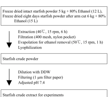
불가사리 추출물 (starfish crude extract, SCE)의 추출과정 은 Fig. 1과 같다. 조직손상을 전혀 주지 않은 분말 불가사리 와 조직 손상 후 8일이 경과한 분말 불가사리를 각각 80%
에탄올 용매로 40℃, 15 rpm에서 6시간 동안 추출한 후, 추 출물을 400 mesh, filter press로 여과하고 진공회전 증발기 (Laborota 4000, Heidolph, Japan)로 50℃, 15 rpm에서 1시간 동안 농축하였다. 농축물을 동결 건조기 (Neocool, Yamato, Japan)로 용매를 완전히 제거하여 분말화하였다. 그 후 영 하 20℃에 저장한 후, 3차 멸균수로 희석하여 pH를 7.4로 맞춘 후 1 μm filter (Whatman, England)로 여과하여 실험에 사용하였다.
2.4. 담배연기 추출
담배는 KT & G에서 생산한 THIS (타르 8.0 mg, 니코틴 0.8 mg/개비)를 사용하였다. 최대의 담배연기 추출물 (cigarette smoking extract, CSE)을 얻기 위해서 10개비의 담배를 각 각 연소시키면서 콤프레샤를 통해 50 mL의 PBS가 들어 있 는 고무풍선 내로 주입시킨 후 용해시켰다. 이 때 만들어진 담배연기 추출물은 -20℃에서 보관하였으며 0.22 μm filter (Millipore, Bedford, USA)로 여과하여 이를 100%로 정하여 사용하였다.
Freeze dried intact starfish powder 5 kg + 80% Ethanol (12 L), Freeze dried eight days starfish powder after arm cut 6 kg + 80%
Ethanol (15 L)
Extraction (40℃, 15 rpm, 6 h) Filtration (400 mesh, nylon pocket)
Evapolation for ethanol removal (50℃, 15 rpm, 1 h) Lyophilization
Starfish crude powder
Dilution with DDW Filtering (1 μm filter paper) Adjusted pH 7.4
Starfish crude extract for experiments
Fig. 1. Schematic view of the starfish crude extract preparation process for experiments.
2.5. 세포주 및 실험조건
본 실험에 사용한 세포주는 사람유래 정상 섬유아세포인 IMR-90 (human lung fibroblast cell)으로 ATCC (American type culture collection)에서 구입하였다. 세포배양에는 10%
FBS와 1% penicillin/streptomycin이 첨가된 DMEM배지에 서 5% CO₂존재하의 37℃ 항온, 항습배양기에서 배양하였다.
2.6. 실험군 및 실험조건
실험동물은 7주령 된 ICR 마우스 (평균체중 33 g)를 사용하 였으며, 실험 시작 전 폴리프로필렌 상자 (453 × 293 × 247 mm, 19 L)안에 넣어 1주간 적정 사육조건 하에서 적응시켰다 [22].
각각 7마리씩 사용한 실험동물군은 흡연과 SCE를 처리하지 않은 대조군 (Control), 증류수만 처리한 군 (Sham), 조직 손상을 전혀 주지 않은 SCE 처리군 (Int), 조직 손상 후 8일 이 경과한 SCE 처리군 (8d), 조직손상을 전혀 주지 않은 SCE 와 흡연을 처리한 군 (Int + SM), 조직 손상 후 8일이 경과한 SCE와 흡연을 처리한 군 (8d + SM) 그리고 흡연군 (SM) 으로 나누었다 (Table 1). 흡연은 명 상태에서 5분 흡입, 5분 휴식으로 오전, 오후 각 1시간씩 하루 12개비 담배연기를 간접 흡연시켰으며, SCE는 예비실험에서 확인한 적정 농도 (0.5% 희석된 50 mL 증류수)를 one-jet nebulizer (MIDAS, H-30, Mega medical, Korea)를 이용하여 실험실에서 제작 한 흡입용 시스템으로 9시간 명 상태 동안 흡입시켰다.
Table 1. Experimental animals were divided into seven groups as follow
Group Treatment
Control Sham
Int 8d Int + SM 8d + SM
SM
no (only fresh air)
deionized distilled water (50 mL) 0.5% intact starfish crude extract (50 mL)
0.5% eight days starfish crude extract after arm cut (50 mL) 0.5% intact starfish crude extract (50 mL) + smoking 0.5% eight days starfish crude extract after arm cut (50 mL) + smoking
smoking
2.7. In Vitro 실험 2.7.1. MTT 분석
5 × 10⁴cell/mL의 세포를 1% FBS가 함유된 배지에 부유 시켜 seeding하여 16시간 starvation시킨 후, CSE와 SCE 를 처리하였다. 시료 당 각각의 실험군은 96 well plate의 1 column (5-well)을 동일한 조건으로 사용하였으며 37℃, 5% CO₂incubator에서 배양하였다. 여기에 0.1 mg MTT를 각 well에 첨가하여 3시간 배양하고 MTT를 환원시켜 생성 된 포마잔 (formazan)을 DMSO와 EtOH가 1:1 섞인 용액 150 μL을 첨가하여 용해시킨 후 ELISA를 이용하여 540 nm 에서 흡광도를 측정하였다.
2.7.2. 단백질 발현 측정
CSE와 SCE를 시간대별로 전처리하고 세포를 수거하였다.
RIPA lysis buffer로 1시간동안 균질화시킨 후 원심분리하 고 상층액을 획득하였다. 각각의 단백질 시료를 Bradford를 사용하여 정량한 후 MTT 분석과 단백질 발현 실험에 사용 하였다. 10∼20 μg의 단백질을 10% SDS-polyacrylamide gel 전기영동으로 분리한 후, nitrocellulose transfer membrane 에 전이시켰다. 단백질이 전이된 membrane은 상온에서 2시 간 동안 5% skim milk로 blocking 시킨 후, 1차 항체와 반응 시켰다. 1차 항체반응은 TNF-α (1:1000), NF-κB (1:1000), Ik-Bα antibody (1:1000)를 이용하여 4℃에서 하룻밤
동안 수행하였다. 1차 항체반응이 끝난 membrane은 0.1%
Tween 20이 포함된 Tris-saline (TBS-T) 용액으로 세척한 후 2차 항체 (1:2000)로 희석하여 2시간 동안 반응한 후 세척 하여 ECL detection kit로 반응시켜 결과를 검출하였다.
2.8. In Vivo 실험 2.8.1. Catalase 활성측정
10 mM phosphate buffer (pH 7.0)에 0.015 M H2O2를 가한 후 조직에서 추출한 단백질을 첨가하여 240 nm에서 흡광도 를 측정하였다.
2.8.2. 지질과산화 측정
지질과산화에 의해 생성되는 malondialdehyde 측정을 위해 Thiobarvituric acid-reactive substance (TBARS)를 이용하였 다. 1 Volume sample에 2 Volume TBA용액 (0.375% TBA/
15% TCA in 0.25 N HCl)을 가한 후 15∼20분 동안 중탕 하여 식힌 다음 12.000 rpm에서 10분간 원심분리하고 상층 액을 취하여 535 nm에서의 흡광도 (ε = 1.5 × 10⁵ M-1cm-1) 를 측정하여 지질과산화 정도를 나타내었다.
2.8.3. 단백질 발현 측정
In Vitro 실험과 동일하며, 1차 항체로 SP-A (1:2000)를 추 가 반응시켰다.
2.9. 폐의 채취 및 분리
쥐의 복강에 PBS (phosphate-buffered saline), Zoletil, Rumpun 을 12:2:1로 혼합한 마취제를 주사하였다. 마취시킨 후 흉 골 정중절개술로 개흉하고 폐를 적출하여 Surgical blade (아 이리(주), No. 10)로 다듬은 후 구조 관찰용 조직을 일부 취 하고 나머지 조직은 sonication buffer (50 mM Tris-/HCl (pH 7.5), 20 mM HEPES, 1 mM EDTA, 2 mM PMSF, 1%
Triton-X 100)에 옮겨 담아 ultrasonic generator sonication (US-50, Nissei, Tokyo, Japan)을 이용하여 4℃에서 균질화 하여 표본을 얻었다. 그 중 일부는 원심분리 없이 일정량을 취해 단백질을 정량하여 지질과산화 측정용으로 사용하고, 나머지는 4℃에서 15,000 rpm으로 15분간 원심분리 하여 상층액을 -70℃에 보관하여 실험에 사용하였다.
2.10. Hematoxylin & Eosin stain (H&E stain)
폐 조직을 10% 포르말린에 24~48시간 관류 고정한 후 수세 하여 일정한 크기로 잘라서 자동침투기 (Shandon, Japan)를 거친 다음 파라핀 블럭을 만들어 Rotaly Microtom (Shandon, Japan)으로 4 μm 두께로 자른 후 H&E 염색 조건에 따라 염색하였다. 판독 및 사진은 광학현미경을 사용하여 100배 에서 시행하였다.
2.11. 통계 처리
모든 측정치는 평균 ± 표준편차로 표시하였으며, SPSS Ver 11.5 for Window를 이용하여 필요한 통계적 처리를 시행하 였다. 통계학적 의의는 p < 0.05인 경우를 유의한 차이가 있 는 것으로 판정하였다.
3. 결과 및 고찰 3.1. 세포 증식효과
예비실험을 통한 MTT assay에서 CSE와 SCE를 처리한 후 LD50을 측정한 결과 각각 적정 농도가 CSE의 경우는 0.03 g/mL, SCE의 경우는 0.0006 g/mL으로 확인되었다.
Fig. 2는 CSE와 SCE를 처리한 후 MTT assay를 사용하여 세포의 증식 효과를 확인한 결과이다. CSE군은 Control군에 비해 흡광도가 현저히 감소하는 것을 확인하였다 (p < 0.05).
한편 8d + CSE군과 Int + CSE군은 CSE군보다 흡광도가 증 가하였으며 (p < 0.05), 8d + CSE군이 Int + CSE군보다 월등 히 증가하였다 (p < 0.05). 이는 8d 추출물이 Int 추출물보다 담배에 대한 세포방어 작용에 효과적임을 알 수 있다.
Fig. 2. IMR-90 cells (5 × 10⁴/mL) were incubated with SCE for 4 h at 37℃, after treated with CSE. SCE : Starfish crude extract. CSE : Cigarette smoking extract. Int: Intact starfish crude extract. 8d:
8 days starfish crude extract after arm cut. Data are means ± SD of six independent experiments for each point. *p < 0.05 compared with each group.
3.2. 세포내 단백질 활성 측정
Fig. 3은 SCE가 염증관련 전사인자인 TNF-α, NF-κB, Ik-Bα 발현에 미치는 영향에 대한 실험결과이다. 세포에 CSE와 SCE를 각각 처리한 결과, TNF-α와 NF-κB의 발현이 Int군,
Control 8d 8d + CSE Int Int + CSE CSE TNF-α
NF-κB
Iκ-Bα
β-actin
Fig. 3. The expression of TNF-α, NF-κB and Ik-Bα protein from isolated IMR-90. Sample were separated by SDS-PAGE and protein band were visualized by ECL. IMR-90 cells (5 × 10⁴ /mL) were incubated with SCE for 4 h at 37℃, after treated with CSE for incubated times at 37℃, respectively.
Int + CSE군, CSE군은 Control군보다 크게 증가하였으나, 8d군, 8d + CSE군은 거의 변화가 없었다. Ik-Bα의 발현 또 한 Control군, 8d군, Int군에서는 동일하게 나타났다. 하지만 CSE군은 8d + CSE군과 Int + CSE군보다 현저히 감소되었 으며, 8d + CSE군이 Int + CSE군보다 더 많이 발현됨을 확인 할 수 있다. 이는 염증 반응에서 일어나는 TNF-α와 NF-κB 의 발현증가와 Ik-Bα의 발현감소에 대하여 [23] 8d 추출물 이 Int 추출물보다 CSE로 인한 염증작용을 더 효과적으로 억제하거나 완화시키는 것임을 알 수 있다.
3.3. 체중의 변화
Fig. 4는 시간에 따른 실험동물의 체중변화를 나타낸 결과이 다. 비흡연군인 Control군, Sham군, Int군, 8d군에서는 정상 적인 증가를 보였으나, 흡연군에서는 1주 후에 급격히 체중 이 감소되었다 (p < 0.05). SM군은 그 후에도 체중의 증가는 없었으나, 8d + SM군과 Int + SM군은 점차적으로 증가함을 보였다 (p < 0.05). 그 중 8d + SM군의 체중이 Int + SM군보 다 상대적으로 증가되었으나 유의성은 나타나지 않았다.
Fig. 4. After experiment, the weight of each mouse was measured at every with the same time. The ordinate in figure is the relative value which means the ratio of the control value to each group.
Data are means ± SD of seven mice per group experiments for each point. *p < 0.05 compared with each group.
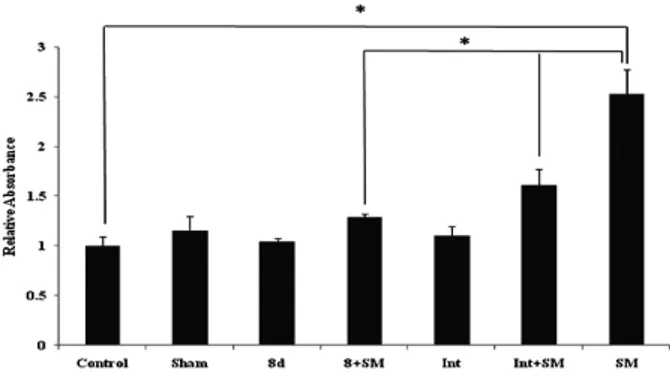
3.4. Catalase 활성
Fig. 5는 항산화 효소인 catalase의 활성을 확인한 결과이다.
Fig. 5. Relative activity of each group in catalase activity. The ordinate in figure is the relative value which means the ratio of the control value to each group. Data are means ± SD of four independent experiments for each point. *p < 0.05 compared with each group.
비흡연군인 Control군, Sham군, 8d군, Int군에서의 활성은 큰 차이가 없었으나, 흡연군인 SM군의 활성은 감소하였다 (p < 0.05). 8d + SM군과 Int + SM군은 SM군보다 활성의 감소가 적었으며 (p < 0.05), 특히 8d + SM군이 Int + SM군에 비해 활성의 감소가 훨씬 적게 나타났다 (p < 0.05). 이러한 결과는 catalase의 활성이 폐질환에 의해 감소되는 이전의 보고에서와 같은 결과를 보여 주었다 [24]. 따라서 8d 추출 물이 Int 추출물보다 항산화 효소의 활성에 더 많은 영향을 미침을 알 수 있다.
3.5. 지질과산화 측정
Fig. 6은 세포막의 지질과산화 정도를 측정한 결과이다. 흡연 을 포함한 기타 외부 자극에 의한 지질과산화의 증가는 [25]
본 실험에서도 흡연군인 SM군이 Control군보다 현저히 증가 되어 나타나는 것으로 확인되었다 (p < 0.05). 반면 8d + SM 군과 Int + SM군은 SM군에 비해 현저한 증가는 나타내지 않았으며 (p < 0.05), 특히 8d + SM군이 Int + SM군보다 지질 과산화가 적게 나타났다 (p < 0.05). 이는 8d 추출물이 Int 추출물보다 흡연으로 인한 지질과산화 작용에 대한 예방효과 및 손상으로부터 세포막을 보호한다는 것을 알 수 있다.
Fig. 6. Absorbance of each group in lipid peroxidation. The ordinate in figure is the relative value which means the ratio of the control value to each group. Data are means ± SD of four independent experiments for each point. *p < 0.05 compared with each group.
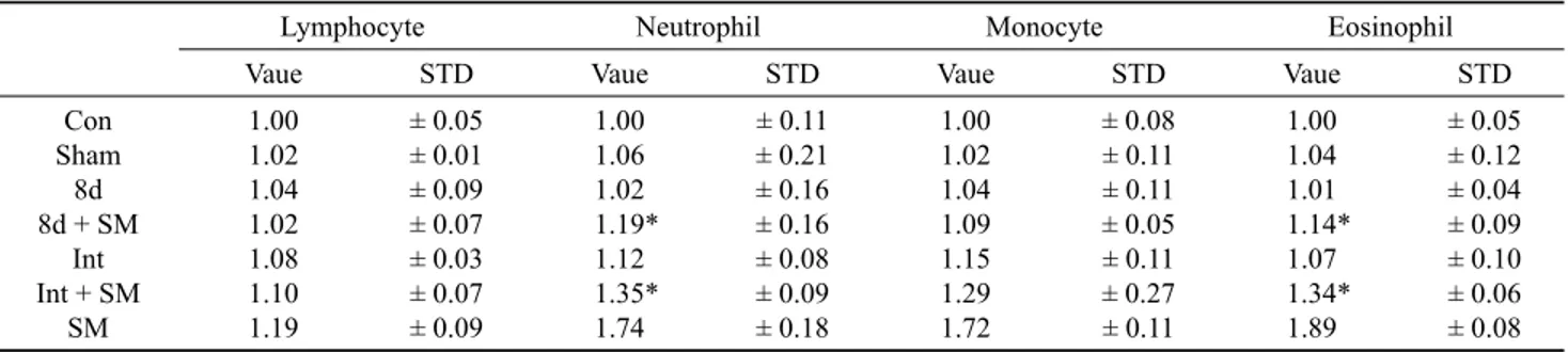
3.6. 혈액성분의 변화
생체의 방어시스템인 염증반응에서 백혈구는 중요한 역할을 한다 [26]. Table 2는 SCE가 백혈구에 미치는 영향을 보여 주는 것으로 lymphocyte, neutrophil, monocyte, eosiniphil 을 분석한 결과이다. 비흡연군인 Control군, Sham군, 8d군, Int군에서는 각 성분의 증가 변화가 없었다. 흡연군인 SM군 은 Control군에 비해 각 성분에서 확연한 증가를 나타내는 반면 (p < 0.05), 8d + SM군과 Int + SM군은 SM군보다 증가 의 폭이 적었으며 (p < 0.05), 특히 8d + SM군이 Int + SM군 에 비해 증가폭이 적음을 알 수 있었다 (p < 0.05). 이는 8d 추출물이 Int 추출물보다 흡연으로 인한 염증관련 neutrophil, eosiniphil의 증가를 억제하여 더 효과적인 항염증 작용을 함을 알 수 있다.
3.7. 단백질 활성 측정
Fig. 7은 폐 조직에서의 TNF-α, NF-κB, Iκ-Bα와 SP-A의 발
Lymphocyte Neutrophil Monocyte Eosinophil
Vaue STD Vaue STD Vaue STD Vaue STD
Con Sham
8d 8d + SM
Int Int + SM
SM
1.00 1.02 1.04 1.02 1.08 1.10 1.19
± 0.05
± 0.01
± 0.09
± 0.07
± 0.03
± 0.07
± 0.09
1.00 1.06 1.02 1.19*
1.12 1.35*
1.74
± 0.11
± 0.21
± 0.16
± 0.16
± 0.08
± 0.09
± 0.18
1.00 1.02 1.04 1.09 1.15 1.29 1.72
± 0.08
± 0.11
± 0.11
± 0.05
± 0.11
± 0.27
± 0.11
1.00 1.04 1.01 1.14*
1.07 1.34*
1.89
± 0.05
± 0.12
± 0.04
± 0.09
± 0.10
± 0.06
± 0.08 The value is the relative value which means the ratio of the control value to each group. Result present the mean ± SD of five separate experiment. Data are means ± SD of four independent experiments for each point. *p < 0.05 compared with SM group.
Table 2. The effects of cigarette smoking and starfish extracts treatment on white blood cell
현정도를 확인한 결과이다. TNF-α, NF-κB 발현이 Sham군, 8d군, 8d + SM군은 Control군과 거의 동일하게 나타났으나, Int군, Int + SM군은 약간 증가하였으며, SM군이 가장 크게 증가하였다. 염증 반응에서 TNF-α와 NF-κB의 발현증가에 대하여 [23] 8d 추출물이 Int 추출물보다 SM으로 인한 염증 작용을 완화시키는 것임을 알 수 있다. Iκ-Bα와 SP-A의 발 현이 비흡연군인 Control군, Sham군, 8d군, Int군에서 각각 동일하였으나, 흡연군인 SM군은 Control군에 비해 현저히 감소되었다. 이는 염증 발생시 Ik-Bα의 발현감소 [23]와 특 히 폐 조직의 염증 발생시 SP-A가 감소한다는 보고 [27]와 일치한다. 한편 흡연군 중 8d + CSE군과 Int + CSE군의 Iκ- Bα의 발현은 Control군과 비슷하게 나타났다. SP-A의 발현 에서 8d + SM군은 Control군보다 감소하였지만 SM군에 비해 증가되어 나타났다. 이는 SCE가 흡연으로 인한 Iκ-Bα 의 감소와 SP-A의 감소를 효과적으로 방어함을 알 수 있다.
Control Sham 8d 8d + SM Int Int + SM SM TNF-α
NF-κB
Iκ-Bα
SP-A
β-actin
Fig. 7. The expression of TNF-α, NF-κB, IkBα and SP-A protein from isolated lung tissue. Increment of cigarette smoking and starfish extracts with inhalation of seaweed extracts combination.
Sample were separated by SDS-PAGE and protein band were visualized by ECL.
3.8. 폐 조직의 변화
Fig. 8은 폐 조직을 H&E 염색한 결과이다. 비흡연군인 Control군 (a), Sham군 (b), 8d군 (c), Int군 (d)의 폐포 구조 는 배열이 규칙적이며, 폐포벽이 전반적으로 얇게 관찰되 었으나, 흡연군 중 SM (g)군은 폐포의 공간이 불규칙하며
폐포벽이 심한 유착으로 두꺼워져 나타났다. 흡연군 중 8d + SM (e)군과 Int + SM (f)군은 폐포공간이 약간 불규칙하게 커지고 폐포벽이 약간 두꺼워져 있을 뿐 다른 구조적 변화 는 관찰할 수 없었다. 이는 SCE가 성분 그 자체로는 조직과 세포내에 영향을 미치지 않으면서, 흡연으로 인한 유해물질 로부터 폐 조직을 보호하는 것에 효과적임을 알 수 있다.
(a) (b)
(c) (d)
(e) (f)
(g)
Fig. 8. The H&E stain of pulmonary structure during 4 week.
Experimental group inhaled according to each condition for 10 h.
The samples were analyzed by analysis method as described in the Materials and Methods section. Magnification is 100×.
4. 결론
본 실험은 불가사리에서 조직손상 후 8일 후 발현되는 성장
인자 [22]와 인삼의 주성분인 사포닌과 화학적 구조, 약리학 적 효능이 유사하다고 알려진 사포닌을 포함한 불가사리 추출 물 흡입이 흡연으로 인한 염증성 폐 손상 [6-8]의 방어에 어떠 한 효과가 있는지를 알아보았다.
담배 추출물과 불가사리 추출물을 IMR-90에 처리한 결과 조직손상 후 8일이 경과한 불가사리 추출물이 손상 받지 않 은 불가사리 추출물보다 세포증식의 증가 및 염증성 세포 전사인자인 TNF-α, NF-κB의 발현 억제와 Ik-Ba 분해를 억 제하는 효과가 나타났으며, 이는 불가시리의 손상 후 발현 되는 전사인자 [22]에 의하여 손상 받지 않은 불가사리 추출 물에서보다 염증의 억제와 완화효과가 향상되었음을 확인하 였다. 또한 사포닌을 포함한 천연 추출물의 흡입에 관한 이 전 실험 [15]에서와 같이 흡연 및 nebulizer를 통해 불가사 리 추출물을 실험동물에 직접 흡입시킨 결과, 체중, 항산화 효과, 지질과산화, 단백질 발현, 조직학적 변화 그리고 혈액 성분과 SP-A의 변화에서 조직손상 후 8일이 경과한 불가사 리 추출물이 손상 받지 않은 불가사리 추출물보다 염증의 억제와 완화효과가 더 좋은 것으로 나타났다. 즉, 불가사리 에 포함된 인삼과 유사한 사포닌 성분과 조직 손상 후 8일이 경과된 불가사리 추출물이 TNF-α, NF-κB의 발현 및 Iκ-Bα 의 분해를 억제하여 흡연 시 생성되는 독성물질들에 의한 폐 의 구조 변화와 손상으로부터 기능과 구조의 항상성을 유지 하는데 효과가 있음을 시사하고 있다. 특히, 조직손상 후 8일 이 경과한 불가사리 추출물이 흡연으로 발생하는 세포의 비 정상적인 형태 변화 및 폐포벽의 두꺼워짐과 폐포 공간의 불규칙적 변화, 세포막의 지질과산화를 억제하여 조직 구조 와 기능의 항상성을 유지시킴으로 염증의 진행을 억제하는 것으로 사료된다. 본 실험의 재료로 선택한 불가사리가 인삼 사포닌과 비슷한 구조와 효과를 지닌 성분을 포함하고 있으 면서도 인삼보다 가격 면에서 저렴하고 추출과정이 복잡하 지 않으며, 특히 환경오염물로서 폐기되고 있는 불가사리를 염증치유에 대한 유효한 물질로 사용함으로서 경제적 부가 가치를 높일 수 있으리라 생각된다.
References
1. Sohn, H. Y., S. K. Kim, and K. H. Kim (1985) Anatomy of the respiratory system. Tuberculosis and Respiratory Diseases 32: 1-18.
2. Song, J. S., Y. K. Kim, K. H. Kim, K. D. Han, H. S. Moon, and S. H. Park (1989) The release of superoxide (O2-) from the neutrophils in pulmonary emphysema patients associated with long-term smoking. The Korean Journal of Internal Medicin 36: 453-459.
3. Kim, K. Y. (1987) Smoking and COPD. Tuberculosis and Respiratory Diseases 34: 185-191.
4. Park, E. M., Y. M. Park, and Y. E. Gwak (1998) Oxidative damage in tissues of rats exposed to cigarette smoke. Free Radical Biology & Medicine 25: 79-86.
5. Notion, D. (2003) The Chemical Constituents in Cigarettes and Cigarette Smoke: Priorities for Harm Reduction (A Report to the New Zealand Ministry of Health) Manager. Environmental Health Effects Programme, ESR.
6. Gobran, L. I. and Rooney (2001) SA. Regulation of SP-B and SP-C secretion in rat type II cells in primary culture. Am. J.
Physiol. Lung Cell Mol. Physiol. 281: L1413-L1419.
7. Bridges, J. P., H. W. Davis, M. Damodarasamy, Y. Kuroki, G.
Howles, D. Y. Hui, and F. X. McCormack (2000) Pulmonary surfactant proteins A and D are potent endogenous inhibitors of lipid peroxidation and oxidative cellular injury. J. Biological Chemistry 275: 38848-38855.
8. Subramaniam, S. J., A. Whitsett, W. Hull, and C. G. Gairola (1996) Alteration of pulmonary surfactant proteins in rats chronically exposed to cigarette smoke. Toxicology and Applied Harmalogy 140: 274-280.
9. Jorge, P. (2001) Inflammatory pathway in atherosclerosis and acute coronary syndrome. Am. J. Cardiol. 88: 10-15.
10. Pikarsky, E., R. M. Porat, I. Stein, R. Abramovitch, S. Amit, S.
Kasem, E. Gutkovich-Pyest, S. Urieli-Shoval, E. Galun, and Y.
Ben-Neriah (2004) NF-kapppaB function as tumor promotor in inflammation-association cancer. Nature 431: 461-466.
11. Kang, J, H. (2004) Studies on the Anti-inflammatory and Anti- metastatic Properties of Soybean Saponins. Master. Thesis.
University of Ulsan, Ulsan, Korea.
12. Yeul, N. K. (2002) Clinical application and efficacy of Korea ginseng (Panax ginseng C.A.Meyer). J. Ginseng Res. 26: 111-131.
13. Shin, J. Y., E. H. Choi, and J. J. Wee (2001) The difference ginsenoside compositions according to the conditions of extraction and fractionation of crude ginseng saponins. Korean J. Food Sci. Technol. 33: 282-287.
14. Lee, D. W., H. B. Sohn, H. B. Lim, and Y. G. Lee (1995) Antioxidant action of ginseng: an hypothesis. Korea J. Ginseng Sci. 19: 31-38.
15. Jung, H., S. K. Kim, S. K. Kim, M. Y. Seong, H. J. Kim, J. F.
Xeu, and Y. Y. Kim (2004) The effects of extracts from ginseng, wormwood and pine needle in pulmonary structure and anti- oxidant enzyme in smoking. Korean J. Biotechnol. Bioeng.
19: 138-142.
16. Hideki, K., H. Kenji, and A. Seiichi (2006) Characteristics of carboxypeptidase B from pyloric ceca of the starfish asterina pectinifera. Food Chemistr. 95: 264-269.
17. Hideki, K. and H. Kenji (2002) Isolation and characteristics of trypsin from pyloric ceca of the starfish. Asterina Pectinifera Bio. and Phy. 132: 485-490.
18. Hideki, K. and A. Seiichi (2006) Characteristics of phospholipase A2 mutant of the starfish asterina pectinifera. Enzyme and Microbial Technology 40: 461-465.
19. Böttner, M., K. Krieglstein, and K. Unsicker (2000) The transforming growth factor-betas: structure, signaling and roles in nervous system development and functions. J. Neurochem.
75: 2227-2240.
20. Dunker, N. and K. Krieglstein (2000) Targeted mutations of transforming growth factor-beta genes reveal important roles in mouse development and adult homeostasis. Eur. J. Bioche.
267: 6982-6988.
21. Linkhart, T. A., S. Mohan, and D. J. Baylink (1996) Growth factors for bone growth and repair: IGF, TGF-, and BMP.
Bone. 19: 1S-12S.
22. Sung, M. Y. (2003) Effect of Injured Starfish Extract on Growth Factors and Cell Proliferation. Master. Thesis. Kyungpook National University, Daegu, Korea.
23. Jobin, C. and R. B. Sartor (2000) The IkB/NF-kB system: a key determinant of mucosal inflammation and protection. Am. J.
Physiol. Cell Physiol. 278: C451-C462.
24. Odajima, N., T. Betsuyaku, K. Nagai, C. Moriyama, D. H. Wang, T. Takigawa, K. Ogino, and M. Nishimura (2010) The role of catalase in pulmonary fibrosis. Respiratory Research 11: 183-196.
25. Negre-Salvayre, A., C. Coatrieux, C. Ingueneau, and R. Salvayre (2008) Advanced lipid peroxidation end products in oxidative damage to proteins. Potential role in diseases and therapeutic prospects for the inhibitors. British Journal of Pharmacology 153: 6-20.
26. Yang, Y. J., Y. C. Hong, S. Y. Oh, M. S. Park, H. Kim, J. H. Lee, and E. H. Ha (2009) Bisphenol A exposure is associated with oxidative stress and inflammation in postmenopausal women.
Environmental Research 109: 797-801.
27. Walker, M. G., J. M. Tessolini, L. McCaig, L. J. Yao, J. F. Lewis, and R. A. Veldhuizen (2009) Elevated endogenous surfactant reduces inflammation in an acute lung injury model. Experimental Lung Research 35: 591-604.