INTRODUCTION
In head and neck, AVM(ateriovenous malformation) rarely occurred. Specially intraosseous AVM of jaw is rare and sometimes mortal because of frequent bleeding tendency. Children are mostly commonly involved, if the movable tooth involved the lesion extracted, it cause massive bleeding. Intraosseous malformations have risk from life-threatening bleeding
1,3).
We report a case of unilateral mandibular AVM in 14- year-old boy treated successfully by combined tech- niques.
CASE REPORT
In March 2005, a 14-year-old boy was referred for inter- mittent bleeding. There was no historic trauma. He had facial asymmetry by left mandibular swelling and facial skin color was pale. The bluish ecchymosis was on left buccal skin. Bruit could be audible by stethoscope over the area, and pulsations were palpable. The consistent oozing point was distal of left mandibular second lower
molar. The patient was smaller than normal, had light 30Kg weight. This could be presumed concerning of AVM.
Panorama and mandible PA view showed lytic expan- sion, septum, coarse trabeculae, root resorption, soap- bubble radiolucency inner lesion. Mandible bony cortex was continuity(Fig. 1).
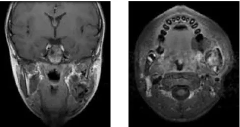
In MRI view, high flow vascular mass existed from left mandibular angle to ramus, included buccal space and masseter muscle. The boundary with pterygoid muscle was not uncertain. The size of lesion was 40×35×45 mm(Fig. 2).
At first day, first angiography was done. By Seldinger method, right femoral artery was punctured, using guide wire, right common carotid artery was selected and approached to Left external carotid artery. High flow vascular mass was inner left mandible. Bone remodeling and some destruction was showed in bony cortex(Fig. 3).
Hypervascular mass supplied by facial artery and max- illary artery was observed on left mandibular angle area.
Facial artery branched to mental artery, submental artery and so on. The diameter of multiple branching artery was lager and showed tortuousity.
The lesion had large nidus and aneurysmal sac. In arte- rial phase, the lesion was early venous shunted to inter- nal jugular vein. Intraoral bleeding was showed at molar area(Fig. 4).
* Corresponding author Mi-Sun Jung
Dept. of OMFS, Kyunghee University Medical center
# 1 Hoegi-dong Dongdaemun-gu, Seoul, 131-702, Korea Tel: 82-2-958-9441 Fax: 82-2-966-4572
E-mail: aiyak@naver.com
A treatment of atreiovenous malformation on mandible
Mi-Sun Jung, Dong-Mok Ryu, Eui-Jong Kim*, Jeong-Hwan Oh
Department of Oral & Maxillofacial Surgery, School of Dentistry, Kyung-Hee University, Korea
*Department of Interventional Radiology, School of Medicine, Kyung-Hee University, Korea
The treatment of intraosseous ateriovenous malformation in the jaw is difficult because of life threatening frequent bleeding tendency.
The surgical resection of AVM may be mortal due to massive blood loss .In the growing pediatric patient, surgery may cause facial deformity and growth disturbance. So currently, the treatment of AVM is only embolization using various material through endovascu- lar access, direct-puncture or embolization in conjunction with surgical resection. We report a case of combined techniques.
Key words
AVM, Ateriovenous malformation, Embolization Abstract
One month later, repeated angiogram and emoboliza- tions was done by trans arterial approach. Angiogram showed AV(arterio-venous) shunting and flow decreased, but still contrast filled vascular mass and
aneurysmal sac was observed. The branches of internal maxillary artery; Inferior alveolar artery, buccal artery, lesser descending palatine artery was dilated(Fig. 5). For one month, many bleeding tendency was occurred, so embolization was decided.
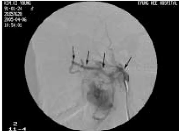
Using microcatheter and microguide wire, above three arterial branches was by turns superselected and con- firmed as feeder by angiography. Embolization was done by contour(250-350 micron)(Fig. 6).
After each artery was embolized, blood filling was sel- dom occurred from this artery. Flow was lower than before, at the same time lower AV shunting and under- flow of contrast media was showed. However still blood filling in vascular mass by unknown small branches was showed(Fig. 7).
Fig. 2. Boderline with soft tissue.
Fig. 1. Lytic expansion in left mandible.
After first embolization, the amount of intraoral bleed- ing was decreased extremely, however intermittent bleeding was sometimes occurred for an week.
We decided to perform repeated embolization by trans- venous approach and transarterial approach.
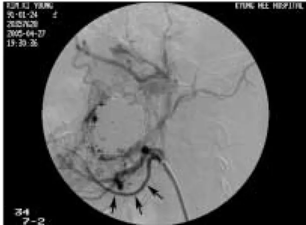
Inflow from internal maxillary artery into aneurysmal sac was decreased remarkably. However still feeding from jugal branch of facial artery and small branches of internal maxillary artery, small AV shunting was noted(Fig. 8).
Because of drain from aneurysmal sac to internal jugu- lar vein, we decided to approach to internal jugular vein and obliterate aneurysmal sac, in which area to intercept flow to internal jugular vein. By this transvenous approach embolization method was selected and guiding
catheter was set into internal jugular vein. Using micro- catheter and microguide wire we approached venous drainage route, but failed for proceeding of jugular bulb, fast flow and formation of venous drain route.
Therefore using microcatheter two jugal branches;
feeder branch of facial artery was embolized by con- tour(Fig. 9).
After performing of transarterial embolization, in angiography, almost flow of large feeder was decreased, too small to be superselected multiple branch filled, instead of many decreasing AV shunting was slightly existed.
Two weeks later, intraoral bleeding was decreased much, but for interval sometimes small intraoral bleed- ing was occurred.
Fig. 5. Redilated internal maxillary artery. Fig. 6. Superselection and embolization with micro- catheter and microguide.
Fig. 7. After embolization reduced filling nidus. Fig. 8. Redilated facial artery.
In angiography, flow from feeder vessel was dropped greatly, still contrast media was fed by small branched and AV shunting was noted.
For impossible to approach by arterial route, percuta- neous approach was selected.
Under ultrasound induce, puncture was tried to near most thin cortical bone; angle area, needle tip was locat- ed in aneurysmal sac. In angiography, from large aneurysmal sac through variated vein fast filling was noted to internal jugular vein.
To prevent to embolic material move for internal jugu- lar vein, inside aneurysmal sac using nine J shaped detachable coil, mesh was made, through Percutaneous route 40 variant size of microcoil was inserted and sac was filled out (Fig. 12~14). Inside sac, there was no
vacant space but still slow and mild AV shunting was exist. Therefore using 20% Glue(mixed with lipiodol) 2 vial, aneurysmal sac was injected(Fig. 11).
Patient had not other abnormal symptom during pro- cedure.
In July 2005, follow-up angiography was performed.
After last embolization during 2 months there was no problem, but recently intraoral bleeding was occurred.
The branch of internal maxillary artery; inferior alveo- lar artery, buccal artery, lesser descending palatine artery, was re-dilated again. Facial artery branches were also redilated a little. Before aneurysmal sac was almost blocked, however in posterior portion filling of contrast media was noted. Entirely comparing last embolization AV shunting was more larger(Fig. 15, 16).
Fig. 11. Insert niddle tip into nidus Fig. 12.Insert microcoil by percutaneous route.
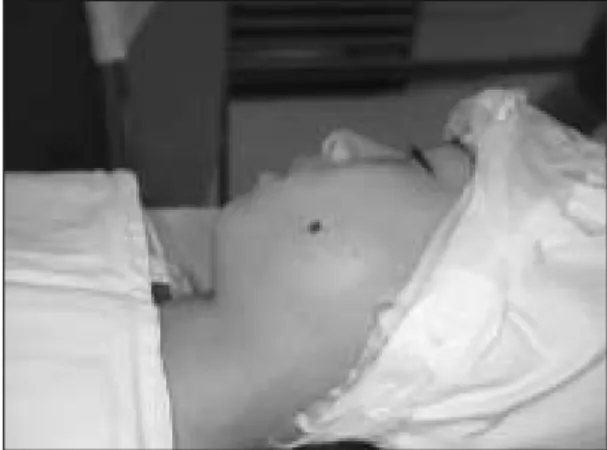
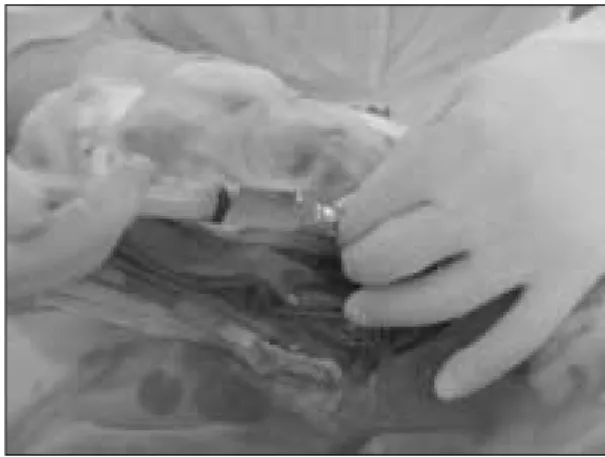
Fig. 9. Embolization with contour. Fig. 10. Marking on puncture site.
Above three branch arteries was superselected by turns, in angiography, feeder was embolized by contour (250-350 micron) therefore that supply was almost
blocked was confirmed.
By dental CT, successful empolization was affirmed (Fig. 18).
Fig. 13. Blocked lesion by microcoil. No more feeding internal maxillary artery.
Fig. 14.No more feeding facial artery.
Fig. 15.Slightly dilated facial artery. Fig. 16.Slightly dilated internal maxillary artery.
Fig. 17.Well-located microcoil in lesion. Fig. 18.Dental CT view.
DISCUSSION
Angiography most be used to diagnose this AVM;
especially feeding vessels, collateral circulation, multiple anastomoses.
The varix is a pool of blood with high pressure sup- plied by many arterial branches. Feeding artery goes directly into the nidus(enuerismal sac) of the lesion. The varix causes intraosseous lytic changes, resorption of tooth roots, and mobile teeth. Mandibular AVMs often drain to the inferior alveolar venous system. The approach to the varix of intraosseous AVM via direct puncture is one of accessible treatment.
The treatment of AVM is two method. One is surgical method; partial resection of the mandible, ligation of the
feeding artery. In 1970’s surgical resection was preferred but this could not cause cosmetic result to child. Ligation of feeding artery is too difficult to control bleeding suc- cessfully. If feeder would be many, this is not effected and after ligation many new feeder could grow into.
From 1980’s transaterial embolization was started, how- ever could not fill nidus enough independently, in high flow case catheter’s accessibility was not good
3). Recently direct injection independently or mixed method with endovascular embolism is good resulted from many lit- erature. However especially to mix with surgical method, after embolization must be operated within 8 days, because of ability to control bleeding
6).
Direct puncture embolization can be peformed in most instances. Direct percutaneous puncture in conjunction
Fig. 19.Microcoil insertion. Fig. 20.Glue injection.
with endovascular therapy is effective and safe
5,6).
In addition the treatment of AVM is considered size, evaluated flow characterics.
High-flow AVM is aterial or ateriovenous type and low-flow AVM is capillary, venous, lymphatic.
Microcatheter location be able to control high blood flow
3). All about this considered, when embolizations being done, proper embolic material is important.
The embolic material be selected for high thrombo- genicity, permanence of vascular blockade. Different emboli were used, gelfoam soaked in thrombotic agent (Avitene), balloon, fiber coils, polyvinyl alcohol polymer
= PVA = large sized particle= Contour(250~350 micron), platinum coil, microcoil, glue (N-butyl-2-cyanoacrylate)
= NBCA, Ethibloc
1,4,6).
Mandible is much denser and thicker than that of the maxilla, so blood pressure in the varix of mandible is higher. More widely diffused involvement in the mandible, difficulty in controlling emergency bleeding in the AVM of the mandible by local pressure
4). If the AV shunting existed, embolic material could passed through the int. jugular vein, resulting in pulmonary embolism
1,7).
So that direct-puncture embolization using microcoil accompanied with endovascular embolization is more effective.
CONCLUSIONS
In our case, patient was growing up and had big nidus with high flow and early AV shunting. In the manage- ment of such patient, catastrophic bleeding is the most severe problem, and this can be achieved by mixed tech- niques.
Transarterial embolization and percutaneous approach
was more successful to blockage nidus and to prevent feeders growing into nidus.
Undoubtedly the boy is growing up, and recurrence rate is high in high-flow type
3). Therefore follow-up check required for a long time.
REFERENCES
1. Kaneko R, Tohnai I, Ueda M, Negoro M, Yoshida J, Yamada Y: Curative treatment of central hemangioma in the mandible by direct puncture and emblisation with n- butyl-cyanoacrylate (NBCA). Oral Oncology 2001;37:605- 608.
2. Yoshiga K, Tanimoto K, Okui T, Kobayashi M: High-flow arteriovenous malformation of the mandible: treatment and 7-year follow-up. British J Oral Maxillofac Surg 2003;
41:348-350.
3. Giaoui L, Princ G, Chiras J, Guilbert F, Bertrand JC:
Treatment of vascular malformations of the mandible: a de- scription of 12 cases. Int J Oral Maxillofac Surg 2003;32:132- 136.
4. Siu WWY, Weill A, Gariepy JL, Moret J, Marotta T: Arterio- venous Malformation of the Mandible: Embolization and Direct Injection Therapy. J Vasc Interv Radiol 2001;
12:1095-1098.
5. Fan X, Zhang Z, Zhang C, Tang Y, Hu Y, Mao Q, etal:
Direct-Puncture Embolization of Intaosseous Arterio- venous Malformation of Jaws. J Oral Maxillofac Surg 2002;
60:890-896.
6. Corsten L, Bashir Q, Thornton J, Aletich V: Treatment of a Giant Mandibular Arteriovenous Malformation With Percutaneous Embolization Using Histoacrylic Glue: A Case Report. J Oral Maxillofac Surg 2001;59:828-832.
7. Persky MS, Yoo HJ, Berenstein A: Management of Vascular Malformations of the Mandible and Maxilla. Laryngoscope 2003;113:1885-1892.
8. Ziyeah S, Schumacher M, Strecker R, Ro¨ssler J, Hochmuth A, Klisch J: Head and neck vascular malfomations: time-re- solved MR projection angiography. Neuroradiology 2003;
45:681-686.
9. Wong IYC, Batista LL, Alvarez H, Lasjaunias PL:
Craniofacial arteriovenous metameric syndrome (CAMS) 3-a transitional pattern between CAM 1 and 2 and spinal arteriovenous metameric syndromes. Neuroradiology 2003;45:611-615.