Abstract (J. Kor. Oral Maxillofac. Surg. 2004;30:323-330)
Ⅰ. 서 론
세포사멸(Apoptosis)은 조직의 생성과 파괴에 기여하는 기전의 하나로 DNA fragmentation을 통하여 궁극적으로 세포의 파괴를 야기하는 다양한 경로를 거치게 된다. 세포사멸 기전이 시작되 는 신호는 TNFα에 의하는데 이는 TNFα수용체를 통하게 된다.
이밖에도 CD95와 Fas, APO1, APO2, TRAIL-R 그리고 APO3/LARD 등의 수용체 등이 작용하는 것으로 알려져 있으며 이런 신호들 은 caspase 등의 단백분해효소와 작용하는 FADD/MORT1 혹은 TRADD 등의 단백질과 관계 있는 다양한 경로를 통해 매개되는 것으로 알려지고 있다. Bcl 2, Bax, Bad 등, caspase를 활성화 시키
는 여러 가지 조절 인자들이 밝혀졌는데 궁극적으로 caspase는 세포사멸을 유발하는 다양한 핵산 분해효소를 활성화시키는 것 으로 알려져 있다
1).
세포사멸을 조절하는 여러 유전자뿐만 아니라 몇 가지 hsp(heat shock protein)도 역시 세포사멸을 방지하는 역할을 하는 것으로 여겨지고 있는데 몇 가지 실험 모델에서 hsp70과 hsp27에 의한 세포사멸 억제가 보고되었다
2,3). 또한 미리 열처리(heat shock)를 시행한 경우에서 역시 hsp의 발현과 이에 따른 세포사 멸의 억제를 보이는 결과가 보고되었으나
4), 흥미롭게도 유사한 실험 모델에서 정반대로 열처리에 의한 세포사멸의 촉진도 보고 되고 있다
5,6). 위의 보고들을 통하여 볼 때 세포에 따른 특이성 혹 은 hsp 특이성에 의하여 세포사멸의 신호화를 촉진, 혹은 억제하 는 것으로 생각된다.
조골세포(osteoblast)의 증식이나 세포사멸 기전에 의해 성장과 발육과정에서 이 세포들의 수가 조절되는데 골조직에서 골세포 (ostecoyte)와 조골세포가 세포사멸 과정을 거쳐 제거된다
7). 골세 포의 세포사멸은 골병변이 발생하였을 때 증가되는 것이 관찰되
는데
8-10), 골 대사조절(bone adaptation) 과정에 관련된 세포들의
조골세포 세포사멸의 Estrogen 조절에 대한 Hsp27의 영향에 관한 연구
장현석∙윤정주∙임재석∙권종진∙최철민 고려대학교 의과대학 치과학교실 구강악안면외과
장 현 석
425-020 경기도 안산시 고잔동 516번지 고려대학교 안산병원 구강악안면외과 Hyonseok Jang
Dept. of OMFS, College of Medicine, Korea University 516, Gojan-dong, Ansan, Kyunggi, 425-707, Korea Tel : 82-31-412-5370 Fax : 82-31-485-5373 E-mail : [email protected]
HSP27 CONTRIBUTES TO ESTROGEN REGULATION OF OSTEOBLAST APOPTOSIS
Hyonseok Jang, Jungju Eune, Jaesuk Rim, Jongjin Kwon, Cheolmin Choi
Dept. of Oral & Maxillofacial Surgery College of Medicine, Korea University,
Estrogen may promote osteoblast/osteocyte viability by limiting apoptotic cell death. We hypothesize that hsp27 is an estrogen- regu- lated protein that can promote osteoblast viability by increasing osteoblast resistance to apoptosis. The purpose of this study was to determine the effect of estrogen treatment and heat shock on TNFα- induced apoptosis in the MC3T3-E1 cell line. Cells were treated with 0 - 100 nM 17βestradiol (or ICI 182780) for 0 - 24 hours before heat shock. After recovery, apoptosis was induced by treatment with 0 - 10 ng/ml TNFα. Hsp levels were evaluated by Northern and Western analysis using hsp27, hsp47, hsp70c and hsp70i - specific reagents. Apoptosis was revealed by in situ labeling with Terminal Deoxyribonucleotide Transferase (TUNEL). A 5 - fold increase in hsp27 protein and mRNA was noted after 5 hours of treatment with 10 - 20 nM 17βestradiol prior to heat shock. Increased abundance of hsp47, hsp70c or hsp70i was not observed. TUNEL indicated that estrogen treatment also reduced (50%) MC3T3-E1 cell susceptibility to TNFα- induced apoptosis. Treatment with hsp27-specific antisense oligonucleotides prevented hsp27 protein expression and abol- ished the protective effects of heat shock and estrogen treatment on TNFα- induced apoptosis. Hsp27 is a determinant of osteoblast apoptosis, and estrogen treatment increases hsp27 levels in cultured osteoblastic cells. Hsp27 contributes to the control of osteoblast apoptosis and may be manipulated by estrogenic or alternative pathways for the improvement of bone mass.
Key words: Heat Shock, Bone, Oligonucleotide, TNF-α, TUNEL
작용에 의한 것으로 생각되고 있다
10). 현재까지 조골세포와 골세 포에서 이런 기전이 명확히 규명되지는 않았지만 MC3T3-E1 세 포주를 이용한 연구에서 NF-κB의 활성화에 의한 세포사멸 기전 이 보고 되어있다
11).
조골세포와 골세포의 세포사멸 과정에서의 estrogen의 작용은 많은 관심을 유발하고 있는데 이는 조골세포의 활성에 영향을 미치는 생리적, 혹은 병적상태의 골조직에서 estrogen에 의해 골 량이 변화될 수 있기 때문인 것으로 생각된다. 실제로 생활력 있 는 조골세포의 수가 골량을 나타내는 중요한 지표로 사용되고 있다. 이와 같이 estrogen은 세포사멸 작용에 관계되어 골내의 조 골세포와 골세포의 수를 조절할 수 있는 것으로 생각되는데 난 소절제 실험동물의 골수에서 세포사멸 과정의 골세포가 다수 관 찰된다
8). 실제 사람에서도 약물과 연관되어 estrogen이 저하된 경 우에 골세포의 세포사멸이 다량 관찰되는데
12)이와 같은 estrogen 에 의한 세포사멸 조절은 다양한 기전을 통하여 이루어지는 것 으로 생각되는데 hsp27이 조골세포의 세포사멸 조절에 작용하는 estrogen에 관련되는 단백질임이 밝혀짐에 따라
13-15)본 연구에서 는 성장중인 백서 경골부에서 면역조직화학 분석을 통하여 hsp27의 발현을 확인하여 보고 MC3T3-E1 세포주를 이용하여 estrogen에 의한 세포사멸 조절에 미치는 hsp27의 작용기전을 규 명하고자 하였다.
Ⅱ. 연구 재료 및 방법
1. 면역조직 화학분석: 200g의 성장중인 Fisher 344 백서 경골부 에서 hsp27의 발현 여부를 확인하기 위하여 면역조직 화학분석 을 시행하였다. 백서 경골부를 채취하여 10% formalin에서 2시간 동안 고정한 후 100 mM phosphate buffer (pH 7.3)에 보관하였다.
10% EDTA를 이용하여 탈회하고 5 μm 파라핀 포매 절편을 제작 하여 toluidine blue (pH 5.5)를 이용하여 각각의 절편을 염색하여 연조직, 연골부 및 골조직부를 확인하였고 normal goat serum으 로 blocking하고 세척한 후 affinity purified anti-hsp27 antibody, nor- mal rabbit serum 혹은 PBS로 1-24 시간 동안 부란하였다. 세척 후 모든 표본을 1:250의 horseradish peroxidase - conjugated goat anti - rabbit antibody (Vector)으로 부란하였고 AEC chromagen (Zymed, South San Francisco, CA)을 이용하여 관찰하였다.
2. 세포배양: 10% FCS와 penicillin/streptomycin 등의 항생제를 포함한 αMEM을 사용하여 MC3T3 - E1 세포를 37℃, 5% CO
2의 조 건에서 배양 하였다
16). 먼저 10% charcoal stripped serum (Hyclone) 과 항생제를 포함한 phenol red free media에서 24시간 동안 계대 배양을 시행하였으며 에탄올에 포함된 10mM stock solution을 이 용하여 배양액에 0 - 100 nM의 농도로 희석하여 17 βestradiol(βE
2, Sigma)을 0-24 시간 동안 투여하였다. 배양용기를 42.5℃ incubator 에서 2시간 동안 열처리(heat shock)를 시행 하였으며 열처리 시 행 후 3시간 동안 세포의 회복기를 준 후 RNA와 protein을 추출하 였다. TNFα를 투여하여 세포사멸을 유도하였는데 0 - 2.0 ng / ml 의 TNFα를 대조군 배지와 열처리 배지에 각각 투여하였다.
3. HSP 발현의 측정: Hsp의 발현을 열처리와 βE
2의 농도 및 TNFα투여에 따라 측정하였는데 총 단백을 RIPA buffer를 이용하 여 추출하여 micro BCA assay를 이용하여 양을 측정하였다. 각 10 μg의 단백질을 10% SDS - PAGE을 이용하여 전기영동을 시행 하였으며 methanol - based buffer를 이용하여 니트로셀루로스 fil- ter에 이전하였다
17). Immunoblotting은 정제된 rabbit anti-hsp27 antibody
18)와 hsp47, hsp70 cognate, hsp70 inducible, hsp90 (StressGen: Vancouver, BC)에 대한 monoclonal antibody를 이용하 여 Blotto를 사용하였으며 Horseradish - peroxidase conjugated sec- ondary antibody (Vector; Burklingame, CA)을 이용하여 immuno- complex(ECL, Amersham, Arlington Heights, IL)의 chemiluminescent dectection을 시행 하였다. 배양세포로 부터 acid phenol 추출법을 이용하여 RNA를 추출하였는데 cold PBS로 3회 세척한 후 guani- dine isothyocyanate buffer에 녹여 시행하였다
19). RNA의 농도를 spectrophotometry를 이용하여 OD 260에서 측정하였다. 10 μg의 RNA를 1.0% agarose formaldehyde denaturing gel에서 전기영동을 시행하였으며 이 gel을 Whatman’s paper에서 건조 시킨 뒤 DEPC H
2O를 이용하여 rehydration 시켰다. Prehydration 후 gel을 hsp27, hsp68과 hsp86의 [
32P]-αdCTP random primer labeled mouse cDNA probe를 이용하여 표지 하였고, 65℃에서 1× SSC를 이용하여 세 척하였으며, 자가방사선 사진을 촬영하여 Scionics PC image analysis software (Scionics PC, NIH, Bethesda, MD)를 이용하여 측 정하였다.
4. Antisense oligonucleotide 처리: Murine hsp27 cDNA의 첫 36bp 에 대한 S - oligos
20)(5′-CGCGGCGCTCGGTCATGT and 5′- GCAGCAGCGAGAAGGGCA)를 합성하였다. 80% 정도 confluent한 상태에서 세포를 PBS로 세 차례 세척하고 0 - 20 nM 농도의 oligonucleotide를 포함한 αMEM을 배양액에 투여하였다. 2시간 경과 후 10% 양의 serum을 투여한 후 밤새 부란하였다. 그 후 세 포를 estrogen과 vehicle 처리하였고 열처리를 시행하였다. 세포 사멸을 유도하기 위해 TNFα를 투여하였다.
5. 세포 생활력의 측정: TNFα처리 후와 hsp 발현 후의 생활력을 유지하는 세포의 수를 trypan blue를 이용하여 직접 계산하였다.
각각의 처리 군에서 5회씩 측정하였으며 평균값을 구하였다.
6. DNA fragmentation의 분석: 세포사멸을 분석하기 위해 DNA
fragmentation을 이용하였는데 biotinylated nucleotides의 terminal
deoxyribonucleotide transferase incorporation(TUNEL, Trevigen,
Rockville MD)을 사용하여 fragmented DNA를 측정하였다. 세포들
을 charcoal stripped serum를 포함한 phenol - red free media를 이
용하여 4 well glass 배양slide를 사용하여 estrogen, vehicle 및 열처
리를 시행한 후 8-16 시간 동안 0 - 2.0 ng/ml TNFα또는 0 - 600 nM
staurosporine로 처리하였다. Coverslips을 cold PBS로 세척한 뒤 고
정하고 fragmented DNA의 enzymatic labeling을 시행하였다. Well
당 전체 세포 수와 양성 반응을 보이는 세포의 수를 직접 계산하
였으며 세포사멸을 보이는 세포의 비율을 계산하였다(n = 5).
Ⅲ. 연구결과
1. 골 주위세포(bone lining cell)와 조골세포의 hsp27의 발현 백서 경골부의 면역조직화학 검사 소견상 골을 형성하는 부위 의 골 주위세포와 조골세포 부위에서 hsp27의 발현을 관찰할 수 있었고 골세포 부위에서는 anti - hsp27 항체의 염색 소견을 관찰 할 수 없었다(Fig. 1).
2. MC3T3-E1 세포에서 hsp27, hsp68, hsp86 mRNA의 발현 Hsp27, hsp68과 hsp86의 [
32P]-αdCTP random primer labeled mouse cDNA probe를 이용하여 hsp27, hsp68과 hsp86 mRNA의 발 현을 관찰하였다. 대조군(C)에서는 hsp의 발현이 관찰되지 않았 지만 열처리를 시행한 경우(HS) hsp27, hsp68과 hsp86 mRNA의 발현이 관찰 되었다(Fig. 2).
3. MC3T3-E1 세포에서 estrogen에 의한 hsp27의 발현 조절 열처리 전 MC3T3-E1 세포에 5시간 동안 0, 2, 20, 100 nM 17β estradiol(βE
2)를 투여하고 42.5℃에서 2시간 동안 열처리를 가하였 을 때 hsp27의 발현이 증가되었다(Fig. 3).
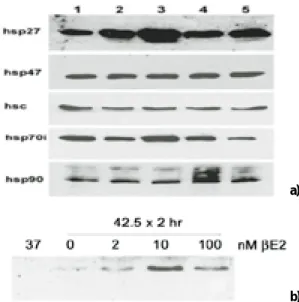
4. Estrogen 투여에 의한 hsp27 및 기타 hsp의 발현
MC3T3-E1 세포에 20 nM의 βE
2을 0(lane 1), 2(lane 2), 5(lane 3), 8(lane 4), 24 시간(lane 5) 동안 투여한 후 42.5℃에서 2시간 동안 열처리를 시행하고 3시간 동안 회복기를 준 뒤 hsp27, hsp47, hsc (heat shock protein 70 cognate), hsp70i (inducible hsp70)와 hsp90의 발현이 관찰되었고(Fig. 4a), 열처리 전 5시간 동안 2 nM 이상의 농도를 투여 하였을 때 열처리에 의한 hsp27의 증가가 뚜렷하였 으며 열처리를 시행하지 않은 경우(37℃) hsp27의 발현이 관찰되 지 않았다(Fig. 4b).
5. 열처리된 MC3T3-E1 세포에서 hsp27 발현에 대한 estrogen antagonist의 영향
세포 배양액에 0 - 100 nM 농도의 ICI182780를 투여하였을 때 20 nM βE
2에 의한 hsp27의 증가가 관찰되었으나 taxmoxifen의 고 농도 투여 시에는 이의 증가가 관찰되지 않았다(Fig. 5).
6. TNF α 나 staurosporine 처리가 MC3T3-E1 세포의 사멸에 미치는 영향 세포배양에서 MC3T3-E1 세포의 사멸을 유도할 수 있는데(Fig.
5), 고농도의 TNFα나 staurosporine 투여 시 잔존 부착 세포의 수 가 현저히 감소되는 것을 관찰할 수 있었으나(Fig. 6a), estrogen 처치는 TNFα나 staurosporine에 의한 세포사멸을 감소시키는 것 이 관찰되었다(Fig. 6b).
7. Hsp27의 발현 억제가 열처리 후 MC3T3-E1 세포의 생활력에 미치는 영향
열처리 전 MC3T3-E1 세포에 대한 antisense S-oligonucleotide처 치로 hsp27의 발현이 현저히 감소 되었으며(Fig. 7a), TNFα처리에 의한 MC3T3-E1 세포의 생활력 감소도 관찰 되었다(Fig. 7b).
Ⅳ. 총괄 및 고찰
본 연구의 MC3T3-E1을 이용한 실험 결과는 백서 두개부의 장 기배양(organ culture)과 SMER 14 세포에서 estrogen 처치에 의한 골 조직과 조골세포에서 hsp27의 발현이 증가 되는 결과와 일치 하였다
14). Estrogen은 단독으로 유방암 세포주에서 hsp27의 발현 을 증가 시키는 것으로 알려져 있으나, 대조적으로 본 연구에서 보면 MC3T3-E1 세포주에서 열처리와 무관한 hsp27의 발현 증가 는 관찰 되지 않았다.
Estrogen antagonist인 ICI 182,780 투여는 estrogen에 의한 열처리 hsp27의 증가를 억제하지는 못 했는데 고농도(100배), 장시간 처 치(4시간)에서도 ICI 182,780는 사멸세포 수의 현저한 증가가 관 찰 되어 본 연구의 이 antagonist 투여에 의한 세포사멸 결과 분석 에 많은 혼란을 가져왔지만 ovarian cancer cell을 이용한 이전의 연구 결과와 일치하였다
22). Tamoxifen은 strain과 연관된 골의 remodeling을 포함한 골 조직에 대한 estrogen의 효과에 반대되는 기전을 보이는데
23), 본 연구에서는 tamoxifen의 투여가 estrogen에 의한 hsp27의 발현 증가를 억제하는 결과를 나타내지만 현재까 지 tamoxifen이 estrogen에 의한 세포사멸의 억제작용을 감소시 킬 수 있는지는 명확하지 않은 것으로 알려지고 있다.
현재까지의 연구 결과는 MC3T3-E1 세포에서 어떤 기전에 의하 여 estrogen이 hsp27의 발현을 증가시키는지는 밝혀지지 않고 있 다. 조골세포 세포주에서 hsp27의 promoter는 estrogen 감수성이 있는 것으로 알려지고 있는데, 20 nM βE
2를 이용하여 5시간 동안 전처치를 시행하였을 때 SMER14 세포에 chimeric reporter con- structs를 transfection 하였을 때 전사의 증가가 관찰되지만 parental ROS17/2.8 세포에서는 증가를 나타내지 않는 것으로 알 려지고 있다. 24 Promotor 상에서 estrogen response element (ERE) 의 절반은 인접한 SP1 부위와 같이 작용하여 estrogen에 의한 전 사조절에 영향을 미치며 또한 여기에 핵기질 단백인 HET1이 작 용하여 전사를 조절한다
26). Hsp27의 발현에 미치는 비유전적인 영향은 특별히 밝혀진 바는 없지만 hsp27의 인산화(phosphoryla- tion)는 hsp27의 기능에 필수적이므로 여기에 미치는 estrogen의 효과에 대한 규명이 필요하리라 생각된다
27).
Hsp27은 caspase 9의 활성화를 억제함으로써 세포사멸 작용을 조절하는 것으로 알려져 있다
3). Estrogen 결핍으로 인한 골세포의 세포사멸이 증가된다는 보고가 있으나
8)골세포에서 hsp27의 발 현이 조골세포나 골 주위세포에 비해 미약하거나 거의 없는 것 으로 보아 골 형성 세포의 생성과 생활력의 유지에 조골세포내 의 hsp27의 유무가 커다란 영향을 미치는 것으로 생각된다.
MC3T3-E1 세포주에서 estrogen의 투여는 후의 TNFα나 stau-
rosporine의 투여에도 불구하고 열처리 세포의 세포 생활력을 증
가시키는 것이 관찰되었는데 다른 hsp에 대한 βE
2의 효과가 없는
것으로 보아 이는 단지 hsp27에 의한 것으로 여겨진다. 또한 열
처리를 시행하지 않은 MC3T3-E1 세포에서, 즉 hsp27이 없는 경우
에는 estrogen의 투여가 세포의 생활력에 영향을 미치지 않은 결
과도 이를 뒷받침하는 것으로 생각된다. 본 연구의 결과에서
MC3T3-E1 세포에 대한 열처리 시행 후 hsp27 뿐만 아니라 hsp68,
hsp 47, hsc, hsp 70i, hsp 90 및 hsp86 mRNA의 발현이 증가되어 있 어 이에 대한 추후 연구가 필요하리라 사료된다.
Estrogen에 의한 세포사멸 조절은 여러 가지 기전을 생각해볼 수 있는데 몇 가지 종류의 세포에서 estrogen 투여가 TNFα의 발 현을 감소시키는 것으로 알려졌으며
28), 다른 기전으로는 estrogen 에 의한 Nitric Oxide(NO) 조절을 들 수 있다. NO는 cytokine의 조 절에 중요한 역할을 하며 또한 배양 조골세포의 mechanical strain 효과에 영향을 미치는 것으로 알려지고 있으며, estrogen의 결핍 은 Nitric Oxide Synthase의 발현을 감소시켜 세포사멸의 유도에 작용하는 것으로 보고되고 있다
29). Estrogen 투여에 의한 혈액 내 NO의 증가와
30)NO에 의한 PC12 세포 사멸 방지 효과에 대한 보 고가 있지만
31), 이와 대조적으로 최근의 다른 연구에서 MC3T3-E1 세포에서는 NO가 세포사멸을 촉진시키는 연구 결과가 보고되 고 있어
32)현재까지 estrogen이 세포사멸의 억제에 미치는 기전에 대해서는 논란이 많은 실정이다.
결론적으로 세포 수 조절에 의한 골량의 조절에 미치는 estro- gen의 역할은 이전의 많은 연구와 같은 결과를 보이고 있다.
Estrogen 투여는 파골세포 에서는 세포사멸을 촉진시키는 것으 로 알려지고 있는데
33), 이는 이와 같은 세포사멸 기전에 의해 조 골세포와 파골세포 수의 생리적 조절이 이루어짐을 보여 준다.
조골세포와 골세포의 세포사멸 작용은 골대사 작용의 다른 조절 기전일 수도 있는 것으로 알려지고 있는데 최근에 와서 PTH의 투여에 의한 골형성 증가는 조골세포의 수명을 증가시켜 이에 따라 세포사멸의 억제에 의한 것이라는 보고가 있다
34). 본 실험 의 결과를 통해 향후 골량의 조절에 있어서 조골세포와 골세포 수 조절 과정에서의 세포사멸에 미치는 estrogen의 역할 및 기능 에 대한 연구에 도움이 되었으면 하는 바램이다.
참고문헌
1. Peter ME, Heufelder AE, Hengarner MO: Advances in apoptosis research. Proc Natl Acad Sci USA 1997;94:12736-12737.
2. Brar BK, Stephanou A, Wagstaff MJ, Coffin RS, Marber MS, Engelmann G, Latchman DS: Heat shock proteins delivered with a virus vector can protect cardiac cells against apoptosis as well as against thermal or hypoxic stress. J Mol Cell Cardiol 1999;31:135-146.
3. Mosser DD, Caron AW, Bourget L, Denis-Larose C, Massie B: Role of the human heat shock protein hsp70 in protection against stress- induced apoptosis. Mol. Cell Biol 1997;17:317-327.
4. Ito H, Shimojo T, Fujisaki H, Tamamori M, Ishiyama S, Adachi S, Abe S, Marumo F, Hiroe M: Thermal preconditioning protects rat cardiac muscle cells from doxorubicin-induced apoptosis. Life Sci.
1999;64:755-761.
5. DeMeester SL, Buchman TG, Qiu Y, Dunnigan K, Hotchkiss RS, Karl IE, Cobb JP: Pyrrolidine dithiocarbamate activates the heat shock response and thereby induces apoptosis in primed endothelial cells.
Shock 1998;10:1-6.
6. Wagstaff MJ, Smith J, Collaco-Moraes Y, de Belleroche JS, Voellmy R, Coffin RS, Latchman DS: Delivery of a constitutively active form of the heat shock factor using a virus vector protects neuronal cells from thermal or ischaemic stress but not from apoptosis. Eur J Neurosci 1998;10:3343-3350.
7. Jilka RL. Weinstein RS. Bellido T. Parfitt AM. Manolagas SC:
Osteoblast programmed cell death (apoptosis): modulation by growth factors and cytokines. J Bone Miner Res 1998;13(5):793-802.
8. Tomkinson A, Gevers EF, Wit JM, Reeve J, Noble B: The role of estrogen in the control of rat osteocyte apoptosis. J Bone Miner Res 1998;13:1243-1250.
9. Ohyama K, Farquharson C, Whitehead CC, Shapiro IM: Further observations on programmed cell death in the epiphyseal growth plate: comparison of normal and dyschondroplastic epiphyses. J.
Bone Miner Res 1997;12:1647-1656.
10. Noble BS, Stevens H, Loveridge N, Reeve J: Identification of apop- totic changes in osteocytes in normal and pathological human bone.
Bone 1997;20:273-282.
11. Kitajima I, Nakajima T, Imamura T, Takasaki I, Kawahara K, Okano T, Tokioka T, Soejima Y, Abeyama K: Maruyama I Induction of apoptosis in murine clonal osteoblasts expressed by human T-cell leukemia virus type I tax by NF-kappa B and TNF-alpha. J Bone Mine. Res 1996;11:200-210.
12. Tomkinson A, Reeve J, Shaw RW, Noble BS: The death of osteo- cytes via apoptosis accompanies estrogen withdrawal in human bone. J Clin Endocrinol Metab 1997;82:3128-3135.
13. Cooper LF, Uoshima K: Differential estrogenic regulation of small M(r) heat shock protein expression in osteoblasts. J Biol Chem 1994;18;269:7869-7873.
14. Kosmidou L, Webb L, Cooper LF: Estrogenic control of hsp 27 expression in calvarial organ culture. J Dent Res (special issue) 1995;74:51.
15. Shakoori AR, Oberdorf AM, Owen TA, Weber LA, Hickey E, Stein JL, Lian JB, Stein GS: Expression of heat shock genes during differ- entiation of mammalian osteoblasts and promyelocytic leukemia cells. J Cell Biochem 1992;48:277-287.
16. Shiina-Ishimi Y, Abe E, Tanaka H, Suda T: Synthesis of colony-stim- ulating factor (CSF) and differentiation-inducing factor (D-factor) by osteoblastic cells, clone MC3T3-E1. Biochem & Biophys Res Comm 1996;134(1):400-406.
17. Towbin H, Staehelin T, Gordon J: Electrophoretic transfer of pro- teins from polyacrylamide gels to nitrocellulose sheets: procedure and some applications. Proc Natl Acad Sci U S A 1979;76:4350-4354.
18. Schneider GB, Hamano H, Cooper LF: In vivo evaluation of hsp 27 as an inhibitor of actin polymerization: hsp 27 limits actin stress fiber and focal adhesion formation after heat shock. J Cell Physiol 1998;177:575-584.
19. Chomczynski P, Sacchi N: Single-step method of RNA isolation by acid guanidinium thiocyanate-phenol-chloroform extraction. Anal.
Biochem 1987;162:156-159.
20. Nakano M, Knowlton AA, Dibbs Z, Mann DL: Tumor necrosis fac- tor-alpha confers resistance to hypoxic injury in the adult mam- malian cardiac myocyte. Circulation 1998;97:1392-1400.
21. Gollapudi L, Oblinger MM: Estrogen and NGF synergistically protect terminally differentiated, ERalpha-transfected PC12 cells from apop- tosis. J Neurosci Res 1999;56:471-81.
22. Ercoli A, Scambia G, Fattorossi A, Raspaglio G, Battaglia A, Cicchillitti L, Malorni W, Rainaldi G, Benedetti Panici P, Mancuso S:
Comparative study on the induction of cytostasis and apoptosis by ICI 182,780 and tamoxifen in an estrogen receptor-negative ovarian cancer cell line. Int J Cancer 1998;76:47-54.
23. Damien E, Price JS, Lanyon LE: The estrogen receptor’s involve- ment in osteoblasts’adaptive response to mechanical strain. J Bone Miner Res 1998;13:1275-1282.
24. Mitchell KB, McAndrew KS, Cooper LF: Structural determinants affecting heat shock inducible transcription of the rat hsp27 gene. J.
Dent Res (special issue) 1997;76:49.
25. Porter W, Wang F, Wang W, Duan R, Safe S. Role of estrogen recep- tor/SP1 complexes in estrogen-induced heat shock protein 27 gene expression. Mol Endocrinol 1996;10:1371-1378.
26. Oesterreich S, Lee AV, Sullivan TM, Samuel SK, Davie JR, Fuqua SA:
Novel nuclear matrix protein HET binds to and influences activity of the HSP27 promoter in human breast cancer cells. J Cell Biochem 1997;67:275-286.
27. Marin R, Landry J, Tanguay RM: Tissue-specific posttranslational
modification of the small heat shock protein HSP27 in Drosophila.
Exp. Cell Res 1996;223:1-8.
28. Srivastava S, Weitzmann MN, Cenci S, Ross FP, Adler S, Pacifici R:
Estrogen decreases TNF gene expression by blocking JNK activity and the resulting production of c-Jun and JunD. J Clin Invest 1996;104:503-513.
29. Berman JR, McCarthy MM, Kyprianou N: Effect of estrogen with- drawal on nitric oxide synthase expression and apoptosis in the rat vagina. Urology 1998;51:650-656.
30. Cicinelli, E, Ignarro LJ, Matteo MG, Galantino P, Schonauer LM, Falco N: Effects of estrogen replacement therapy on plasma levels of nitric oxide in postmenopausal women. Am J Obstet Gynecol 1999;180:334-339.
31. Kim YM, Chung HT, Kim SS, Han J., Yoo YM, Kim KM, Lee GH,
Yun HY, Green A, Li J, Simmons RL, Billiar TR: Nitric oxide protects PC12 cells from serum deprivation-induced apoptosis by cGMP- dependent inhibition of caspase signaling. J Neurosci 1999;19:6740- 6747.
32. Mogi M, Kinpara K, Kondo A, Togari A: Involvement of nitric oxide and biopterin in proinflammatory cytokine-induced apoptotic cell death in mouse osteoblastic cell line MC3T3-E1. Biochem Pharmacol 1999;58:649-655.
33. Hughes DE, Dai A, Tiffee JC, Li HH, Mundy GR, Boyce BF:
Estrogen promotes apoptosis of murine osteoclasts mediated by TGF-beta. Nat Med 1996;2:1132-1136.
34. Jilka RL, Weinstein RS, Bellido T, Roberson P, Parfitt AM, Manolagas SC: Increased bone formation by prevention of osteoblast apoptosis with parathyroidhormone. J Clin Invest 1999;104:439-446.
사진부도 설명
Fig. 1. Immunohistochemical localization of hsp27 in bone. Adult male rat tibia was decalcified and prepared for immunohistochemical localiza- tion of hsp27 using rabbit polyclonal anti -hsp27 antiserum (1:50 dilution) and AEC chromagenic identification via a biotinylated-conjugat- ed secondary antibody. Hsp27 is localized within cells lining bone (large arrows), but absent from osteocytes in bone (small arrows).
Fig. 2. Expression of heat shock protein mRNAs in MC3T3-E1 cells. 15 μg aliquots of total RNA extracted from MC3T3-E1 cells grown at 37℃
(C) or heat shocked for 2 hours at 43℃ followed by 3 hour recovery at 37℃ (HS) were separated by denaturing agarose gel (1.2%) elec- trophoresis and transferred to nitrocellulose membranes. Hsp27, hsp68 and hsp86 mRNA was revealed by probing with [
32P]-labeled cDNAs encoding hsp27, hsp68 and hsp86. For each transcript, a constitutive absence was revealed and induction was observed follow- ing heat shock.
Fig. 3. Effect of βE
2treatment on the steadystate abundance of hsp27 in heat shocked MC3T3-E1 cells. 10 μg aliquots of total RNA from cells pre- treated for 5 hours with 0, 2, 20 or 100 nM βE
2(as indicated above lanes) were separated by electrophoresis in a 1.0% agarose formalde- hyde denaturing gel. The gel was stained with ethidium bromide and the UV transilluminated image was photographed (bottom panel).
The gel was dried, rehydrated, prehybridized and hybridized with 0.5×10
6cpm / ml [
32P]dCTP - labeled mouse hsp27 cDNA. After wash- ing in 0.5X SSC at 65℃, hybridized RNA was identified by autoradiography. A 24 hour exposure on Kodak XAR film is represented here.
Fig. 4. Heat shock protein expression in MC3T3-E1 cells following estrogen treatment.
a) 10 μg of total protein lysates were separated on 10% SDS PAGE, transferred to nitrocellulose and probed with antibodies specific for hsp27, hsp47, hsc (heat shock protein 70 cognate), hsp70i (inducible hsp70) and hsp90. MC3T3-E1 cells were heat shocked for 2 hours at 42.5℃ and allowed three hours recovery before lysing. Prior to heat shock cells were pre treated with 20 nM βE
2for 0 hours (lane 1), 2 hours (lane 2), 5 hours (lane 3), 8 hours (lane 4), and 24 hours (lane 5).
b) Estrogen enhancement of hsp27 expression is maximal between 2 and 10 nM βE
2. Cells were pretreated with 0 - 100 nM βE
2for 5 hours preceding heat shock. Unstressed cells (37℃) do not produce hsp27. When pre-treated with βE
2, hsp27 abundance was increased.
Fig. 5. Effect of estrogen antagonists on hsp27 and hsp70c abundance in βE
2treated MC3T3-E1 cells. 10ug total protein lysates prepared in RIPA buffer were separated by SDS-PAGE (10% total acrylamide), transferred to nitrocellulose and immunoblotted with anti-hsp27 or anti- hsp70c antibodies. Chemiluminescent (3 - 4 minutes) images are shown. Cells were treated for 5 hours preceding heat shock with vehi- cle (O), βE
2, ICI182,780 (ICI), βE
2+ ICI, Tamoxifen (Txf) or Txf + βE
2(as indicated above lanes) and subsequently heat shocked for 2 hours at 42.5℃ and allowed to recover for 3 hours at 37℃.
Fig. 6. Cells cultured in the presence or absence of 20nM βE
2were subsequently heat shocked and after 3 hour recovery the cells were chal- lenged with 0 - 10 ng/ml TNFαor 0 - 600 nM staurosporine. In the absence of TNFαor staurosporine, confluent cultures were largely devoid of DNA fragmentation revealed by TUNEL (a). Either inducing agent resulted in significant loss of adherent cells and the presence of DNA fragmentation in the nuclei of adherent cells (b). βE
2treatment (20 nM) limited apoptosis following exposure to 400 nM stau- rosporine; At 600 nM, the loss of adherent cells prohibited meaningful assessment of cell death.
Fig. 7. a) Effect of antisense oligonucleotides on hsp27 expression in heat shocked MC3T3 cells. Cells were treated with antisense oligos in serum free media for 2 hours, then overnight in complete media and subsequently heat shocked for 2 hours at 37℃ (lanes 1 - 4) or 42.5℃ (lanes 5 - 8) and allowed to recover for 3 hours at 37℃. Cells were lysed into RIPA buffer and 10 μg total protein lysates were evaluated by immunoblotting of hsp27 as described in Figure 2. Cells were treated with 0 nM (lanes 1 and 5), 1 nM (lanes 2, and 6), 5 nM (lanes 3 and 7), or 20 nM (lanes 4 and 8) antisense oligonucleotide. The arrow identifies the hsp27 band.
b) Effect of antisense oligonucleotides on heat shock protection of TNFαmediated MC3T3-E1 cell killing. Antisense oligonucleotides were
added at 0 or 20 nM as described above and viability of cells measured by counting of trypan blue excluding cells after 24 hours
growth in 0 or 1 ng/ml TNFα(n = 4 or 5 wells / point). Heat shock alone killed cells, however, relative protection (28,000 vs 12,000
viable cells) from TNFαmediated killing was noted. Antisense oligonucleotides had no effect on unstressed MC3T3 - E1 cells which
do not express hsp27. Antisense olignucleotides reduced heat shock protection of TNFαmediated killing (28,000 vs. 17,000 viable
cells). Asterisk indicates significant differences (p < 0.05) evaluated by pair-wise comparison.
사진부도 ①
Fig. 1. Immunohistochemical localization of hsp27 in bone
Fig. 2. Expression of heat shock protein mRNAs in MC3T3-E1 cells.
Fig. 3. Effect of βE2treatment on the steadystate abun- dance of hsp27 in heat shocked MC3T3-E1 cells.
Fig. 5. Effect of estrogen antagonists on hsp27 and hsp70c abundance in βE2treated MC3T3-E1 cells.
Fig. 4.Heat shock protein expression in MC3T3-E1 cells following estrogen treatment.
a)
b)
사진부도 ②
Fig. 6.Cells cultured in the presence or absence of 20nM βE2were subsequently heat shocked and after 3 hour recovery the cells were challenged with 0 - 10 ng/ml TNFαor 0 - 600 nM staurosporine.
Fig. 7.
a) Effect of antisense oligonucleotides on hsp27 expression in heat shocked MC3T3 cells.
b) Effect of antisense oligonucleotides on heat shock protection of TNFα-mediated MC3T3-E1 cell killing.
a) b)