서 론
치아 골이식재의 학술적 근거는 오래 전부터 제시되어 왔 다. 즉 인간의 치아에서 골형성유도 단백질이 추출되었고 탈 회된 상아질은 골유도 능력을 가진다고 보고되었다(Yeomans
& Urist, 1967; Morris, 1967; Bessho et al., 1991). 국내에서 1993 년 이후 발표된 치아회분말 실험 및 논문을 통해 고온에서 회 화시킨 동종 및 이종 치아회분말의 안전성 및 골전도성 치유
능력이 밝혀진 바 있다. 당시 치아회분말은 면역거부반응을 억제할 목적으로 1,250oC 고온에서 태우는 방법으로 제조하 였음에도 불구하고 생체적합성이 있는 골이식 재료임이 입증 되었다(Kim et al., 1993; Kim et al., 1997; Kim et al., 1999). 자 가치아 골이식재는 2008년부터 실용화되어 임상에서 사용되 기 시작하였으며 무기질과 유기질 성분을 모두 함유하고 있 기 때문에 매우 우수한 골유도 및 골전도에 의한 골치유를 보 인다. 또한 조직학적 소견에서도 우수한 골치유를 보이는 것 이 확인되었다(Kim et al., 2010). 2009년 자가치아 골이식재의 임상적 안전성에 대한 논문이 발표되었는데, 수술 직후 빠른 골치유를 보이며 창상이 일부 벌어지면서 노출되더라도 감염 에 대한 저항성이 우수하고 2차 치유가 잘 이루어지는 장점이 있다고 언급되었다(Kim & Lee, 2009). 분당서울대학교병원 치과에서는 2008년 10월부터 자가치아 골이식술이 활발하게 시행되었으며 수많은 임상증례들이 확보되었다. 이에 저자들
자가치아 골이식술의 후향적 관찰연구
이지영ᆞ김영균*
분당서울대학교병원 치과 구강악안면외과
Retrospective cohort study of autogenous tooth bone graft
Ji-Young Lee, Young-Kyun Kim*
Department of Oral and Maxillofacial Surgery, Section of Dentistry, Seoul National University Bundang Hospital, Seongnam, Korea
ABSTRACT
Purpose: The purpose of this retrospective study is to evaluate the clinical effi cacy of autogenous tooth bone graft material.
Materials and Methods: This study included 37 patients (54 implants) who underwent autogenous tooth bone graft between
Oct 2008 and Dec 2009. The mean follow up period was 31 months. Medical records and dental radiography were used in evaluation of postoperative complications and marginal bone status around the implants.Results: Development of wound dehiscence and hematoma was observed in seven patients (eight implants). Failure of
osseointegrationwas observed in two patients (four implants). These complications were well managed through conservative treatment and re-implantation. Mean peri-implant marginal bone loss at one year after implant placement was 0.33±0.63 mm.Conclusion: Through a two-year retrospective study, autogenous tooth bone graft was confirmed to be a safe procedure,
resulting in excellent bony healing.Key Words: Autogenous tooth bone graft, Graft material
Received Feb 21, 2012; Revised version received Mar 6, 2012 Accepted Mar 7, 2012
Corresponding author: Young-Kyun Kim
Department of Oral and Maxillofacial Surgery, Section of Dentistry, Seoul National University Bundang Hospital, 300 Gumi-dong, Bundang-gu, Seongnam 463-707, Korea
Tel: 82-31-787-7541, Fax: 82-31-787-4068
E-mail: [email protected]
은 자가치아 골이식술의 임상적 유효성을 확인하기 위해 자 가치아 골이식술에 대한 후향적 관찰연구를 시행하였다.
재료 및 방법
2008년 10월부터 2009년 12월 31일까지 자가치아 골이식술 이 시행되었던 환자들을 대상으로 의무기록과 치과 방사선검 사 소견을 근거로 하여 환자의 성별, 나이 등의 인적사항과 자 가치아 골이식술의 종류, 타 골이식재 사용 유무, 술 후 합병 증 및 식립된 임플란트 주변 변연골의 상태를 평가하였다. 의 무기록이 미비하고 방사선 촬영이 누락된 환자들은 연구대상 에서 제외되었다. 평가자는 의무기록지를 분석하여 경과관찰 기록지에 데이터를 기록하고 디지털 치근단방사선 사진을 분 석하여 임플란트 근원심측의 치조정골 흡수량을 측정하였다.
발치 후 자가치아 골이식재(AutoBT; Korea Tooth Bank Co., Seoul, Korea)를 이용하여 골이식을 시행하고 임플란트를 식 립한 37명(54개 임플란트)이 연구대상에 포함되었다. 이번 연 구는 분당서울대학교병원 생명윤리심의위원회의 승인을 받 은 후 시행되었다.
결 과
환자들의 나이는 18세부터 76세까지로 평균 50.9세였다. 남 자가 23명, 여자가 14명이었고 54개 임플란트가 식립되었다.
골이식술 후 경과 관찰기간은 최소 24개월부터 최대 35개월 까지로 평균 31개월이었다. 동반된 외과적 술식은 골유도 재 생술 29예, 치조정 접근법을 이용한 상악동 거상술 7예, 측방 접근법을 이용한 상악동 골이식술 14예, 치조능 증대술 4예였 다(Table 1). 35예에서 골이식과 동시에 임플란트가 식립되었 고, 19예에서는 지연 식립되었다. 자가치아 골이식재는 대부 분의 환자들에서 분말형이 사용되었고 2명의 환자들에서는 블록(치조능 증대술 1명, 상악동 골이식술 1명), 3명의 환자들 에서는 블록과 분말(상악동 골이식술 1명, 치조능 증대술 1명, 발치창 골이식술 1명)이 함께 사용되었다(Table 2). 27명의 환
자들에서는 자가치아 골이식재가 단독으로 사용되었고 10명 의 환자들에서는 타 골이식재들과 혼합되어 사용되었다. 13 명의 환자들에서 차단막이 사용되지 않았으며 24명의 환자들 에서는 다양한 종류의 차단막이 사용되었다.
보철 기능 6개월 후 임플란트 변연골 흡수량은 평균 0.17±
0.37 mm였고 1년 경과 시에는 평균 0.33±0.63 mm였다. 기능 1년 후 1 mm 이상의 골소실은 4명의 환자(5개 임플란트) 주위 에서 발생하였다. 이들 중 3명의 환자에게 식립된 4개 임플란 트는 술 후 창상열개가 발생한 증례들이었다. 7명의 환자(8개 임플란트)에서 혈종 및 창상 열개 등 술 후 합병증이 발생하였 지만 모두 보존적 치료를 통하여 양호하게 치유되었다. 골유 착 실패는 2명의 환자들에서 4개가 발생하였다(Table 3). 57세 남자 환자에서 상악동 골이식 및 골유도 재생술과 동시에 6개 임플란트가 식립되었다. 고혈압이 있었지만 투약으로 잘 조 절되었고 흡연은 하지 않았다. 임플란트 식립 후 치유 기간 중 에 임시 총의치를 장착하였고 4개월 후 2차 수술 시 이들 중 3 개(#23, 25, 27) 임플란트가 초기 골유착에 실패하여 제거되었 다. 다른 환자는 23세 여자 환자로서 건강하였고 흡연은 하지 않았다. #25 부위에 치조정 접근법을 통한 상악동 골이식과 동 시에 임플란트를 일회법으로 식립하고 4개월 후 보철치료를 위해 인상을 채득하는 도중에 통증 및 과민반응이 발생하여 제거되었다. 임플란트 제거 후 골이식 부위의 상태는 양호하 여 임플란트를 다시 식립한 후 보철치료를 완성할 수 있었다.
임플란트 실패 원인은 치유기간 부족 및 임시의치에 의한 과 부하로 추정되었으며 감염 등 골이식재와 연관된 소인은 없 었다.
증례: 건강한 59세 남자 환자가 상악 우측 제1대구치 유동 성을 주소로 내원하였다. 초진 파노라마 방사선 사진에서 제1 대구치 주변의 골소실이 심한 것이 관찰되었다. 유동성 치아
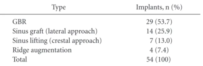
Table 1. Types of Surgery
Type Implants, n (%)
GBR
Sinus graft (lateral approach) Sinus lifting (crestal approach) Ridge augmentation
Total
29 (53.7) 14 (25.9) 7 (13.0) 4 (7.4) 54 (100) GBR: guided bone regeneration.
Table 2. Types of AutoBT
Types Patients, n (%)
Powder Block
Powder + block Total
32 (86.5) 2 (5.4) 3 (8.1) 37 (100)
Table 3. Types of Complications
Types Implants, n
Wound dehiscence Hematoma
Osseointegration failure Total
7
1
4
12
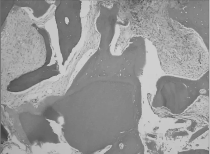
를 발치하여 자가치아골이식재를 제작한 후 상악동골이식과 임플란트 지연식립을 고려하였다. 2009년 6월 29일 자가치아 골이식재 1.3 cc를 이용하여 상악동골이식술을 시행하였고 4 개월 후 임플란트(Implantium Superline 5D/12L, Dentium Co, Suwon, Korea)를 식립하였다. 임플란트 식립 전에 trephine bur로 조직시편을 채취하였다. 2010년 3월 19일 최종 보철물 이 장착되었으며 최근까지 특별한 문제점 없이 정상적인 기 능을 유지하고 있다. 4개월 후 조직검사 결과 잔존 치아골이식
재와 직접 유합되는 신생골들이 관찰되었고 염증 반응은 발 견되지 않았다(Figs. 1-5).
고 찰
최근 연구들을 통해 자가치아 골이식재는 우수한 골유도 및 골전도성 치유를 보인다고 발표되었다(Kim et al., 2010;
Murata et al., 2011). Kim 등(2011)은 자가치아 골이식재의 무 기성분과 표면구조를 분석하였다. 그 결과 치근 부위는 저결 정성 인산칼슘, 치관부위는 고결정성 인산칼슘으로 구성되 어 있어 골전도 및 유도능력에 차이가 있을 것이라고 언급하 였다. 자가치아 골이식재의 결정구조분석을 위한 X-선 회절 평가 결과가 발표되었다. 임상에서 사용되고 있는 다양한 골 이식 재료들을 대조군인 자가피질골과 비교한 결과 AutoBT dentin과 irradiated cancellous bone (ICB)의 X-ray diff raction
Fig. 1. Initial panoramic radiograph. Bone destruction around right
maxillary fi rst molar and sinus pneumatization were observed.
Fig. 2. Preoperative panoramic radiograph. Maxillary fi rst molar was extracted 1 month ago. Powder-type graft material (1.3 cc) was fabri- cated.
Fig. 3. Postoperative panoramic radiograph. Sinus bone graft was performed.
Fig. 4. Histologic fi nding 4 months after bone graft. Direct union be- tween autogenous tooth bone graft material and newly-formed bone was observed(H&E stain, original magnifi cation ×200).
Fig. 5. Panoramic radiograph 1 year after final prosthetic delivery.
Implants were installed 4 months after sinus bone graft.
(XRD) pattern이 자가골과 가장 유사한 소견을 보이는 것으로 확인되었다(Kim et al., 2011). 인간에서 채취한 자가치아 골 이식재의 조직형태계측학적 분석을 통해 자가치아 골이식재 를 이용한 임플란트 주변 소규모 골이식술과 상악동 골이식 은 2-4개월 후에 우수한 골전도성 치유를 보이는 것이 확인되 었다(Kim et al., 2011). Kim 등(2011)은 자가치아 골이식재를 이용한 골유도 재생술에 관한 증례를 보고하였다. 모든 증례 들에서 양호한 임상성적을 보였고 6개월 후 채취한 조직학적 검사에서 우수한 골전도성 치유가 확인되었다. Lee 등(2011) 은 상악동 골이식술에 사용되는 다양한 골이식재와 자가치 아 골이식재의 효율성을 비교하기 위해 조직형태계측학적 연구를 시행하였다. 사용된 골이식재의 종류에 따라 3그룹으 로 분류하였는데, 1군은 자가치아 골이식재, 2군은 Orthoblast II (Integra Co., Irvine, USA)+Biocera (Osscotec, Cheonan, Korea), 3군은 DBX (Synthes, West Chester, PA, USA)+BioOss (Geistlich Pharm AG, Wolhusen, Switzerland)을 사용한 그룹 이었다. 신생골 형성 비율은 1군 52.5±10.7%, 2군 52.0±23.4%, 3군 51.0±18.3%였고 통계적으로 유의성 있는 차이는 없었다.
신생골과 잔존 골이식재의 비율은 1군 81.3±10.4%, 2군 72.5±
28.8%, 3군 80.3±24.0%였다. 4개월의 치유기간 후에 모든 군에 서 양호한 신생골 형성이 이루어졌으며 자가치아 골이식재는 상악동 골이식에 사용될 수 있는 새로운 골이식 재료라고 언 급되었다. Kim 등(2011)은 자가치아 골이식재를 이용한 발치 창 보존 및 재건술 증례를 보고하였다. 분말형과 블록형 재료 모두 우수한 발치창 보존 및 골치유 효과를 보였으며, 생체적 합성이 우수한 자가치아 골이식재를 이용한 발치창 보존 및 재건술은 임상적으로 우수한 효과를 얻을 수 있으며 치유기 간이 현저히 단축될 가능성이 있다고 언급하였다. 자가치아 골이식재를 이용한 치조능 수직 및 수평 증대술에 관한 임상 증례보고가 발표되었는데 치조능 수직 및 수평 증대술에서 자가치아 골이식재는 자가골 이식의 대체수단이 될 수 있으 며, 양이 부족할 경우엔 타 골이식재와 혼합하여 사용한다면 임상에서 매우 유용하게 사용할 수 있다고 언급되었다(Kim et al., 2011). 그 외에도 여러 학자들에 의해 골유도 재생술 및 상 악동 골이식술에 대한 임상연구가 시행되었으며 골전도에 의 한 우수한 골치유가 이루어지는 것이 확인되었다(Kim, 2011;
Lee et al., 2011, Jeong et al., 2011). Jeong 등(2011)은 자가치아 골이식재를 동반한 상악동 골이식과 임플란트 식립에 관한 임상연구를 통해 96.15%의 임플란트 생존율을 보고하였으며, 자가치아 골이식재는 서서히 흡수되면서 골유도 및 골전도성 치유를 보이는 우수한 재료라고 언급하였다. 치아이식술 후 주변 결손부에 자가치아 골이식재를 이식하여 안정성을 얻고 10개월 경과를 관찰한 결과 재부착이 이루어지는 것이 관찰
됨으로써 자가치아 골이식재의 골유도성 치유능력을 제시한 증례도 발표된 바 있다(Kim & Choi, 2011).
이번 연구는 자가치아 골이식술이 시행된 후 2년 이상 경과 를 관찰한 결과 감염 등과 같은 이물 거부반응은 전혀 관찰되 지 않았다. 술 후 창상열개, 혈종 등과 같은 합병증은 일반적인 골이식술 후에도 빈발하는 합병증이다. 그러나 자가치아 골 이식술 후 창상열개는 2차 치유가 잘 이루어지면서 골이식재 의 상당량이 유지되는 특성을 보였다. 임플란트 골유착 실패 역시 자가치아 골이식재와 연관성이 없었으며 치유기간 부족 및 임시 보철물에 의한 조기 과부하가 원인으로 관여하였다.
실패한 임플란트 제거 후 골이식 부위의 치유가 양호하였기 때문에 즉시 임플란트를 다시 식립한 후 보철치료를 종료할 수 있었다.
자가치아 골이식재는 대부분 분말형이 사용되었으나 선택 적으로 블록형을 사용하면 좋은 임상결과를 얻을 수 있고, 골 이식재의 양이 부족한 경우엔 다른 골이식재와 혼합하여 사 용하면 비교적 큰 결손부 재건에도 좋은 임상결과를 보일 것 으로 생각된다. 후향적 연구의 한계점으로 인해 다양한 변수 들을 표준화하지 못한 문제점이 있기 때문에 다양한 전향적 임상연구가 필요하다고 생각된다.
참 고 문 헌