201 http://dx.doi.org/10.4196/kjpp.2014.18.3.201
ABBREVIATIONS: AChE, acetylcholine esterase; AD, Alzheimer's disease; BSA, bovine serum albumin; DMSO, dimethyl sulfoxide;
DTNB, 5,5-dithiobis (2-nitrobenzoic acid); ELT, escape latency time;
GSH, reduced glutathione; p.o., periorbital; MWM, Morris water maze; CMC, carboxymethylcellulose; NED, Naphthylethylenediamine;
TBARS, thiobarbituric acid reactive substances; TSTQ, time spent in target quadrant.
Received January 6, 2014, Revised March 17, 2014, Accepted April 12, 2014
Corresponding to: Nirmal Singh, Pharmacology Division, Depart- ment of Pharmaceutical Sciences and Drug Research, Faculty of Medicine, Punjabi University, Patiala (Punjab) 147002, India. (Tel) 91-9815129884, (Fax) 91-175-2283073, (E-mail) nirmal_puru@
rediffmail.com
This is an Open Access article distributed under the terms of the Creative Commons Attribution Non-Commercial License (http://
creativecommons.org/licenses/by-nc/3.0) which permits unrestricted non-commercial use, distribution, and reproduction in any medium, provided the original work is properly cited.
Ameliorative Effect of a Selective Endothelin ET A Receptor
Antagonist in Rat Model of L-Methionine-induced Vascular Dementia
Gautamjeet S Mangat, Amteshwar S Jaggi, and Nirmal Singh
Pharmacology Division, Department of Pharmaceutical Sciences and Drug Research, Faculty of Medicine, Punjabi University, Patiala (Punjab) 147002, India
The present study was designed to investigate the efficacy of selective ET
Areceptor antagonist, am- brisentan on hyperhomocysteinemia-induced experimental vascular dementia. L-methionine was admi- nistered for 8 weeks to induce hyperhomocysteinemia and associated vascular dementia in male rats.
Ambrisentan was administered to L-methionine-treated effect rats for 4 weeks (starting from 5
thto 8
thweek of L-methionine treatment). On 52
ndday onward, the animals were exposed to the Morris water maze (MW M) for testing their learning and memory abilities. Vascular endothelial function, serum nitrite/nitrate levels, brain thiobarbituric acid reactive species (TBARS), brain reduced gluta- thione (GSH) levels, and brain acetylcholinesterase (AChE) activity were also measured. L-methi- onine-treated animals showed significant learning and memory impairment, endothelial dysfunction, decrease in/serum nitrite/nitrate and brain GSH levels along with an increase in brain TBARS levels and AChE activity. Ambrisentan significantly improved hyperhomocysteinemia-induced impairment of learning, memory, endothelial dysfunction, and changes in various biochemical parameters. These effects were comparable to that of donepezil serving as positive control. It is concluded that ambri- sentan, a selective ET
Areceptor antagonist may be considered as a potential pharmacological agent for the management of hyperhomocysteinemia-induced vascular dementia.
Key Words: Ambrisen, L-Methionine, Morris water-maze, Vascular dementia
INTRODUCTION
Dementia is a syndrome due to disease of the brain, usu- ally of a chronic or progressive nature, in which there is disturbance of multiple higher cortical and neuropsycholo- gical functions including memory, thinking, orientation, and comprehension, calculation, learning capacity, langu- age and judgement [1]. Dementia of vascular origin (VaD) has gained much attention in the past years for being the second most common type of dementia after Alzheimer’s disease (AD) [2,3]. It was estimated that 35.6 million people lived with dementia worldwide in 2010, with numbers ex- pected to almost double every 20 years, to 65.7 million in 2030 and 115.4 million in 2050 [4]. VaD in turn has in- creased the risk of recurrent stroke, dependent living and death [5]. As the incidence rate of dementia increases rap-
idly with advancing age, a large increase in the number of patients is expected as a result of continuous aging of the population [6].
There is substantial evidence from observational studies that conventional risk factors such as hypertension [7], dys- lipidemia [8], smoking [9], hyperhomocysteinemia [10] and diabetes [11] play a key role in the development of VaD and targeting these risk factors will minimize the burden.
Our research group has recently reported that VaD can be induced in rats with the help of hyperhomocysteinemia, diabetes, experimental hypertension and hyperlipidemia [12,13].
Endothelin (ET) and nitric oxide (NO) are well known
mediators produced by endothelial cells to maintain hemo-
dynamic responses [14]. There are three main endothelial
isoforms: ET-1, ET-2 and ET-3, of which ET-1 is the most
potent vasoconstrictor agent. ET-1 binds to two receptors,
endothelin A (ET
A) and endothelin B (ET
B) which are respo-
nsible for a variety of physiological functions, primarily
blood flow control [15]. A key event in endothelial dysfunc-
tion is the reduction in bioavailability and biological activ-
ity of NO. Studies have demonstrated endothelial dysfunc-
tion in hyperhomocysteinemia leading to increased sensi- tivity to endothelin-1 and decreased relaxation in basilar artery [16]. Reduced levels of NO contribute to increased vascular tone, inflammation, platelet aggregation and oxi- dative stress which all are central features of athero- sclerosis and hyperhomocysteinemia [17]. Endothelin re- ceptor antagonists including ambrisentan are noted to exert their anti-inflammatory actions along with reduction in re- active oxygen species (ROS) generation which are sub- sequently responsible for endothelial dysfunction [18,19].
ET receptor antagonists have also been shown to provide a beneficial effect in various cerebrovascular disorders such as moyamoya disease [20], ischemic stroke [21] and sub- arachnoid hemorrhage [22]. Furthermore it has been re- cently reported that these antagonists have potential for the treatment of AD [23]. However, the potential of endo- thelin receptor antagonists in VaD is still unexplored.
The present study has been undertaken to investigate the efficacy of ambrisentan, a selective ET
Aendothelin receptor antagonist in a rat model of L-methionine-induced VaD.
METHODS Animals
Adult male albino Wistar rats, weighing 200∼250 g were employed in the present study and were housed in animal house with free access to water and standard chow (Kisan Feeds Ltd, Mumbai, India). The animals were exposed to 12 h light and 12 h dark cycle. The experiments were con- ducted between 9.00 and 18.00 h. The animals were acclim- atized to laboratory conditions five days prior to behavioral study and were maintained in the laboratory until the com- pletion of the study. The protocol of the study was duly approved by the Institutional Animal Ethics Committee (Approval No. 2012/14) and care of the animals was taken as per the guidelines of the Committee for the Purpose of Control and Supervision of Experiments on Animals (CPCSEA), Ministry of Environment and Forests, India (Reg No.
107/1999).
Drugs and reagents
All the drug solutions were freshly prepared before use.
Donepezil was obtained as a gift sample from Wokhardt Ltd, Baddi, Himachal Pradesh, India. Ambrisentan was purchased from Atra Drugs Pvt Ltd, Hyderabad, India.
Folin-Ciocalteu’s Phenol and Acetylthiocholine were pur- chased from Merck Limited, Mumbai, India. L-methionine was purchased from SD fine chemicals limited, Mumbai, India. 5,5’, dithiobis (2-nitrobenzoic acid) (DTNB), reduced glutathione (GSH), bovine serum albumin (BSA), sulfanila- mide, N-naphthylethylenediamine (NED) and thiobarbi- turic acid were obtained from Loba Chem, Mumbai, India.
Sodium nitroprusside was purchased from SD fine chem- icals limited, Mumbai, India. Phenylephrine was obtained as a free sample from Aarti industries, Dombivli (East), Maharashtra, India. All the agents including ambrisentan, L-methionine and donepezil were administered orally.
Ambrisentan and L-methionine were suspended in 0.5%
w/v of CMC (Carboxymethylcellulose) whereas donepezil was dissolved in saline.
L-Methionine induced vascular dementia
L-methionine (1.7 g/kg/day, p.o.) was administered for 8 weeks to produce hyperhomocysteinemia induced vascular dementia in rats [12]. Body weight of rats was monitored weekly. Rats were subjected to Morris water maze test for the evaluation of their learning and memory status after a span of 8 weeks. L-methionine treatment was continued during acquisition trials on the Morris water maze. Anal- ysis of serum homocysteine concentration was conducted on 1
stday of the study before L-methionine treatment and on the 52
ndday of the study before exposure on the Morris water maze. Behavioral and other assessments were done on the 52
ndday of L-methionine treatment [3]. HPLC sys- tem was additionally used to measure levels of homo- cysteine.
Assessment of learning and memory by Morris water maze (MWM)
Memory was tested by practicing Morris water maze which is one of the most commonly used animal models to assess memory [24,25]. It is based on a principle where rats are placed in a large pool of water and their tendency to escape from the water is accomplished by finding an escape platform as animals dislike swimming. MWM consists of a large circular pool (150 cm in diameter, 45 cm in height) filled to a depth of 30 cm. The pool contained water main- tained at a temperature of 22±2
oC and 1 kg of powdered skim milk to make the water opaque. Two threads, fixed at right angle to each other on the rim of the pool were used to divide the tank into four equal quadrants. A sub- merged white platform (15 cm in diameter) was fixed at 1.5 cm below the surface of the water in one of the four locations, approximately 50 cm from the side walls [26].
There was no alteration in the position of platform through- out the training session. Four consecutive trials were con- ducted daily on each rat with a gap of 5 min. The rat was placed centrally between the quadrants, facing the wall of the pool with the drop location changing for each trial, and was allowed a time period of 120 s to locate the submerged platform. Later, 20 s was accustomed to each rat while stay- ing on the platform. If the rat failed to find the platform within 120 s, then it was guided gently onto the platform and allowed to remain there for 20 s. Escape latency time (ELT) was noted as an index of acquisition or learning to locate the hidden platform in the water maze was. The ani- mal was subjected to acquisition trials for four consecutive days. Randomized daily starting positions were exercised and were not repeated in any of the quadrants. Quadrant (Q4) was maintained as a target quadrant in all of the ac- quisition trials.
Day1 Q1 Q2 Q3 Q4
Day2 Q2 Q3 Q4 Q1
Day3 Q3 Q4 Q1 Q2
Day4 Q4 Q1 Q2 Q3
The platform was removed and each rat was allowed to ex- plore the pool for 120 s on the fifth day. Mean time spent in all four quadrants i.e. Q1, Q2, Q3 and Q4 were recorded.
Index of retrieval was noted at the mean time spent with
the animal in target quadrant i.e. Q4 in search of the hid-
den platform. Later, every rat was subjected to four such
trials and each trial was started from a different quadrant.
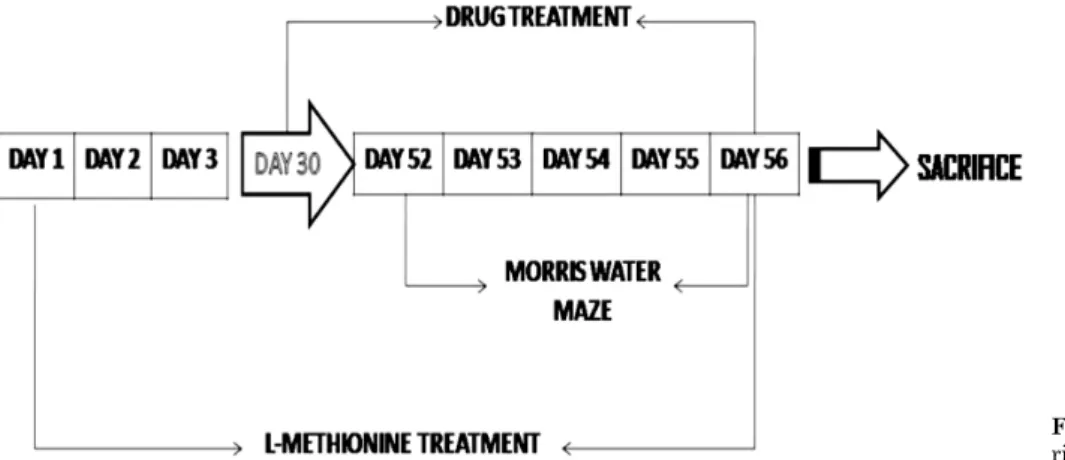
Fig. 1. Schematic presentation of expe- rimental protocol.
Same position was exercised by the experimenter during all trials while performing the MWM test. Prominent visual clues were not disturbed during the total duration of study as care was taken not to alter the relative location of the Morris water maze with respect to other objects in the laboratory. The proportion of time spent searching for the platform in the training quadrant, i.e. the previous location of the platform, was used as a measure of memory retention.
Biochemical assays
Blood samples for biochemical estimations were collected via retro-orbital bleeding just before sacrificing the animals.
They were kept at room temperature for a period of 30 min followed by centrifugation at 4000 rpm for 15 min done to separate serum which was later used for estimation of ho- mocysteine and nitrite/nitrate levels. The animals were sac- rificed by cervical dislocation; thoracic aorta and brain tis- sue were carefully removed. Thoracic aorta was used to es- timate endothelium dependent and independent relaxa- tions, whereas brain was subjected to various biochemical estimations. Clear supernatant was first obtained after ho- mogenizing the brain tissues in phosphate buffer (pH 7.4, 10% w/v) using teflon homogenizer followed by centri- fugation at 3000 rpm for 15 min. This clear supernatant was then removed carefully from the centrifugation tube and it was later used for different biochemical estimations of TBARS, GSH, AChE, nitrite and proteins.
Determination of homocysteine was carried out using HPLC (Varian Inc., CA, USA) attached with fluorescent HPLC detector according to the method of Dimitrova et al.
[27]. Serum nitrite concentration [28], whole brain AChE activity [29], TBARS levels [30], reduced glutathione (GSH) content [31] and total protein content [32] was assayed us- ing standard methods by spectrophotometry (DU 640B spec- trophotometer, Beckman Coulter Inc., CA, USA).
Assessment of vascular endothelial function using isolated rat aortic ring preparation
The rats were decapitated and descending thoracic aorta was removed. Thoracic aorta was cut into a ring of 3∼5 mm width and was mounted in the tissue bath chamber containing Krebs-Henseleit solution (NaCl, 119 mM; KCl, 4.7 mM; NaHCO
3, 25 mM; MgSO
4, 1.0 mM; Glucose, 11.1
mM; KH
2PO
4, 1.2 mM and CaCl
2, 2.5 mM) maintained at 37.8ºC and bubbled with carbogen (95% O
2and 5% CO
2).
The ring was held with the help of two opposite parallel L-shaped stainless steel loops in opposite directions. One loop was connected with force-displacement transducer (FT- 2147) which was further coupled to physiograph (INCO, Ambala, India) whereas second loop was joined with a hold- er acting as an anchor submerged in bath chamber. The preparation was allowed to be stretched with 1.5 g tension followed by equilibration for 90 min with continuous wash- ing with bath solution after every 10∼15 min. The iso- metric contractile force was measured with the force-dis- placement transducer. The aortic ring preparation in- cubated with Krebs-Henseleit solution was primed with 80 mM KCl to check its functional integrity and to improve its contractility. The aortic ring preparation was stimulated with phenylephrine (3×10
-6M) until the contractile re- sponse reached a steady tension. Cumulative dose re- sponses of acetylcholine (ACh; 10
-8to 10
-4M) and sodium nitroprusside (SNP; 10
-8to 10
-4M) were recorded with in- tact or denuded endothelium, respectively at 30 minute in- terval. The internal layer of aortic ring was rubbed gently with a moistened filter paper for 30 s to obtain an endothe- lial free preparation. The loss of ACh 10
-6M-induced relaxa- tion confirmed the absence of endothelial layer.
Experimental protocol
In total eight groups have been employed in the present study and each group consisted of six rats (Fig. 1).
1. Group I − Control group
Normal untreated rats were subjected to acquisition trial, conducted from day 1 to day 4 and retrieval trial, conducted on day 5 using MWM test.
2. Group II − Vehicle control group
Rats were administered 0.5% w/v CMC (10 ml/kg/day, p.o.) for 4 weeks and then subjected to MWM test. The treatment was continued during acquisition (from 24
thto 27
thday) and retrieval trials (on 28
thday) on MWM.
3. Group III − L-Methionine treatment group
Rats were administered L-methionine (1.7 g/kg/day, p.o.)
for 51 days and then subjected to MWM test. The treatment
of L-methionine was continued during acquisition (from
Table 1. Effect of pharmacological interventions on escape latency time (ELT) using Morris water maze
GROUP DOSE Day 1 ELT (Sec) Day 4 ELT (Sec)
Control 107.4±3.5 34.1±2.7
aVehicle (CMC) 10 ml/kg, p.o. 112.5±3.6 39.1±2.3
aL-Methionine 1.7 g/kg, p.o. 117.5±2.5 68.9±2.4
a,bAmbrisentan 10 mg/kg, p.o. 108.6±3.6 38.19±3.2
aL-Methionine+Ambrisentan low dose 1.7 g/kg, p.o.+5 mg/kg, p.o. 111.2±3.0 55.8±2.8
a,cL-Methionine+Ambrisentan high dose 1.7 g/kg, p.o.+10 mg/kg, p.o. 108.8±3.4 50.5±2.6
a,cDonepezil per se 1 mg, p.o. 108.3±3.2 36.3±3.1
aL-Methionine+donepezil 1.7 g/kg, p.o.+1 mg, p.o. 114.6±3.4 48.3±3.0
a,cEach group (n=6) represents mean±standard deviation. Two way ANOVA followed by Bonferroni post hoc test. F (1, 40)=5210.38 for days, p<0.0001 and F (7, 40)=59.32 for treatment, p<0.0001.
ap<0.01 versus Day 1 ELT in the respective group.
bp<0.05 versus Day 4 ELT in the control group.
cp<0.05 versus Day 4 ELT in L-Methionine treated group.
Fig. 2. Effect of pharmacological interventions on mean time spent in the target quadrant (TSTQ) using Morris water-maze test.
L-MET, L-Methionine; Amb LD, Ambrisentan low dose; Amb HD, Ambrisentan high dose; DON, Donepezil; CMC, Carboxymethyl- cellulose. Each group (n=6) represents mean±standard deviation.
Two way ANOVA followed by Bonferroni post hoc test.
ap<0.05 versus mean time spent in other quadrants in control;
bp<0.05 versus mean time spent in the target quadrant in control group;
c
p<0.05 versus mean time spent in the target quadrant in L- Methionine treated group.
52
ndto 55
thday) and retrieval trials (56
thday) on the MWM.
4. Group IV − Ambrisentan
Rats were administered ambrisentan (10 mg/kg/day, p.o.) for 4 weeks followed by exposure to the MWM; the rest of the procedure was same as described in group II.
5. Group V − L-Methionine and Ambrisentan low dose Ambrisentan (5 mg/kg/day, p.o.) was administered to L-methionine (1.7 g/kg/day, p.o.) treated rats, starting from the 30th day of L-methionine treatment followed by expo- sure to MWM on the 52nd day of L-methionine administ- ration. The treatment was continued during acquisition (from 52
ndto 55
thday) and retrieval trials (on 56
thday) on the MWM..
6. Group VI − L-Methionine and Ambrisentan high dose Ambrisentan (10 mg/kg/day, p.o.) was administered to L-methionine (1.7 g/kg/day, p.o.) treated rats, starting from the 30
thday of L-methionine treatment followed by ex- posure to MWM on the 52
ndday of L-methionine admini- stration. The treatment continued during acquisition (from 52
ndto 55
thday) and retrieval trials (on 56
thday) on the MWM.
7. Group VII − Donepezil
Rats were administered donepezil (1 mg/kg/day, p.o.) for 4 weeks followed by exposure to MWM; the rest of the pro- cedure was same as described in group II.
8. Group VIII − L-Methionine and Donepezil
Donepezil (1 mg/kg/day, p.o.) was administered to L-me- thionine (1.7 g/kg/day, p.o.) treated rats, starting from the 30
thday of L-methionine treatment followed by exposure to MWM on the 52
ndday of L-methionine administration.
The treatment continued during acquisition (from 52
ndto 55
thday) and retrieval trials (on 56
thday) on the MWM.
Statistical analysis
Statistical analyses were done using Sigma stat 3.5. All results were expressed as means±standard deviation. Data for isolated aortic ring preparation were statistically ana- lyzed using repeated measures of analysis of variance (ANOVA) followed by Newman Keul's test. Data obtained from various groups was statistically analyzed using two-way ANOVA followed by Bonferroni Multiple Range
test in case of ELT and TSTQ. The rest of the data obtained from various groups was statistically analyzed using one- way ANOVA followed by Tukey’s test. p<0.05 was consid- ered to be statistically significant.
RESULTS
Effect on escape latency time (ELT) and time spent in the target quadrant (TSTQ), using the Morris water maze (MWM)
Control rats showed a downward trend in their ELT.
There was a significant fall in day 4 ELT, when compared
to day 1 ELT of these rats (Table 1). Further on day 5 a
significant rise in TSTQ was observed, when compared to
Fig. 3. Effect of pharmacological interventions on Ach-induced endo- thelium-dependent relaxation using an aortic ring preparation.
L-MET, L-Methionine; Amb LD, Ambrisentan low dose; Amb HD, Ambrisentan high dose; DON, Donepezil; CMC, Carboxymethyl- cellulose. Each group (n=6) represents mean±standard deviation.
Responses are expressed as percentage of precontraction induced by 3×10
-6M phenylephrine. Repeated measure ANOVA followed by Newman Keul's test.
ap<0.05 versus control;
bp<0.05 versus L-Methionine treated group.
Fig. 4. Effect of pharmacological interventions on endothelium inde- pendent relaxation using an aortic ring preparation. L-MET, L-Methionine; Amb LD, Ambrisentan low dose; Amb HD, Ambri- sentan high dose; DON, Donepezil; CMC, Carboxymethylcellulose.
Each group (n=6) represents mean±standard deviation. Responses are expressed as percentage of precontraction induced by 3×10
-6M phenylephrine. Repeated measure ANOVA followed by Newman Keul's test.
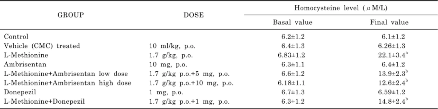
Table 2. Effect of various pharmacological interventions on serum homocysteine level of animals
GROUP DOSE Homocysteine level (μM/L)
Basal value Final value
Control 6.2±1.2 6.1±1.2
Vehicle (CMC) treated 10 ml/kg, p.o. 6.4±1.3 6.26±1.3
L-Methionine 1.7 g/kg, p.o. 6.83±1.2 22.1±3.4
aAmbrisentan 10 mg, p.o. 6.3±1.1 6.4±1.2
L-Methionine+Ambrisentan low dose 1.7 g/kg p.o.+5 mg, p.o. 6.6±1.2 13.9±2.3
bL-Methionine+Ambrisentan high dose 1.7 g/kg p.o.+10 mg, p.o. 6.18±1.1 12.6±2.4
bDonepezil 1 mg, p.o. 6.7±1.3 6.59±1.2
L-Methionine+Donepezil 1.7 g/kg p.o.+1 mg, p.o. 6.3±1.2 14.8±2.4
bEach group (n=6) represented by mean±SD.
One way ANOVA followed by Tukey's multiple range test. F (7, 40)=28.955.
ap<0.05 versus basal values in L-Methionine group.
b