Gastrointestinal Tract Perforation:
MDCT Findings according to the Perforation Sites
Our objective is to describe the characteristic CT findings of gastrointestinal (GI) tract perforations at various levels of the gastrointestinal system. It is beneficial to localize the perforation site as well as to diagnose the presence of bowel perfora- tion for planning the correct surgery. CT has been established as the most valu- able imaging technique for identifying the presence, site and cause of the GI tract perforation. The amount and location of extraluminal free air usually differ among various perforation sites. Further, CT findings such as discontinuity of the bowel wall and concentrated free air bubbles in close proximity to the bowel wall can help predict the perforation site. Multidetector CT with the multiplanar reformation images has improved the accuracy of CT for predicting the perforation sites.
astrointestinal (GI) tract perforations can occur due to various causes, and most of these perforations are emergency conditions of the abdomen that require early recognition and timely surgical treatment (1). The mainstay of treatment for bowel perforation is surgery. Endoscopic, laparoscopic and laparo- scopic-assisted procedures are now being increasingly performed instead of conven- tional laparotomy (2, 3). Moreover, if any signs and symptoms of generalized peritoni- tis are absent and the perforation site has sealed spontaneously, then a perforated duodenal ulcer can be treated nonoperatively (4). Owing to these various therapeutic options, the site and cause of GI tract perforation are major questions that should be answered by performing imaging studies. CT has been established as the most valuable imaging technique for identifying the presence, site and cause of GI tract perforation (1, 3, 5-10). Several researchers have reported on the direct and indirect CT findings of bowel perforation, and free extraluminal air has been regarded as a major imaging finding for making the diagnosis of the GI tract perforation (3, 6, 8, 10). CT is highly sensitive for detecting free extraluminal air. It can display extraluminal, intraperitoneal or retroperitoneal air more sensitively than can plain radiography. In addition to determining the presence of perforation, CT can also localize the perforation site. The overall accuracy of CT for predicting the site of bowel perforation has been reported to range between 82% and 90% (3, 10, 11). The CT findings of GI tract perforation may be different according to the perforation site, and the various CT findings can help predict the perforation site (3, 5). Moreover, the recent introduction of multide- tector-row CT has improved the accuracy of CT for predicting the site of GI tract perforation (3, 7, 10). In this article, we illustrate the characteristic CT findings of GI tract perforation at various levels of the gastrointestinal tract.
CT Technique
The entire abdomen from the dome of the diaphragm to the pelvic floor should be Sung Hwan Kim, MD1
Sang Soo Shin, MD2 Yong Yeon Jeong, MD2 Suk Hee Heo, MD2 Jin Woong Kim, MD3 Heoung Keun Kang, MD2
Index terms :
Gastrointestinal tract, CT Gastrointestinal tract, perforation MDCT, acute abdomen
MDCT, perforation
DOI:10.3348/kjr.2009.10.1.63
Korean J Radiol 2009 ; 10 : 63-70 Received February 19, 2008; accepted after revision October 9, 2008.
1Department of Radiology, Chonnam National University Hospital, Gwangju 501-757, Korea; 2Department of Radiology, Chonnam National University Medical School, Gwangju 501-757, Korea; 3Department of Radiology, Chonnam National University Hwasun Hospital, Jeollanam-do 519-809, Korea
Address reprint requests to : Sang Soo Shin, MD, Department of Radiology, Chonnam National University Medical School, 8 Hack-dong, Dong-gu, Gwangju 501-757, Korea.
Tel. (8262) 220-5746 Fax. (8262) 226-4380 e-mail: [email protected]
G
scanned. Contiguous axial images less than 5 mm thick are obtained and thinner sections and multiplanar reformations may be applied when necessary. After the precontrast images have been acquired, CT scanning is initiated 70 to 80 seconds after the intravenous injection of contrast material (300 to 370 mgI/mL; 100 to 150 mL at a rate of 3 to 4 mL/s). In our hospital, we prefer CT scanning without oral contrast material for evaluating bowel perforation.
Although extraluminal leakage of oral contrast material has been reported to be a specific finding for bowel perforation, several authors have raised doubts about the added benefit of oral contrast material (6, 9, 12). These reports cite safety issues (i.e., the risk of aspiration and the subsequent complications), the potential delay in the
diagnosis and the lack of substantial added benefit for detecting bowel perforation. Further, the slow progression of the oral contrast material in the GI tract of a patient suffering from paralytic ileus and the rapid sealing of the perforation site may preclude extraluminal leakage of oral contrast material in patients with GI tract perforation.
CT Findings of Gastrointestinal Tract Perforation The diagnosis of GI tract perforation is based on the direct CT findings, such as discontinuity of the bowel wall and the presence of extraluminal air, and on the indirect CT findings, such as bowel wall thickening, abnormal bowel wall enhancement, abscess and an inflammatory mass adjacent to the bowel (1, 3, 5-8).
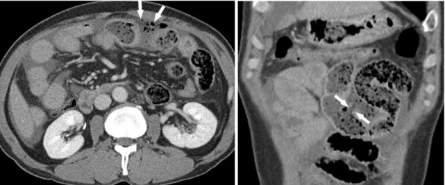
Fig. 1. 46-year-old man with traumatic small bowel perforation.
A. Transverse CT image shows concentrated free air bubbles (arrows) in vicinity of jejunal loop.
B. Coronal reformation CT image demonstrates discontinuity of jejunal wall (arrows), which is not clearly seen on transverse CT image.
A B
Fig. 2. 47-year-old man with traumatic small bowel perforation.
Transverse CT image shows small air bubble (arrow) at anterior abdominal surface. Presence of air is more clearly seen on CT image with wide window setting.
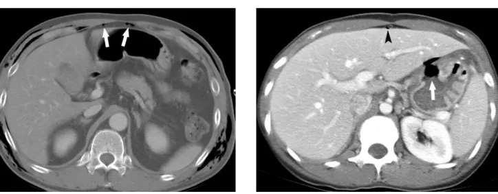
Fig. 3. Traumatic duodenal perforation in 53-year-old woman.
Transverse CT scan demonstrates extraluminal free air (arrows) surrounding 3rd portion of duodenum (D) and retroperitoneal fluid (arrowheads).
Direct visualization of the discontinuity of the bowel wall can specify the presence and site of GI tract perfora- tion, which is marked by a low-attenuating cleft that usually runs perpendicular to the bowel wall on CT (10).
However, this cleft has been reported to be observed less frequently than free air on CT, and a cleft is usually seen in less than 50% of the patients with GI tract perforation (1, 3, 8, 10). The relatively infrequent detection of this finding is partly due to the small size of the lesion (8). MDCT with multiplanar reformation images may help identify disconti- nuity of the bowel wall, and especially when the axial CT images are indeterminate (Fig. 1).
Although extraluminal air is highly specific for making the diagnosis of GI tract perforation, the sensitivity of plain radiograph to detect this is not high (5, 10). In contrast, CT is highly sensitive for detecting free extraluminal air. When tiny bubbles of free air are embedded in the omentum and mesentery, or if they are located in places such as the anterior peritoneal surface of the liver and among the bowel loops, CT images with the wide window setting can be useful for enhancing the sensitivity to detect this free air (Fig. 2) (5). In addition to the presence of GI tract perfora-
tion, concentrated free air bubbles in close proximity to the bowel wall may help determine the site of perforation because free air bubbles tend to be in the vicinity of the bowel wall from which they arises, and especially when the amount of air is small (Fig. 3) (3). The amount and location of free air could be different according to the perforation site (Table 1).
On the contrary, air confined to the inner layer of the abdominal wall and external to the parietal peritoneum should not be interpreted as pneumoperitoneum (Fig. 4) (8). It should be noted that extraluminal intra- or retroperi- toneal air can occur without GI tract perforation. Various causes can produce air, such as mechanical ventilation and pulmonary barotraumas, peritoneal lavage that’s
performed prior to CT, pneumothorax, chest injury and entry of air via the female genital tract (8). Thus, additional CT findings that are indicative of GI tract perforation intensify the significance of extraluminal free air.
CT Findings according to Sites of the GI Tract Perforation Gastroduodenal Perforation
Among various disease entities, peptic ulcer disease is a
Table 1. Summary of Amount and Location of Free Air according to Perforation Sites Free Air Perforation Site
Amount Location
Stomach/duodenum Abundant Around liver and stomach
Post-bulbar duodenum Right anterior pararenal space
Small bowel Small Mesenteric folds, around liver
Appendix Small/absent Around appendix
Large bowel Variable Pelvis, mesenteric folds, retroperitoneal space
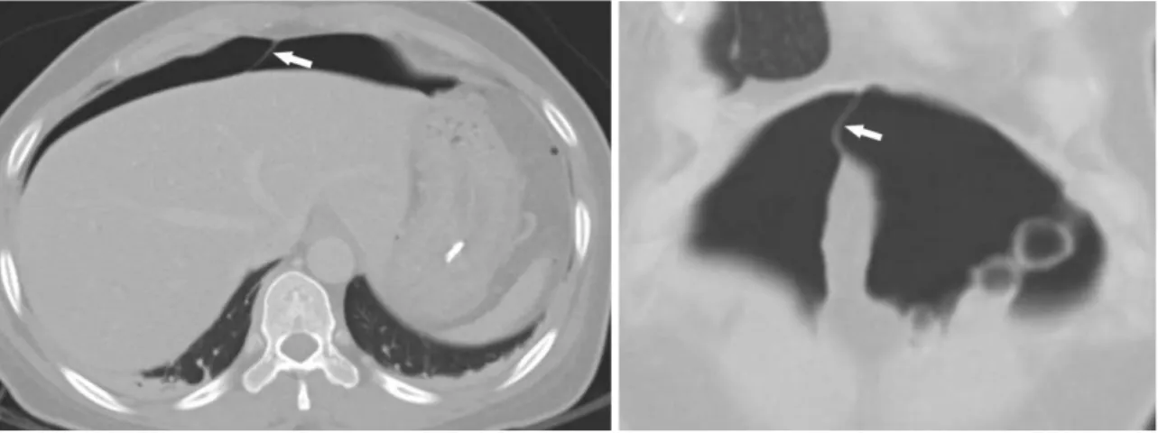
Fig. 4. Pseudopneumoperitoneum in 53-year-old man.
Transverse CT image shows free air bubbles (arrows) between transversalis fascia and parietal peritoneum, which could be misinterpreted as pneumoperitoneum.
Fig. 5. 37-year-old woman with perforated gastric ulcer.
Focal defect in lesser curvature of gastric body is caused by deep ulcer (arrow) associated with surrounding mural thickening. Note small air bubble (arrowhead) on anterior peritoneal surface of liver.
major cause of gastroduodenal perforation, followed by necrotic or ulcerated malignancies, iatrogenic injuries and traumatic injuries (5). Gastroduodenal perforation associ- ated with peptic ulcers is commonly found in the gastric antrum and duodenal bulb, whereas the descending and horizontal segments of the duodenum are the common sites of perforation caused by blunt trauma because of their firm attachment, the acutely angled flexures and the compression against the vertebral column (13).
Perforation sites can be frequently demonstrated by the CT findings, such as ulceration or focal defect of the gastro- duodenal wall (Fig. 5), air bubbles in contact with the stomach or the duodenum (Fig. 6), abrupt wall thickening associated with adjacent “dirty fat” density and local fluid between the duodenum and the pancreatic head (6).
Extraluminal free air is the most common and consistent finding of gastroduodenal perforation, although it may be
absent in gastroduodenal perforation, and especially at the onset of symptom (14). It is usually abundant and it is noted around the liver and stomach (5). Free air or an air- fluid level crossing the midline and an accentuating falciform ligament (the “falciform ligament sign”) and free air confined in the intrahepatic fissure for the ligamentum teres (the “ligamentum teres sign”) have been considered to be useful findings in patients with perforation of the duodenal bulb or stomach (Fig. 7) (1, 5). If free air is found in the lesser sac, then the likely site of perforation is the posterior wall of the stomach or duodenum (Fig. 8) (9).
Gastric perforation is seldom correlated with air trapped in the mesenteric or sigmoid recess, in contrast to perforation of the colon and small bowel (9). Extraluminal free air in the right anterior pararenal space is the reliable CT finding for diagnosing duodenal perforation beyond the bulbar segment (15).
Fig. 6. 74-year-old man with perforated gastric ulcer.
A. Transverse CT image shows concentration of extraluminal air bubbles (arrows) in close proximity to gastric antral wall.
B. Coronal reformation CT image demonstrates close relationship between extraluminal air bubbles (arrows) and gastric antral wall (arrowheads).
A B
Fig. 7. 41-year-old woman with perforation of gastric body that complicates endoscopic submucosal dissection.
Transverse (A) and coronal reformation (B) CT images with wide window setting show that free air crossing midline accentuates falciform ligament (arrow).
A B
Small Bowel Perforation
Whereas gastroduodenal perforation usually results in massive pneumoperitoneum, small bowel perforation accounts for the majority of the cases in which the amount of extraluminal air is too small to be detected by conven- tional radiography or even CT. According to Grassi et al.
(16), free air was detected with using CT in only approxi- mately 50% of the cases of small bowel perforation. Thus, a small amount of free air in the anterior peritoneal surfaces of the liver and mid-abdomen and among the peritoneal folds, as well as such indirect CT findings as mural thickening and abnormal enhancement of the small
bowel, mesenteric fluid and mesenteric stranding, should be scrutinized in patients with suspected small bowel perforation (Fig. 9). When the CT findings are inconclusive and any obvious clinical manifestations of perforation are absent, close follow-up with repeated CT scanning could be helpful. Postoperative perforation or anastomotic leakage should be suspected when free air and ascites are present or these problems are increased in amount the first week after surgery (5).
Appendiceal Perforation
Given that the primary pathogenic event in acute appendicitis is luminal obstruction (17), it is not surprising
Fig. 8. 68-year-old woman with perforation of posterior wall of gastric body and this complicates endoscopic submucosal dissection.
Transverse CT image shows free air (arrow) in lesser sac. Large quantity of free air (arrowheads) anterior to stomach is also seen.
Fig. 10. 49-year-old woman with acute perforated appendicitis.
Transverse CT image demonstrates focal defect (arrowhead) in inflamed appendiceal wall and periappendiceal inflammatory stranding (arrows).
Fig. 9. 47-year-old man with small bowel perforation due to blunt trauma.
Transverse CT image shows tiny air bubble (arrowhead) around perforated small bowel with thick enhancing wall (arrows).
Fig. 11. 74-year-old man with perforation of sigmoid colon.
Transverse CT image shows extraluminal air-fluid level (arrows) adjacent to sigmoid colon with mural thickening and enhance- ment (arrowheads).
that the amount of extraluminal air for this condition is generally small or absent, usually no more than 1 or 2 mL, and a free pneumoperitoneum is very rare in patients with perforated appendicitis (5). In one study (18), the five specific CT findings of extraluminal air, extraluminal appendicolith, abscess, phlegmon and a defect in the enhancing appendiceal wall allowed excellent diagnostic accuracy for the diagnosis of perforated appendicitis (Fig.
10).
Large Bowel Perforation
The perforation sites of colonic loops can frequently be correlated with their causes (19). Malignant neoplasm, diverticulitis (in western countries), blunt trauma and ischemia are common causes of perforation on the left side
of colonic loops (Fig. 11). Yet inflammatory bowel disease, diverticulitis (in eastern countries) and penetrating trauma tend to be observed on the right side of colonic loops. The cecum is prone to perforate in patients with mechanical colonic obstruction. Iatrogenic injuries commonly involve the rectum and sigmoid colon. Depending on the perfora- tion site, free air may be detected in intraperitoneal or retroperitoneal spaces (Fig. 12). When the perforation occurs due to diverticulitis or colorectal malignancy without bowel obstruction, the quantity of extraluminal air is usually small and the bubbles of extraluminal air tend to be concentrated in close proximity to the involved colon (5). In order to confidently diagnose perforation of the large bowel loop, the presence of extraluminal air, phlegmon and/or abscess, an extraluminal collection of
A B
Fig. 13. 81-year-old man with spontaneous perforation of sigmoid colon.
A. Transverse CT image shows concentration of extraluminal air bubbles (arrows) in close proximity to sigmoid wall.
B. Transverse CT image obtained at upper level demonstrates extraluminal feces, which display unique appearance of mass with mottled air bubbles (arrows).
A B
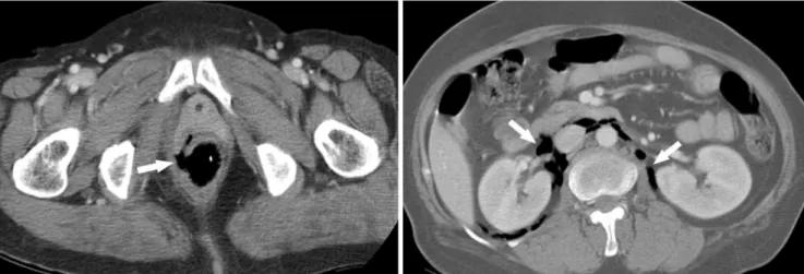
Fig. 12. 60-year-old woman with iatrogenic rectal perforation.
A. Transverse CT image shows focal defect (arrow) in right side of rectal wall.
B. Transverse CT image demonstrates free air (arrows) in both perirenal spaces.
feces and the underlying colonic abnormality should be carefully evaluated on CT images (Fig. 13).
Stercoral perforation of the colon is a distinct clinical and pathologic entity (20). It occurs due to pressure necrosis of the colonic wall from a rock-hard fecaloma. Because performing an immediate, optimal surgical procedure is mandatory to reduce the mortality rate from stercoral perforation, it is important to be familiar with the CT findings of stercoral perforation. The combination of a fecaloma protruding through the colonic wall or lying freely in the abdominal cavity and free air must lead to a suspicion of stercoral perforation of the colon (Fig. 14).
CONCLUSION
It is crucial to make a prompt and correct diagnosis of GI tract perforation with respect to the presence, site and cause of perforation, and this helps the physician choose the optimal therapeutic option. CT allows radiologists not only to detect intraabdominal free air, but also to precisely determine the anatomic site of GI tract perforation. CT findings such as discontinuity of the bowel wall, concen- trated bubbles of extraluminal air in close proximity to the bowel wall and abrupt bowel wall thickening with or without an associated phlegmon or abscess are useful for assessing the site of perforation.
References
1. Yeung KW, Chang MS, Hsiao CP, Huang JF. CT evaluation of gastrointestinal tract perforation. Clin Imaging 2004;28:329-333 2. Siu WT, Chau CH, Law BK, Tang CN, Ha PY, Li MK. Routine
use of laparoscopic repair for perforated peptic ulcer. Br J Surg 2004;91:481-484
3. Hainaux B, Agneessens E, Bertinotti R, De Maertelaer V, Rubesova E, Capelluto E, et al. Accuracy of MDCT in predicting site of gastrointestinal tract perforation. AJR Am J Roentgenol 2006;187:1179-1183
4. Donovan AJ, Berne TV, Donovan JA. Perforated duodenal ulcer: an alternative therapeutic plan. Arch Surg
1998;133:1166-1171
5. Furukawa A, Sakoda M, Yamasaki M, Kono N, Tanaka T, Nitta N, et al. Gastrointestinal tract perforation: CT diagnosis of presence, site, and cause. Abdom Imaging 2005;30:524-534 6. Ghekiere O, Lesnik A, Hoa D, Laffargue G, Uriot C, Taourel P.
Value of computed tomography in the diagnosis of the cause of nontraumatic gastrointestinal tract perforation. J Comput Assist Tomogr 2007;31:169-176
7. Miki T, Ogata S, Uto M, Nakazono T, Urata M, Ishibe R, et al.
Multidetector-row CT findings of colonic perforation: direct visualization of ruptured colonic wall. Abdom Imaging 2004;29:658-662
8. Brofman N, Atri M, Hanson JM, Grinblat L, Chughtai T, Brenneman F. Evaluation of bowel and mesenteric blunt trauma with multidetector CT. Radiographics 2006;26:1119-1131 9. Maniatis V, Chryssikopoulos H, Roussakis A, Kalamara C,
Kavadias S, Papadopoulos A, et al. Perforation of the alimen- tary tract: evaluation with computed tomography. Abdom Imaging 2000;25:373-379
10. Imuta M, Awai K, Nakayama Y, Murata Y, Asao C, Matsukawa T, et al. Multidetector CT findings suggesting a perforation site in the gastrointestinal tract: analysis in surgically confirmed 155 patients. Radiat Med 2007;25:113-118
11. Kim HC, Shin HC, Park SJ, Park SI, Kim HH, Bae WK, et al.
Traumatic bowel perforation: analysis of CT findings according to the perforation site and the elapsed time since accident. Clin Imaging 2004;28:334-339
12. Stuhlfaut JW, Soto JA, Lucey BC, Ulrich A, Rathlev NK, Burke PA, et al. Blunt abdominal trauma: performance of CT without oral contrast material. Radiology 2004;233:689-694
13. Meyers M. The extraperitoneal spaces: normal and pathologic anatomy. In: Meyers MA, eds. Dynamic radiology of the Fig. 14. 90-year-old man with stercoral perforation of sigmoid colon.
A. Transverse CT image shows localized hard fecal mass (arrows) connected with sigmoid colon.
B. Transverse CT image obtained at upper level demonstrates extraluminal gas (arrowhead) adjacent to localized hard fecal mass (arrows), which is indicative of perforation.
A B
abdomen, 5th ed. New York: Springer-Verlag, 2000:333-492 14. Grassi R, Romano S, Pinto A, Romano L. Gastro-duodenal
perforations: conventional plain film, US and CT findings in 166 consecutive patients. Eur J Radiol 2004;50:30-36
15. Kunin JR, Korobkin M, Ellis JH, Francis IR, Kane NM, Siegel SE. Duodenal injuries caused by blunt abdominal trauma: value of CT in differentiating perforation from hematoma. AJR Am J Roentgenol 1993;160:1221-1223
16. Grassi R, Pinto A, Rossi G, Rotondo A. Conventional plain-film radiology, ultrasonography and CT in jejuno-ileal perforation.
Acta Radiol 1998;39:52-56
17. Birnbaum BA, Wilson SR. Appendicitis at the millennium.
Radiology 2000;215:337-348
18. Horrow MM, White DS, Horrow JC. Differentiation of perforated from nonperforated appendicitis at CT. Radiology 2003;227:46-51
19. Ghahremani GG. Radiologic evaluation of suspected gastroin- testinal perforations. Radiol Clin North Am 1993;31:1219-1234 20. Rozenblit AM, Cohen-Schwartz D, Wolf EL, Foxx MJ, Brenner
S. Case reports. Stercoral perforation of the sigmoid colon:
computed tomography findings. Clin Radiol 2000;55:727-729