Seongbuk ENE dental clinic
Abstract
Today’s oral implantology tends to evolve into a less time-consuming, a more aesthetic and a less invasive way to restore a lost dentition. A flapless implant surgery is easy to perform for the surgeon and beneficial for patients. In the past, the sur- geon used to decide on the chosen implant position once the flap was raised, the bone exposed and with the template as a direction indicator. As a consequence, an extended flap was needed to visualize the bone sufficiently in order to avoid perfo- rations of critical anatomical structures. Minimizing the surgical flap can have advantages for soft tissue healing and patient comfort. If a surgeon wants to conduct flapless surgical procedures, an exact transfer of the anatomical information obtained via the CT images to the intra-oral situation during surgery is necessary. In my case, dental implants were installed without open flap. There was about 8-9 mm horizontal bone volume which was analysed by 3D digital CT before surgery.
In most cases, surgical stent is usually necessary for ideal position & angulation of the implants. In conclusion, flapless implant surgery is an effective treatment option for the patient who has sufficient horizontal and vertical ridge volume.
Key Words : flapless implant surgery, 3D digital CT, surgical stent
Flapless implant surgery with 3D digital dental CT; A case report
Myung-Jin Jang
Digital CT를 활용한 flapless implant surgery
장명진 성북ENE치과
좋은 임프란트 수술법이란 무엇인가? 임프란트 수 술법은 길지 않은 세월동안 많은 변화화 시행착 오를 통하여 발전을 거듭하고 있다.. 초기에는 수 술시 가능한 조직피판을 넓게 열고 수술 시야를 잘 확보한 상 태에서 임프란트를 식립한뒤 덮고 골유착을 얻을때까지 기다 린 후다시 열고 보철 수복할 것을 권하였다. 표면처리 기술의 발달로 골유착 기간이 상당히 줄어든 이후 1회법 수술방법이 도입되었으며 골량 부족시에도 여러 골이식재의 발달과 더불 어 임프란트 시술이 가능하게 되었고 치아 발치 즉시 임프란 트 식립, 더 나아가 식립 즉시 보철물 장착후 기능부하를 줄 정도로 발전하고 있다. 초기 구강내 방사선 사진이나 파노라 마 사진에 의존한 2차원적인 수술부위 진단에서 3차원 치과 용 컴퓨터 단층영상촬영이 임프란트 시술에 사용 가능하게 됨에 따라 조직 피판을 열지 않아도 골두께에 대한 진단이 상 당히 정확하게 되어 과거의 개념인 광범위한 개방성 피판형 성 수술법보다는 가능한 조직피판을 적게 열고 식립하는 방 법이 선호되고있고 더 나아가 골두께가 충분한 경우 무피판
형성 수술법이 가능하게 되었다. 이에 상하악 구치부 임프란 트 식립의 예에서 시술전 디지탈 3차원 컴퓨터 단층영상촬영 으로 골두께가 충분하다고 판단되어 무절개피판 임프란트 식 립법을 사용하여 환자측면에서나 술자 측면에서 어떠한 장단 점이 있는지 살펴보고자 한다.
((FFiigg.. 11~~2233))
2006년 디지털 컴퓨터 단층 영상 촬영술을 활용하여 처음 으로 무피판절개 수술법으로 임프란트 수복을 행한 증례이 다. 초진시 70세 남성으로 상하 부분 틀니를 사용중이었지만 정상적인 음식물 저작이 곤란하였다. 칠순 기념으로 자식들 이 돈을 모아와서 임프란트 시술받기를 원하였고 디지털 3차 원 컴퓨터 단층영상촬영을 활용한 술전 검사시 상하악 모두 협설측 골폭이 충분하여 정밀한 수술용 스텐트 제작후 무피 판절개 수술법으로 임프란트 식립을 하였다. 술후 부종 및 출 혈, 불편감이 광범위한 개방성 피판형성 수술법에 비해 현저 히 적었고 더불어 약제 사용량도 적었으며 연조직 치유과정 도 3~4주가량 단축됨을 관찰하였다. 또한 술자도 연조직을
Ⅱ I
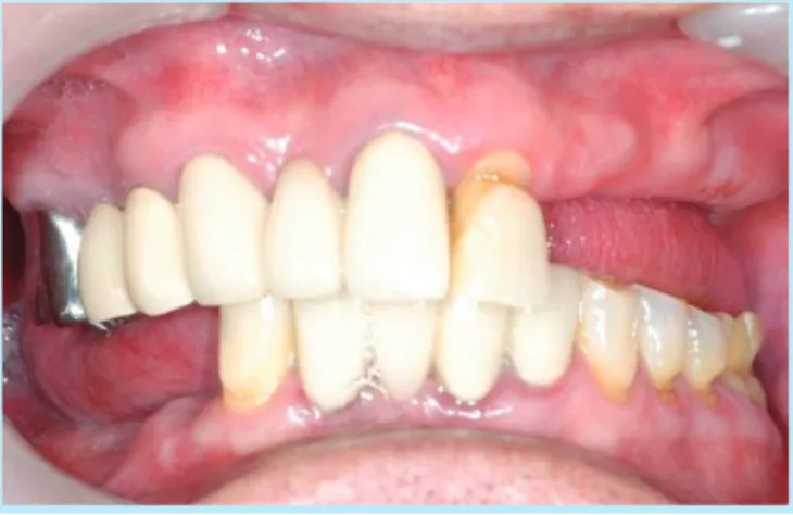
Fig. 1. Anterior view-Before treatment.
Myung-Jin Jang: Flapless implant surgery with 3D digital dental CT; A Case Report.
Implantology 2009
Fig. 2. Panoramic view-Before treatment.
Myung-Jin Jang: Flapless implant surgery with 3D digital dental CT; A Case Report.
Implantology 2009
case report
Fig. 3. Upper left 2ndanterior, canine, 1stpremolar, 2nd premolar, 1stmolar & 2ndmolar teeth loss.
Myung-Jin Jang: Flapless implant surgery with 3D digital dental CT; A Case Report.
Implantology 2009
Fig. 4. Lower right 1stpremolar, 2ndpremolar, 1stmolar
& 2nd molar teeth loss.
Myung-Jin Jang: Flapless implant surgery with 3D digital dental CT; A Case Report.
Implantology 2009
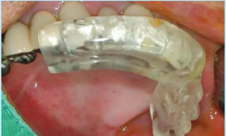
Fig. 5. Surgical stent adaptation before surgery.
Myung-Jin Jang: Flapless implant surgery with 3D digital dental CT; A Case Report.
Implantology 2009
Fig. 6. 3mm twist drill insertion at the 2ndanterior tooth portion.
Myung-Jin Jang: Flapless implant surgery with 3D digital dental CT; A Case Report.
Implantology 2009
Fig. 7. Digital standard x-ray view-3mm twist drill insertion : Check the drill path & direction.
Myung-Jin Jang:
Flapless implant surgery with 3D digital dental CT; A Case Report.
Implantology 2009
Fig. 8. Implant insertion at the upper left 2nd anterior tooth portion : diameter 3.75mm, length 13mm.
Myung-Jin Jang: Flapless implant surgery with 3D digital dental CT; A Case Report.
Implantology 2009
Fig. 9. Implant insertion
at the upper left canine tooth portion(D: 4mm, L: 13mm) at the upper left 1stpremolat tooth portion (D: 4mm, L: 13mm) at the upper left 2ndpremolar tooth portion(D: 4mm, L: 10mm) - Bicortical fixation
Myung-Jin Jang: Flapless implant surgery with 3D digital dental CT; A Case Report.
Implantology 2009
Fig. 10. Flapless subcrestal sinus graft with chisel &
mallet .
Myung-Jin Jang: Flapless implant surgery with 3D digital dental CT; A Case Report.
Implantology 2009
Fig. 11. X-ray view : Upper left 1st molar tooth portion after insertion of the osteotome.
Myung-Jin Jang: Flapless implant surgery with 3D digital dental CT; A Case Report.
Implantology 2009
Fig. 12. Surgicel : For bleeding control & preventing the membrane perforation
Allograft : DFDB = 1 : 1 . Subcrestal sinus bone graft
Myung-Jin Jang: Flapless implant surgery with 3D digital dental CT; A Case Report.
Implantology 2009
Fig. 13. Implant insertion at the upper left 1st molar tooth portion : diameter 4mm, length 10mm after bone insertion.
Myung-Jin Jang: Flapless implant surgery with 3D digital dental CT; A Case Report.
Implantology 2009
case report
Fig. 14. Five implant fixtures were installed with flapless approach.
Myung-Jin Jang: Flapless implant surgery with 3D digital dental CT; A Case Report.
Implantology 2009
Fig. 15. Healing caps were connected.
Myung-Jin Jang: Flapless implant surgery with 3D digital dental CT; A Case Report.
Implantology 2009
Fig. 16. Occlusal view : 5unit PFG bridge construction.
Myung-Jin Jang: Flapless implant surgery with 3D digital dental CT; A Case Report.
Implantology 2009
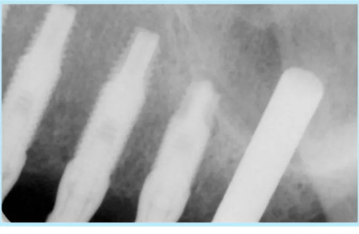
Fig. 17. Lower right molar portion : Pre-OP CT view.
- Enough bone width & height
Myung-Jin Jang: Flapless implant surgery with 3D digital dental CT; A Case Report. Implantology 2009
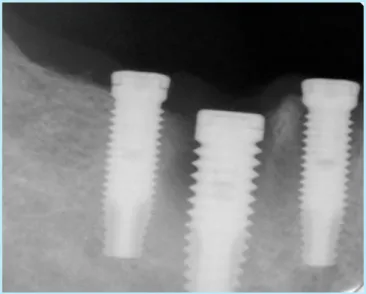
Fig. 18. 3 implants were inserted : Lower 1stpremolar(D;3.75mm, L;13mm), 2ndpremolar(D;5mm, L;13mm),
1st molar(D;4mm, L;13mm)
Myung-Jin Jang: Flapless implant surgery with 3D digital dental CT; A Case Report.
Implantology 2009
Fig. 19. Abutment connection.
Myung-Jin Jang: Flapless implant surgery with 3D digital dental CT; A Case Report.
Implantology 2009
Fig. 20. Digital CT view: after lower right 3unit PFG bridge setting.
Myung-Jin Jang: Flapless implant surgery with 3D digital dental CT; A Case Report.
Implantology 2009
Fig. 21. Occlusal view-after lower right 3unit PFG bridge setting.
Myung-Jin Jang: Flapless implant surgery with 3D digital dental CT; A Case Report.
Implantology 2009
Fig. 23. Panoramic view after prosthesis setting.
Myung-Jin Jang: Flapless implant surgery with 3D digital dental CT; A Case Report.
Implantology 2009
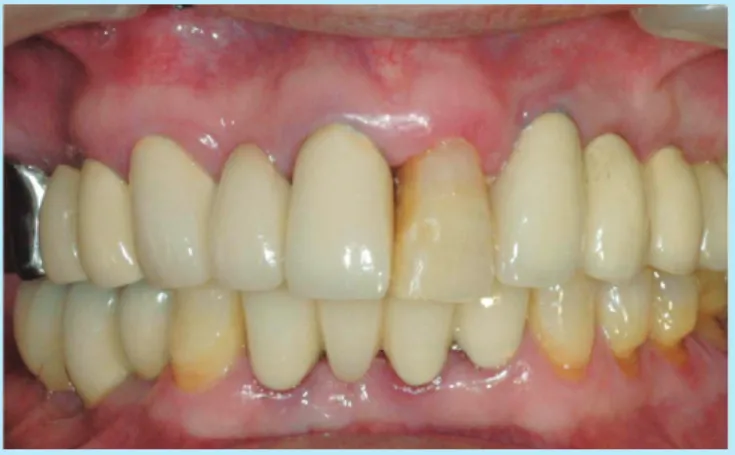
Fig. 22. Anterior view : Aftrer treatment.
Myung-Jin Jang: Flapless implant surgery with 3D digital dental CT; A Case Report.
Implantology 2009
case report
열고 닫는 시간을 줄일 수 있었고 2차 수술을 간단히 시행하 였으며 환자분의 수술에 대한 만족도가 높고 술후 연조직 변 화등의 부작용도 적게 발생하여 환자관리가 용이하였다.
치아를 발치후 임프란트를 구강내에 식립하는 과정은 먼저 구강점막의 절개, 각종 드릴에 의한 치조골의 분쇄, 열발생에 의한 골괴사, 혈류 폐쇄등의 물리적 자극과, 주위조직의 pH의 변화, 기타 화학적 자극등에 의한 구강내 연조직과 경조직에 동시에 창상을 야기하는 과정이다. 임프란트 식립 후 손상받 은 여러 주변 조직의 치유과정을 거치게 된다 조직피판을 열 고 임프란트 식립시 절개부에 열개가 발생되어 염증반응이 심 해지고 장기화되며 다량의 반흔을 남기게 된다. 반면 무피판 절개 수술법은 봉합이 없어 절개부 열개가 발생되지않고 임프 란트 면이 점막과 긴밀한 접촉을 이루어 염증반응 단계가 경 미하고 신속히 경과되어 반흔 조직이 매우 적게 형성되면서 치유된다. 임프란트 주변 점막 두께에 미치는 영향은 피판 절 개 수술후 시간이 경과시 두께는 감소하지만 1년이 경과하여 도 여전히 약 1mm 더 두꺼워진 상태로 유지된다.1)반면 무피 판절개 수술후 부종으로 약 0.7mm 두꺼워지지만 3개월후 원 래 점막 두께보다 약 0.1mm 정도만 두꺼운 상태로 된다. 피판 절개 임프란트 수술후 점막 두께 증가는 골표면의 상방으로 뿐만 아니라 골쪽으로도 두꺼워져서 골소실을 일으키는 원인 이 되지만 무피판절개 수술법은 임프란트 주변 점막의 두께를 원래의 형태대로 유지하는데 매우 유리한 방법이다. 임프란트 주변 열구 깊이에 미치는 영향을 살펴보면 식립 약 3개월후 열 구 깊이는 피판절개 수술시 평균 1.7mm, 무피판절개 수술시 평균 1.0mm로 임프란트 주변 열구의 깊이를 줄여주어 술 후 유지 관리에 유리한 환경을 제공한다.2)임프란트 주변 연조직 의 조직학적 형태에 미치는 영향을 살펴보면 피판절개 수술시
임프란트 주위 연조직 부착부위 약 2mm 높이의 상피층과 약 1mm 높이의 결합조직층이 형성된다.3),4)무피판절개 수술시 에는 접합상피세포가 치근단 쪽으로 적게 증식되어 내려가서 상피층이 더 짧게 만들어지고 열구의 깊이가 더 얕아진다. 임 프란트 주변 연조직 혈관에 미치는 영향은 피판절개 수술시 임프란트 주변 결합조직층은 혈관이 희박한 반흔조직으로 치
유되나5),6)무절개피판 수술시 임프란트 주변 결합조직층은 더
많은 혈관이 분포하는 반흔조직으로 치유되어 치유 및 방어능 력이 향상된다. 무피판절개 수술시 초기 골치유 단계에서 피 판절개 수술법보다 더 적은 골 소실이 일어난다.7),8),9)
아날로그 시대의 임프란트 식립방법은 대개가 환자의 인상 채득과 파노라마 촬영후 대강의 해부학적 중요 구조물을 확 인후 2차원적인 수준에서 하치조 신경이나 상악동등을 손상 시키지 않는 범위에서 행하였다. 그러나 디지털 3차원 컴퓨터 단층영상촬영술을 활용하게 됨에 따라 술전 환자 골두께 및 주요 해부학적 구조물까지의 거리를 정확히 파악할 수 있게 되었고 무피판절개 수술이 가능하게 되어 술후 술자나 환자 입장에서 양호한 결과를 얻을 수 있다.
무피판절개 수술의 장점은 치유기간의 단축, 출혈의 감소, 술후 부작용의 최소화 및 시술후 연조직 변화 의 최소화, 환자 의 불편감 감소, 술후 약물 사용의 최소화 등이 있고 단점으로 는 디지털 3차원 컴퓨터 단층영상촬영을 위한 고가의 장비를 갖추어야하고 시술전 수술용 스텐트를 제작해야 하며 시술중 각 단계를 이동형 디지털 구내 방사선 사진으로 확인해야 하 고 초보자인 경우 연조직 두께를 술전에 정확히 측정하기 어 려워 수직 식립 위치를 정확히 정하기 어렵다는 점이다.
임프란트 식립을 위한 골삭제는 통상의 방법을 사용하며, 최종 단계의 드릴까지 충분히 하여, 임프란트의 식립으로 협 측 피질골이 파절되는 현상을 피하도록 한다. 임프란트의 수 직 식립 높이는 치조정에서 0.5~1mm 깊이까지 하여 만약에 있을 수도 있는 변연골 소실을 대비해야하고 치조제의 상태, 골질 등에 따라 시술 결과가 다른 술식보다 술자의 술기에 영 향을 많이 받는다,
Ⅲ
1. Cardaropoli G. etc. . Tissue alterations at implant supported single tooth replacement : a 1-year prospective clinical study. Clin Oral Implants Res 2006;17:165-171.
2. Becker W, Becker BE, Newman MG. Clinical & microbiologic find- ings that may contribute to dental implant failure. Int J Oral Maxillofac Impl 1990;5:31-38.
3. Berglundh T, Lindhe J. Dimension of the peri-implant mucosa.Biological width revisited. J Clin Periodontol 1996;23:971-973.
4. Berglundh T, Lindhe J, Ericsson I, Marinella CP, Liljenberg B, Thomsen P. The soft tissue barrier at implants & teeth. Clin Oral Implants Res 1991;2:81-90.
5. Berglind T, Lindhe J, Jonsson K, Ericsson I. The tomography of the vas- cular systems in the periodontal & peri-implant tissues in the dog. J Clin Periodontol 1994;21:189-193.
6. Moon IS, Berglundh T, Abrahamsson I, Linder E, Lindhe J. The barrier between the keritinized mucosa & the dental implant. An experimental study in the dog. J Clin Periodontol 1999; 26:658-663.
7. Pham AN, Fiorellini JP, Paguette DP, Williams RC, Weber HP,.
Longitudinal radiographic study of crestal bone levels adjacent to non- submerged dental implants. J Oral Impl 1994;10:26-34.
8. Hermann JS, Cochran DL, Nummikoski PV, Buser D. Crestal bone changes around titanium implants. A radiographic evaluation of unload- ed non-submerged & submerged implants in the canine mandible. J Periodontol 1997;68.
9. Jeong SM, Choi BH, Li J, Kim HS, Ko CY, Jung JH, Kim YH,Lee SH, Engelke W. Flapless implant surgery : an experimental study. Oral Surg Oral Med Oral Pathol Oral Radiol Endod 2007:104;24-28.
교신저자 : 장명진
우편번호 : 136-800, 서울특별시 성북구 길음3동 20-1 현대백화점 10층 성북ENE치과
전자우편 : [email protected] 원고접수일: 2009. 7. 31
1차수정일: 2009. 11. 30 게재확정일: 2009. 12. 1