대흉외지 2003;36:277-279 □ 증례보고 □
- 277 - CASE
Hamartomas, or benign developmental errors, are conventionally defined as focal overgrowths of tissue nor- mally present in the affected organ but in improper proportions1). Primary cardiac hamartomas in children are rare and in the majority of patients, the diagnosis is not usually made until the life-threatening cardiac arrhythmia or ongoing heart failure occurs2). We report the case of young boy with myocardial hamartoma involving the ventricular septum and causing left ventricular out flow tract obstruction.
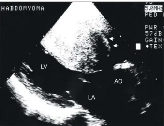
A 15 year-old boy who had a 2 year history of progressive shortness of breath and exertional dyspnea presented with incidentally detected heart murmur in February 1995. His medical history was unremarkable. Physical examination revealed a grade II/VI systolic murmur at the apex and left lower sternal border. An exercise electrocardiographic test was normal, and 24-hour Holter monitoring presented a normal finding except for rare premature ventricular contractions. An echocardiogram showed multiple solid mass shadows in the interventricular septum, as well as in the free wall of both ventricles. The narrowest subaortic dimension
심실중격을 침범한 심근이형종
이정렬*․황호영*․배은정**․김종재***
Myocardial Hamartoma Involving the Interventricular Septum
Jeong Ryul Lee, M.D.*, Ho Young Hwang, M.D.*, Eun Jung Bae, M.D.**
Chong Jai Kim, M.D.***
A 15 year-old boy was referred to us because of mild dyspnea on exertion and incidentally found heart murmur.
On echocardiography, a mass involving mainly interventricular septum and causing left ventricular outflow tract obstruction was detected. Cardiac catheterization demonstrated a transaortic pressure gradient of 20 mmHg. Partial excision of the septal mass was performed via aortotomy under cardiopulmonary bypass. The pathologic diagnosis revealed myocardial hamartoma. The lesion was mainly composed of mature, severely hypertrophic myocytes and intervening fibrosis. During the 5 year of follow-up after the surgery, no evidence of arrhythmia or tumor recurrence was documented.
(Korean J Thorac Cardiovasc Surg 2003;36:277-279) ꠏꠏꠏꠏꠏꠏꠏꠏꠏꠏꠏꠏꠏꠏꠏꠏꠏꠏꠏꠏꠏꠏꠏꠏꠏꠏꠏꠏꠏꠏꠏꠏꠏꠏꠏꠏꠏꠏꠏꠏꠏꠏꠏꠏꠏꠏꠏꠏꠏꠏꠏꠏꠏꠏꠏꠏꠏꠏꠏꠏꠏꠏꠏꠏꠏꠏꠏꠏꠏꠏꠏꠏꠏꠏꠏꠏꠏꠏꠏꠏꠏꠏꠏꠏꠏꠏꠏꠏꠏꠏꠏꠏ Key words : 1. Hamartoma
2. Heart septum
*서울대학교병원 어린이병원 흉부외과, 서울대학교 의과대학 흉부외과학교실, 서울대학교병원 임상의학연구소
Department of Thoracic and Cardiovascular Surgery, Seoul National University Children's Hospital, Seoul National University College of Medicine, Seoul National University Hospital Clinical Research Institute
**서울대학교병원 어린이병원 소아과, 서울대학교 의과대학 소아과학교실
Department of Pediatrics, Seoul National University Children's Hospital, Seoul National University College of Medicine
***서울대학교병원 병리과, 서울대학교 의과대학 병리과학교실
Department of Pathology, Seoul National University Hospital, Seoul National University College of Medicine 논문접수일:2002년 12월 10일, 심사통과일:2003년 1월 27일
책임저자:이정렬 (110-744) 서울특별시 종로구 연건동 28번지, 서울대학교병원 어린이병원 흉부외과 (Tel) 02-760-2877, (Fax) 02-765-7117, E-mail: [email protected]
본 논문의 저작권 및 전자매체의 지적소유권은 대한흉부외과학회에 있다.
대흉외지
2003;36:277-279
- 278 - was 6mm in diameter. A small amount of aortic regurgitation was present as well (Fig. 1). Cardiac catheterization demon- strated a transaortic pressure gradient of 20 mmHg.
At operation, cardiopulmonary bypass was established with cold blood cardioplegia. Through oblique aortotomy, a septal mass causing left ventricular outflow tract stenosis was excised as much as possible. Grossly, the mass was simply not distinguishable with native myocardial tissue, rather much like muscle hypertrophy. The greatest dimension of the
excised septal mass was about 3 centimeters. After one day of intensive care, the patient was transferred to the cardiac ward without an episode of cardiac arrhythmia. Immediate postoperative echocardiography showed no left ventricular outflow tract pressure gradient. Additionally, preexisting systolic murmur disappeared. The pathologic diagnosis revealed myocardial hamartoma. The lesion was mainly composed of mature, severely hypertrophic myocytes and intervening fibrosis. The myocytes showed disarray with slightly enlarged and hyperchromatic nucleoli. The collagen fibers surrounding individual myocytes often formed dense collagenous plaques. The lesion was distinguishable with surrounding slightly hypertrophic myocytes which were thought to be normal myocardium (Fig. 2).
On the 11th postoperative day the patient was discharged.
During the 5-year annual follow-up, no cardiac arrhythmia or remarkable abnormalities including the growth of the remnant tumor in echocardiogram, was noted (Fig. 3).
DISSCUSSION
The usual type of hamartoma occurring in the heart is a firm white nodule, most frequently in the ventricle, consisting of varying amounts of fat, fibrous tissue, striated muscle, nerves, and blood vessels
1,3). In this case, discrete mass Fig. 1. Preoperative echocardiogram: A huge septal mass led
to an obstruction of the left ventricular out flow tract. The narrowest dimension of the left ventricular outflow tract was 6mm in diameter.
LV
LA
AO
Fig. 2. Histologic examination of the cutting edge, showing disarrayed, mature, and hypertrophic myocytes with prominent hyperchromatic nuclei and surrounding dense collagenous plaques (H&E, ×200).
Fig. 3. Postoperative echocardiogram 5 year after the oper-
ation: There were no signs of remnant tumor growth or left
ventricular outflow tract stenosis.
이정렬 외
심근이형종
- 279 - consisted of primarily normal myocardium lacking of other components. It is feasible to give a diagnosis of rhabdomyoma to this patient, but rhabdomyoma itself is thought to be a myocardial hamartoma rather than a true neoplasm. The question of congenital malformation vs.
neoplasm of these cardiac lesions is difficult to resolve.
However, the increased incidence of these lesions in infants and children favors that they are developmental abnor- malities
2). Clinically, in most reported cases detected early in the patients life, there were intractable cardiac arrhythmias, or sudden cardiac arrests
4). In this particular case, myocardial hamartoma was attributed to left ventricular outflow tract obstruction accompanying a clinical exacerbation of the exertional dyspnea, which eventually led him to surgery.
Although this case appeared sporadically, rhabdomyoma or myocardial hamartoma is associated strongly with tuberous sclerosis, a hereditary disorder characterized by hamartomas in various organs, epilepsy, mental deficiency, and sebaceous adenomas.
During the 5-year follow-up since partial excision of the tumor, the patient has been in good health and a serial chest X-ray and follow up echocardiography showed no evidence of cardiomegaly or growth of remnant tumor. As mentioned in the previous reports, surgical excision of the primary myocardial tumor is known to be the most successful method
of treatment with a low mortality and morbidity
5,6). In this case, although multiple parts of the heart were involved, with palliative resection of the interventricular septal mass, successful control of the disease and excellent mid-term follow up results were achieved.
REFERENCES
1. Burke AP, Ribe JK, Bajaj AK, Edwards WD, Farb A, Virmani R. Hamartoma of mature cardiac myocytes. Hum Pathol 1998;29:904-9.
2. Koponen MA, Siegel R. Hamartomatous malformation of the
left ventricle associated with sudden death. J Forensic Sci
1995;40:495-8.3. Wedemeyer AL, Breitfeld V. Cardiac neoplasm, tachyarrhy-
thmia, and anasarca in an infant. Am J Dis Child 1975;
129:738-41.
4. Gharagozloo F, Porter CJ, Tazelaar HD, Danielson GK.
Multiple myocardial hamartomas causing ventricular tachy- cardia in young children: combined surgical modification and medical treatment. Mayo Clin Proc 1994;69:262-7.
5. Takach TJ, Reul GJ, Ott DA, Cooley DA. Primary cardiac
tumors in infants and children: immediate and long-tem operative results. Ann Thorac Surg 1996;62:559-64.
6. Beghetti M, Gow RM, Haney I, Mawson J, Williams WG, Freedom RM. Pediatric primary benign cardiac tumors: a