대한임상병리사회지
:제 24 권 1 호
1992첼분을 함유한 형질세포의 혈액학적 소견
대구 가톨릭대학병원 임상병리과
조창호·문호범
Key words : Iron containing plasma cell, Marrow iron storages,
Alcoholism, Liver function abnormality.
1 .
서
료로‘-
골수검사에서 철분을 함유한 형질세포는 드물게 나타나며 대부분 alcoholism과 관련되어 있고 1-4)
그외에는 hemochromatosisl,5) , 거대적아구성 빈혈 1 , 5),I하I‘actory anemia ( 이 하 R.A.) 1,3,5), porphyria cuta- nea tarda5>, refractory anemia with excess blast(
이
하 RAEB)6), 재생불량성 빈혈 6), dysglobulinemia 7),다발성 골수종8),
inflammatory lesion of spider bite9)둥에서 보고된 예가 있으며 아직까지 철분이 형질 세포 내로 들어가는 기전은 밝혀져 있지 않다.
형질세포 내의 철분함유는 1938년 Jaffe
lO)가 발 견한 이래 국외에서는 산발적으로 가끔씩 보고되 고 있으나 국내에서는 1990년 박 동6)에 의한 3 예 보고 이외는 거의 찾아 볼 수 없었다.
저자들은 최근 2 년 동안 골수상에서 42 례를 경 험한 바 철분함유 형질세포의 의의를 알아보고자 하여 관찰 당시의 병리학적 소견, 특히 말초혈액 및 골수검사의 성적, 혈청 생화학적 검사 성적을 중심으로 보고하고자 하는 바이다.
n . 관찰대상 및 방법
1.관찰 대상
1989 년 9월부터 1991 년 8월까지 2 년동안 말초 혈액검사에서 비정상을 보여 골수검사를 시행한 총 605 명의 환자 중 철분을 함유한 형질세포를 나 타내는 42 례를 대상으로 하였다.
2.
방 법
골수검사를 할 당시의 혈액을 채취하여
CBC,생
화학 및 혈청학적 검사를 실시하였으며 CBC는
Co-ulter사의 T540으로 분석 하고 혈 액 도말은
Wright염색을 하여 세포 백분율과 형태학적 관찰을 하였다.
생화학적 검사 중 aspartate aminotransferase (AST), alanine aminotransferase(ALT), alkaline phosphatase(ALP), lactic acid dehydrogenase (LDH), gamma-glutamyl transferase(GGT) 를 Ab- bott Spectrum
자동분석 기 로,
ferritin,비 타 민
BI2(Vit. Bd,
염산옳
RIA방법으로, 혈청철 (Fe) , 총 철결합능 (TIBC) 을 수동방법으로 실시하였으며, B 형 간염 표면항체 및 항원에 대한 간염표식자 검 사를 EIA 방법으로 실시하였다.
골수도말표본 두 장을
Wright염색하여 세포 200 개를 세어서 형질세포의 백분율 계산과 골수 소견을 관찰하였다. 도말표본 한 장을
prussian blue염색하여
Gale둥의 기준에 의거 저장철을
O-6+로 구분하였으며, 형질세포 50 개를 관찰하여 철함유 형질세포의 백분율을 환산하였다.
임상적 소견은 환자 병록지를 참고하였고, 음주 여부는 과거력상 음주경험 여부로 구분하였다.
ID. 결 과
1.임상소견 및 진단벌 분포
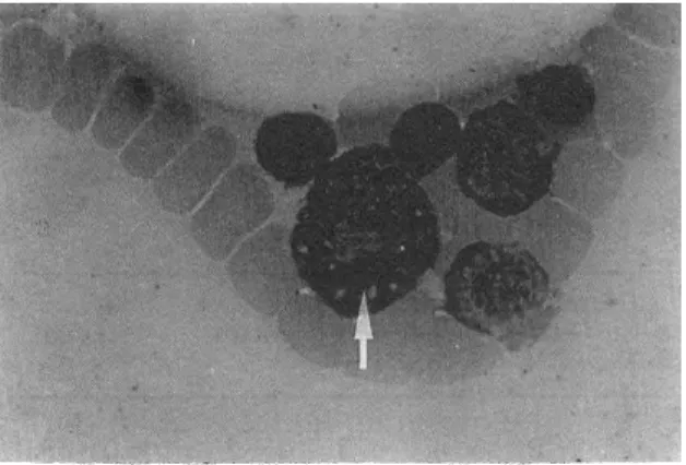
철분을 함유한 형질세포(그림
1,2) 를 가진
42례에서의 성별 및 연령분포는 표 1 과 같았다(평균 연령 46.5세).
진단별로는 표 2 에서 보는 바와 같이 간경변증
및 간염이 13 례 (31 %)로 가장 많았으며 다음으로
골수 이형성 증후군 8 혜 (20%) , 급성 백혈병 및
림프종에서 각각 5 례(1 2%) , 종양에서 2 례 (5%)
순이었다.
Fig.1. Iron containing plasma cell in marrow shows coarse brown black granules.
(Wright stain, X 1000)
Fig.2. Iron containing plasma cell in marrow shows coarse blue granules.
(Prussian blue stain, X 1000)
Table 1. Distribution of study subjects by sex and age
Age Male Female Total
No. % No. % No. %
10-19 20-29 30-39 40-49 50-59 60-69 70-79
I니 Aaτ
nU
A4
‘ RV
AμI
IJ 14
1ι
1i
nι
1l4
2 6 4 6
끄
6 2
1 2
2 7 4 8
퍼
6 2 2
2 5 5
Total 37 88 5 12 42 100
음주경험이 있는 례가 34 명 (81
%),그렇지 않은 경우가 8 명(1 9%) 으로 음주경험이 있는 례가 훨씬
많았다.수혈 여부는 받은 경우가 22 례 (52%) , 그렇지 않은 경우가 20 례 (48%) 로 거의 만반이었다.
철분 함유 형질세포의 백분율은
Wilson질환에 서 90% 로 가장 높았고, 간질환, 종양, 특발성 혈소 판 감소성 자반증, 거대적아구성 빈혈 및 진성 다 혈증에서 약
50%정도로써 다른 질환에 비해 높
았다.T able 2. Distribution of alcohol, blood transf usion and iron containing plasma cells by diagnosis
Diagnosis
Case ICP1)
No. %
Alcohol Trans- Yes No fusion (%)
Acute myelocytic leuke- mla
Non Hodgkin’s lymphoma Myelodysplastic
syndrome Liver cirrhosisjHepatitis Carcinoma2)
ITp3)
Chronic renal failure Wilson’s disease Megaloblastic anemia Aplastic anemia Hemolytic anemia Polycythemia vera Tuberculosis Pneumonia
5 12 12 3 2 5
8
3 7
2 6 2 1 12
20 24 39
퍼
2 1 1 1 1 1 1 1 1 -
왜 왜 많
M
mm 邱잃
mω
많mω
m
5 2
m
5 2 2 2 2 2 2 2 2 2
n/ι
1’4
1li
1li
---ι
Total 42
100魔:]
34 8 22※ 1) Iron containing plasma cell 2) Lung Ca. and bladder Ca.
3) Idiopathic thrombocytopenic purpura 5
m m
패
MU
M 5
2.
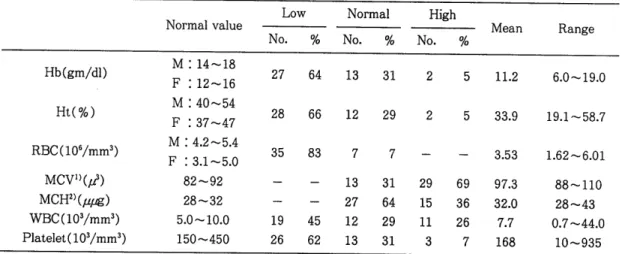
말초 혈액검사 소견
표 3 에서 보는 바와 같이 혈색소가 낮은 경우가 27 례 (64%) 로 가장 많았고 오히려 증가한 경우가 2 례, 평균이
11.2 gm/dl,범위는
6.0-19.0gm/dl로 다양하게 분포하고 있었다. MCV는 높은 경우가 29 례 (69%) 였고, MCH는 정상범위를 보인 경우가
27 례 (64%) 였다.혈소판은 평균이
168,000/mm3,범위는
10,000- 935,000mm
3로 감소한 경우가 26 례 (62%) 로 많았
으며 18 례 (43%) 에서는 거대혈소판이 관찰되었다. 44,000/mm
3이고, 독성과립
6 례,좌방이행
5 례,호 백혈구는 증가한 경우가 11 례 (26%
),감소한 경우 산구증다증 2 례, 과분절 호중구 1 례가 관찰되었다.
가 12 례 (29%) , 평균 7,700/mm
3로서 범위는
700-Table 3. Findings of peripheral blood
Low Normal High
Mean Range
No. % No. % No. %
Normal value
Hb(gm/dl) M : 14-18
F : 12-16
Ht(%) M : 40-54
F : 37-47 RBC(106/mm3) M : 4.2-5.4 F : 3.1-5.0
MCV))(,d) 82-92
MCH2)
(μ짧)
28-32 WBC(103/mm3) 5.0-10.0 Platelet(103/mm3) 150-45027 64 13 31 2 5 11.2 6.0-19.0
28 66 12 29 2 5 33.9 19.1-58.7
35 83 7 7 3.53 1.62-6.01
nJ nJ
7 8
”I
?‘
7.
FL
Qν
nJ
rj
1j
%
%ω %ω
7
%
πu
n
3 E
없 m섭 인싸
퍼 mμ mμ 냐ω
왜 없
m생 잊ω
88-110 28-43 0.7-44.0
10-935
※ 1) Mean corpuscular volume 2) Mean corpuscular hemoglobin
3.
골수검사 소견
골수도말 표본을
prussian blue염색한 것에서 철함유 형질세포는 2-90% 까지 다양하게 분포되 어 있었 A며 평균 36% 였다. 저장철은 1-6+로 역
시 넓게 분포되어 있었고 4+ 이상이 27 례 (64%) 로관찰되었으며, 형질세포도 0-18% 까지 넓은 분포 현상을 보였으며
5%이상 증가된 경우는 8 례(1 9
%)로서 평균 2.9% 였다.
Fig. 3. Megaloblastic change in marrow (Wright stain, X 1000)
골수의 세포충실도는 21 례 (50%) 에서 정상, 감 소가 14 례 (33%
),증가가 7 례(1 7%) 였다. 거핵세포 도 정상 32 례 (76%
),감소 9 례 (22%
),증가 1 례를 보였으며,
differentialpattern은 정상이 10 례 (23
%),
적혈구계 증식이 24 례 (57%
),골수계 증식이 9 례 (20%) 로 각각 나타났다.
형태학적 이상소견은 거대적아구성(그림
3)소 견 26 례 (62%
),지환형 철적아구(그림
4)소견
7례(1 7%) 에서 나타냈다(표
4).티g. 4. Ringed sideroblastic change in marrow (Prussian blue stain, X 1000)
T able 4. Findings of bone marrow by iron containing plasma cells ICP(%)l)
Total( %) Mean Range
0-29 30-59 60-99
Case 21(50%) 14(33% ) 7(17%) 42(100) 36% 2-90%
1-3+ 6 7 2 15(36)
Hemosiderin 4+ 1-6+
4-6+ 15 7 5 27(64)
Plasma cell <5 17 11 6 34(81)
(%/200 marrow cell) 르 5 4 3 1 8(19) 2.9% 0-18%
Cellularity
Normocellularity 10 8 3 21(50)
Hypercellularity 2 4 1 7(17)
Hypocellularity 9 2 3 14(33)
Megakaryocyte
10 4 32(76)
Adequate 18
1 l( 2)
Increased
3 3 9(22)
Decreased 3
Differential pattern2)
Normal 3 2 5 10(23)
Myeloid hyperplasia 7 1 8(20)
Erythroid hyperplasia 11 11 2 24(57)
Megaloblastoid normoblast 13 9 4 26(62)
Ringed sideroblast 3 3 7(17)
※
1) Iron containing plasma cell( %/50 plasma cell) 2) Myeloid: Erythroid=3-4 : l(normal)4.
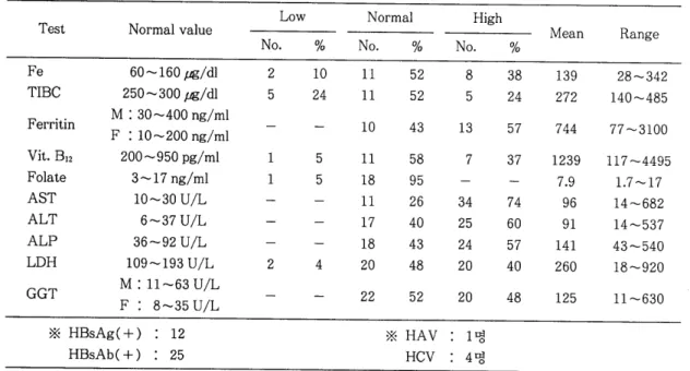
생화학적 검사 및 간염 표식자 소견 Fe와 TIBC는 42 명 중 21 명을 검사하였으며,
Fe는 정상이 11 례
(52%),증가 8 폐
(38%),감소 2 례
(10%),펑균이
139j.tl5/dl 이었고, 범위가 28-342μ g/dl 이였으며, TIBC는 정상이 11 례 (52%) , 감소와 증가가 각각 5 혜 (24%
),평균이
272j.tl5/dl,범위가 140-485j.tl5/dl로 분포되 어 있었다. Ferritin은 42 명 중 23 명을 검사하여서 감소된 예는 없고 증가가 13 례 (57%
),정상은 10 례 (43%) 이었다.
Vit.
B12와 엽산은 각각 11 례 (58%) , 18 례 (95%) 에서 정상, 감소가 각각 1 례씩 있었다.
AST, ALT와 ALP는 각각 31 례 (74%) , 25 례 (60%) , 24 혜 (57
%)에서 높았다. 간염 표식자 검사는 B형 간염 표
변항원이 양성인 경우 12 혜 (29%) , 표면항체에서 25 혜 (60%) 가 양성으로 나타났으며 항원 항체 음 성인 환자에서
HAV1 명, HCV 4 명이 양성을 보였
다(표 5).N. 고 찰
일반적으로 형질세포는 면역계에 관여하는 셰포 로 알려져 있으나 탑식작용을 보이는 사혜가 가끔 씩 보고되고 있으며 Abramson
l1)과
Pavelkal2)품 은 다발성 공수종 환자에게서 적혈구, 백혈구, 혈 소판 동을 탐식한 경우를 보고한 바 있다.
철분을 함유한 형질세포는 1938년 Jaffe
10) 가 보
고 한 이 래
hemochromatosis,거 대 적 아구 성 빈 혈,
Table 5. Findings of biochemical and hepatitis markers Test Normal value Low
No. %
Fe 60 -160 ,ug/dl 2 10
TIBC 250-300
t횡/dl
5 24Ferritin M : 30-400 ng/ml F : 10-200 ng/ml
Vit. B12 200-950 pg/ml 1 5
Folate 3-17 ng/ml 1 5
AST 10-30 U/L
ALT 6-37 U/L
ALP 36-92 U/L
LDH 109-193 U/L 2 4
GGT M : 11-63 U/L F : 8-35 U/L
※ HBsAg( +) 12 HBsAb( +) : 25
RA, RAEB, porphyria cutanea tarda, 재생불량성 빈혈, 다발성 골수종, 간경변증2, 3), 알콜성 간염 2,3,4),
결핵 2) 둥 다양한 질환에서 보고되었으나 주로
al-coholism과 관련되어 나타난다고 하였으며, 아직까 지 확실한 기전은 밝혀져 있지 않다.
1952 년 Koszewkjl)는
hemochromatosis,거 대 적 아 구성 빈혈 환자에서 저장철의 축적이 증가된 9 례 에서 철함유 형질세포를 관찰하였으며 모두
alcoh-lism과 관련되어 있었음을 보고하였다.
Goodman9.} Ha1l5) 은 hemochromatosis, RA, por- phyria cutanea tarda, 거 대 적 아구성 빈혈 4 례 의 골
수에서 전자 현미경 관찰로 형질세포 내의 철분이
Golgi 체 와 내 피 망상계 사이 에 있는 single-walled vesicle내에 있으며 이것은
transferrin으로부터 철 분이 표면 홉수되는 것이라고 하였다.
Lerner
동7)은 1 례의
dysglobulinemia환자 골수 에서 철함유 형질세포의 전자현미경 관찰에서
ves-icle 이 lysosome 일 것 이 라고 추정 했으며 , radioacti-
vity
59Fe를 정맥주사하고 1 일 내에 형질세포 내의
radioactivity가 발견되는 것으로 보아 탐식작용보 다는 직접적인 기전에 의한 것이라고 제시했으며,
Shan-mugathasa
동13) 은 형질세포와 철분을 함유
한 조직구 사이에 bridge가 있다고 주장하였다.
Karcioglu와 Hardison
2) 의 보고에 의 하면 대 상자 21 례는 전부 남자로서, 만성 alcoholism과 빈혈을,
그리고 16/21 레에서 간기능 이상을 보이는 환자로
Normal High
Mean Range
No. % No. %
11 52 8 38 139 28-342
11 52 5 24 272 140-485
10 43 13 57 744 77 -3100
11 58 7 37 1239 117 -4495
18 95 7.9 1.7 -17
11 26 34 74 96 14-682
17 40 25 60 91 14-537
18 43 24 57 141 43-540
20 48 20 40 260 18-920
22 52 20 48 125 11-630
※ HAV 1 명
HCV 4 명
서 거대적아구성과 지환형 철적아구의 변화를 보 인 경우가 각각 5 례, 3 례, 저장철은 12 례에서 증가 를, 형질세포는
1-12%,철함유 형질세포는
2-80%로 넓은 분포 양상을 관찰하였으며, 결론으로 철 분함유 형질세포는 저장철의 과잉과 관련된 질환,
만성감염, 만성염증 진행, 만성 alcoholism과 관련 되어 나타난다고 주장하였다.
1982 년 Cook와 Madden
3) 이 13( 남
: 6,여
:7) 례
를 보고한 바에 의하면 alcoholism 과 관련된 경우가 9 례, 저장철은 다양하고, 형질세포는
0-12%, 철함유 형질세포 22-94%, 혈색소는 모두 저하를보여주며, 형태학적 관찰에서는 거대적아구성과 지 환형 철적아구의 변화를 보여주었다고 하며, 전자 현미경 관찰에서 형질세포의 손상된
mitochondria내부에 철분이 축적되었으며 이것은
alcoholism환 자에서
heme합성과
pyridoxine대사의 장애 때문 이 라고 주장하였고, Romslo
l4) 와 Flatmark
l5) 도 철 분 의 축적은
respiratorychain에 관여하는 모든 조직 세 포의 mitochondria의 역 할이 며
,이 가설 이 간 세 포나 다른 조직에서의 철분의 존재를 설명할 수 있다고 했다.
McCurley
동4)은 남자 53 례를 보고하면서 47 명
이 alcoholism과 관련되 어 있고 다-양한 질병 에서
나타났고 50 례에서 혈색소가 감소되었고, 백혈구와
혈소판의 감소가 각각 6 례, 17 례로서 분포가 다양
했으며 간기능검사의 이상을 보여주는 경우가
35all Y. sol 9i 1:1-. t§ Ell '¢"}
~ ~
::G_.g..~
-T~ ~
-T%, 7-1 Lll~~-T%, ~~-T
l.{i.Q.l :g-:>t.,7-l~t§ ~~
0~-Tg_l
1(! §~ -~- _l]l_ O:j T :d _Q_ ~ ::z-j 7J ~ .Q.l -~- ~ 0 l q- 0d' "61- 111
1985 \::! Blom8l.g_.. t§ ~All _:;t_ Lll .Q.] ~~.g. vesicle l.{i 011 % ol 91 7] 5:_ "61· 7-1l{}, All !I
~
l.f1 01]~ ~
S:j ol~o]
"6]·111:tl-%~ ~_Q_.£ ~A~
tn]~ ~
5/5,~ ~
~
3/5,~4=-
o] t§A~
.:g=-.q:.-;r 2/8all 01P·-l j-1%~4=
AJ-011~ ~ ~*
t§~A-jJ!t_% ~
4=-~<;j.Q.L-j- ~Jqf;k]j
){j_li --'5=- All ~4=-i3J.~011J.-] :Q-71~ o]AJ~ %~-5}0:]
:tl-%S:J.C
~_Q_£ _l]l_O~ ~~* t§~All.:£:~ :j-7]~
0 l "'J I4- ~ {) ~ ::tl-7-1]7} 91 ~ ~ _Q_.£ _l]l_ O:j 7-] Pi -6J=+
91 _Q_
~,
mitochondria 1-li 01]J.-H:~
:tl-~-
W <T i1i 9i 1:]-~
c-] fi: .g.~
-T7~ ~
.R W~
_Q_.£A~
Zf~
-c}.J?.. -T-7J -6]-
~
_Q_ Y., o 1~
-g_-~
!=§ W 4=- 91~-
7}~
.g.;kJ] 1-] -5]-7-] *~ c]-.
::z-] 7-]-%.Q.l :tl-%01JA-l 5:. 1:]-0J~ ~ ~ OJ"'J-~ _l]l_O:j T
9i _Q_
~ ~
ii] alcohol 1l~
34 t:Jl( 81 % ) , Zl-75 1(! 3JZ}-~~7-1-7} 13t:Jl(31%)~_Q_~ ~~7-JAJ- :j-~_Q_.£
~#S:17-1 ~<V;_Q_L-j- :Q-71~
o]"-J%_l]l_o]~
75-9-%~-51-1{! 272:]](64%).£ fi;<V;-c}. o]-c Koszewkil), Ka- rcioglu-2} Hardison2l, Cook-2} Madden3l, McCurley
%4l~- l:l] x~ ~I4--~- _l]l_O:j .2f'-9i_Q_l-]-, hemochroma- tosis-2}
q-~Aa ~4=-% ~.A]-~
i]i 9i q.~
4=- ::z-J 7J~ ~ ~
7a s:.~-
1~
6 + P1a -rr :
4 +) • t§~
-A1l.:£:Q.] 1
!~~-&.g. 0~18%(S!JTI-:
2.9%),~~*
t§~-A1l.:£: ~ 2~90% ( srJ -IT : 36% ).£ o] 5:. Karcioglu .2} Hardison2l, Cook-2} Madden3l, McCurley %4li4-
%J.}ii]-71l
q-
0d'~ ~¥.% _l]l_~_Q_L-j- ~ ~ ~~ .:G-~
7} 27t:ll(64%), 7-a-"J-ol 13t:Jl(31%), ..2.ii1Pl
%-7}~
75-9-7} 22:]](5%).£ q~ -"Jo1~ j}o1-~- _l]l_O:j -T9i
q.
o1~ q]AJ~~~ ~¥.E.]
j}o1IIJl~~ ~_Q_.£
.A~Zf~-c}. ~~.3(1-.g. .:G-~7} 262:]](62%).£ f,§<V;_Q_
o:"j 18t:ll(43%)011J.-l
7-J"CJ1~~Jtl-o1
:t!-%s:19i-c}.~Ell "Qf~
:rl%011 J.-j £ Cook-2} Madden3l, McCurl- ey %4l011J.-]_2} %J.}-5}7117-1Ll1~o~-TA~ ~ 7-1~~
~ ~ o}-=j1-Q.j 1(!§1-% _l]l_O:j -"'f=-9i_Q_o=L ~.A} ~ u175 :tl-
%-g. {Jl-1 -5}7-1 *-5}~ q.
v.~
~~%-g. ~4=-~~ ~~% ~*~ ~~~¥_~
:tl-%~
42t:l101l Ll1iil-O:]~).1-~
l:l}-ct%~- ~.g. ~
""a~
.g. \:f.::zl-71- 37 /42t:JH88% ),~ ~ ~ ~¥_
011 J.-l~ 50~59-A-117}
13/422:]](31% ).£f,§~q-. ~~ ~
~i3J.l-01]J.-]~ ~~~ .:G-~71-
272:]1(64%), %-7} 2t:Jl(5%)~J?.., MCV~ 3E.g. 75-9-7} 292:]](69%), ~
~
.3(1-.g..:G-~ ~
75-9-7~
26 t:Jl( 62%) .£ :tl-% S:19i q-.~4=-~~
01]A-l~ ::Z-]
7J-~ %-7~
272:]1(64% ),~ 0~-T
7-J1
%-~
242:]1(57%)_2}C-J~ol
7-1LJ1~O~TAa
26t:J1 (62%) ~ 7-1 ~t§ ~ ~ O}-=j1-Aa 7t:J1 (17%) 9.1 1(!§}7}:tl-%s:19ic1-.
A~§l-~r~ i3J.l-011J.-1~
ferritin 13/23t:J1 (57%), AST 74%, ALT 60%, ALP 57%.£ ZfZf%-7}~ ~% ~
4=-~9i
q.~~~ ~¥.011J.-]~
%-T~ Z!~~:i!} :tl-TC'!~
75-9- 7}zrzr
81%, 31%&.. f:J-<V;_Q_n:l~-o-1 ~~7-JAJ- :u-
~_Q_&.. ~#s:l.A1 ~<V;_Q_t.f Z}-71~
01-"J-%_l]l_~ ~
%
~w
75+ 64%&..J.-l~~% ~*~ t§~-A-11 ¥.~
%-T
~ Z}~~:i!} ~{)~ ~:tro1 ~.:C ~_Q_.£ .A~
Zfs:19i 71011 i3 J.j-A~ ~% --'5=-{J _Q__£ _l]l_j]_ "6}-E- l:l~O]Lj-.
Hematologic Findings in Iron Containing Plasma Cell
Cho, C. H., Moon, H. B.
Dept. of Clinical Pathology, Taegu Catholic University Hospital
ABSTRACT tients with iron containing plasma cells in marrow at Catholic hospital over a two-years period.
There have been a limited number of reports of Iron containing plasma cells were associated most iron containing plasma cells. We studied clinical fea- commonly with alcoholism and occurred in marrow tures, laboratory findings, and reviewed the medical with increased and normal iron storages, and the records and marrow aspirates of 42 consecutive pa- marrow changes of megaloblastoid normoblasts and
ringed sideroblasts.
Previous reports were reviewed. The presence of iron containing plasma cells is suggestive of alcohol-
ism and its complication, and disease associated with liver function abnormality. The mechanism of entry of iron into plasma cells is unknown.
REFERENCES
1. Koszewski BJ : The occurrence of megaloblastic erythropoiesis in patients with hemochroma- tosis. Blood 7 : 1182-1195, 1952.
2. Karcioglu GL, Hardison JE: Iron-containing plasma cell. Arch Intern Med 138 : 97, 1978.
3. Cook MK, Madden M: Iron granules in plasma cells. J Clin Pathol 35 : 172, 1982.
4. McCurley TL, Cousar JB, Graser SE, Glick AD, Collins RD :Plasma cell iron-Clinical and mor- phologic features. Am J Clin Pathol 81 : 312, 1984.
5. Goodman JR, Hasll SG : Plasma cells contain- ing iron. An electron micrograh study. Blood 28
: 1-83, 1966.
6.
~cr!~, ~_7J
7-}, ~ ~ lf- : ~~% ~%~
1fJ~A~l
¥., ~~~C1~ 78.£-\:J:C1 18:295, 1990.
7. Lerner RG, Parker JW : Dysglobulinemia and iron in plasma cells. Arch Intern Med 121 : 284, 1968.
8. BJorn J : The ultrastructural localization of iron in human bone marrow plasma cells. Acta Path Microbiol Immunol Sect A 93 : 223, 1985.
9. DeLellis RA :Iron containing plasma cell in a healed recluse spider bite. Arch Pathol 92 : 254
-256, 1971.
10. Jaffe RH :The reticuloendothelial system. In handbook of hematology, Edited by H Downey, New York, Hoeber, Vol 2 : 1170, 1938.
11. Abramson N, Kapff CV, Ginsburg AD: The phagocytic plasma cells, N Engl J Med 282 : 248 ... 250, 1970.
12. Ludwig H, Pavelka M :Phagocytic plasma cell in a patient with multiple myeloma. Blood 121
: 284-287, 1968.
13. Shanmugathasa M, Sobel HJ, Marquet E : Plas- ma cells with inclusions containing iron. Arch Pathols Lab Med 103 : 577, 1979.
14. Romslo I : Energy-dependent accumulation of iron by isolated rabbit reticulocyte mitochon- dria. Biochim Biophys Acta 357 : 34, 1974.
15. Flatmark T, Romslo I: Energy-dependent accu- mulation of iron by isolated rat liver mitochon- dria. J Bioi Chern 16 : 6433, 1975.