433 http://www.jchestsurg.org
JCS
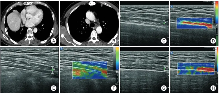
Journal of Chest SurgeryReal-Time Pleural Elastography: Potential Usefulness in Nonintubated Video-Assisted Thoracic Surgery
Federico Tacconi, M.D. 1 , Fabrizio Chegai, M.D. 2 , Tommaso Perretta, M.D., Ph.D. 2 , Vincenzo Ambrogi, M.D., Ph.D. 1
1