as causes of secondary hypereosinophilia [5, 6]. Particularly, ana- plastic large cell lymphoma (ALCL) is one of the T-cell lympho- mas accompanied by secondary eosinophilia. Here, we present a patient with marked proliferation of eosinophils in the peripheral blood and bone marrow (BM) with a complex karyotype, leading to initial misdiagnosis as chronic eosinophilic leukemia (CEL). Af- ter further evaluation of metaphase cytogenetics (MC) and inter- phase fluorescence in situ hybridization (iFISH) at diagnosis and follow-up, the patient was finally diagnosed with ALCL masked by eosinophilia.
A 42-year-old man with a history of pulmonary hypertension due to atrial septal defect and atopic dermatitis had complained of vesicles and bullae on both thighs and the belly for one month. In the initial work-up, his peripheral blood test showed leukocytosis (49.7×10⁹/L) with severe eosinophilia (34.8×10⁹/L) (Table 1). A peripheral blood smear showed mainly mature eosinophils with- out immature cells or abnormal lymphocytes (Fig. 1). Total immu- Primary eosinophilia is a disorder involving clonal proliferation
of eosinophils, whereas secondary eosinophilia can be caused by a variety of factors such as allergic disorders, parasitic and fungal infection, endocrine disorders, toxins, autoimmune diseases, or tumors. Secondary eosinophilia associated with tumors may be related to a cytokine-derived reactive phenomenon secreted by tumor cells [1-4].
Hodgkin or T-cell lymphomas have been commonly described
진단초기 만성호산구백혈병으로 오진한 호산구증가증과 복합핵형을 나타낸 역형성큰세포림프종
Anaplastic Large Cell Lymphoma with Massive Eosinophilia and Complex Karyotype Initially Misdiagnosed as Chronic Eosinophilic Leukemia
소민경1·박설희1·조민선2·문영철3·허정원1
Min-Kyung So, M.D.1, Sholhui Park, M.D.1, Min-Sun Cho, M.D.2, Yeung Chul Mun, M.D.3, Jungwon Huh, M.D.1
이화여자대학교 의과대학 진단검사의학1, 병리학2, 내과학3
Departments of Laboratory Medicine1, Pathology2, and Internal Medicine3, College of Medicine, Ewha Womans University, Seoul, Korea Vol. 8, No. 2: 56-61, April 2018
https://doi.org/10.3343/lmo.2018.8.2.56 진단혈액학
Corresponding author: Jungwon Huh
Department of Laboratory Medicine, College of Medicine, Ewha Womans University, 1071 Anyangcheon-ro, Yangcheon-gu, Seoul 07985, Korea Tel: +82-2-2650-5320, Fax: +82-2-2650-5091, E-mail: [email protected] Received: September 11, 2017
Revision received: October 24, 2017 Accepted: November 14, 2017
This article is available from http://www.labmedonline.org 2018, Laboratory Medicine Online
This is an Open Access article distributed under the terms of the Creative Commons Attribution Non-Commercial License (http://creativecommons.org/licenses/by-nc/4.0/) which permits unrestricted non-commercial use, distribution, and reproduction in any
We report a patient with massive eosinophilia and a complex karyotype that was initially misdiagnosed as chronic eosinophilic leukemia (CEL), but later diagnosed as anaplastic large cell lymphoma (ALCL) masked by massive eosinophilia. The complex karyotype observed at initial diagnosis re- mained unchanged later, after the evidence of bone marrow involvement of ALCL was obtained. At diagnosis, genetic aberrations corresponding to metaphase cytogenetics were not identified by interphase fluorescence in situ hybridization, although abnormal results were noted at follow-up.
Together, these observations indicate that the complex karyotype at initial work-up has been derived from a low proportion of lymphoma cells with high mitotic ability that were not identified by microscopy, rather than from massive eosinophils. These findings suggest that our patient had ALCL with secondary eosinophilia rather than CEL since initial diagnosis.
Key Words: Hypereosinophilia, Metaphase cytogenetics, Interphase fluorescence in situ hybridization, Anaplastic large cell lymphoma, Chronic eosino- philic leukemia
2017-03-16 https://crossmark-cdn.crossref.org/widget/v2.0/logos/CROSSMARK_Color_square.svg
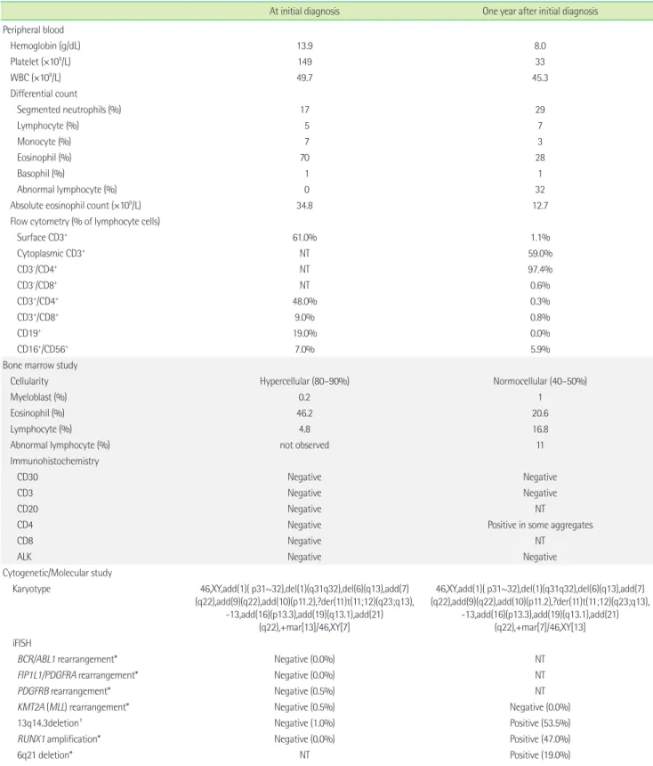
Table 1. Laboratory characteristics at initial diagnosis and follow-up study
At initial diagnosis One year after initial diagnosis Peripheral blood
Hemoglobin (g/dL) 13.9 8.0
Platelet (×109/L) 149 33
WBC (×109/L) 49.7 45.3
Differential count
Segmented neutrophils (%) 17 29
Lymphocyte (%) 5 7
Monocyte (%) 7 3
Eosinophil (%) 70 28
Basophil (%) 1 1
Abnormal lymphocyte (%) 0 32
Absolute eosinophil count (×109/L) 34.8 12.7
Flow cytometry (% of lymphocyte cells)
Surface CD3+ 61.0% 1.1%
Cytoplasmic CD3+ NT 59.0%
CD3-/CD4+ NT 97.4%
CD3-/CD8+ NT 0.6%
CD3+/CD4+ 48.0% 0.3%
CD3+/CD8+ 9.0% 0.8%
CD19+ 19.0% 0.0%
CD16+/CD56+ 7.0% 5.9%
Bone marrow study
Cellularity Hypercellular (80–90%) Normocellular (40–50%)
Myeloblast (%) 0.2 1
Eosinophil (%) 46.2 20.6
Lymphocyte (%) 4.8 16.8
Abnormal lymphocyte (%) not observed 11
Immunohistochemistry
CD30 Negative Negative
CD3 Negative Negative
CD20 Negative NT
CD4 Negative Positive in some aggregates
CD8 Negative NT
ALK Negative Negative
Cytogenetic/Molecular study
Karyotype 46,XY,add(1)( p31~32),del(1)(q31q32),del(6)(q13),add(7) (q22),add(9)(q22),add(10)(p11.2),?der(11)t(11;12)(q23;q13),
-13,add(16)(p13.3),add(19)(q13.1),add(21) (q22),+mar[13]/46,XY[7]
46,XY,add(1)( p31~32),del(1)(q31q32),del(6)(q13),add(7) (q22),add(9)(q22),add(10)(p11.2),?der(11)t(11;12)(q23;q13),
-13,add(16)(p13.3),add(19)(q13.1),add(21) (q22),+mar[7]/46,XY[13]
iFISH
BCR/ABL1 rearrangement* Negative (0.0%) NT
FIP1L1/PDGFRA rearrangement* Negative (0.0%) NT
PDGFRB rearrangement* Negative (0.5%) NT
KMT2A (MLL) rearrangement* Negative (0.5%) Negative (0.0%)
13q14.3deletion† Negative (1.0%) Positive (53.5%)
RUNX1 amplification* Negative (0.0%) Positive (47.0%)
6q21 deletion* NT Positive (19.0%)
7q31 amplification* Negative (0.5%) Positive (30.0%)
*cutoff: 1.5%; †cutoff: 3.7%.
Abbreviations: ALK, anaplastic lymphoma kinase; FIP1L1/PDGFRA, FIP1-like-1–platelet-derived growth factor receptor-alpha; iFISH, interphase fluorescence in situ hybridiza- tion; MLL, myeloid lymphoid leukemia, NT, not tested; PDGFRB, platelet-derived growth factor receptor-beta; RUNX1, Runt-related transcription factor.
Fig. 1. Morphology, cytogenetic studies, and interphase fluorescence in situ hybridization (iFISH) at diagnosis (A) and follow-up (B). (A1) Peripheral blood smear showing eosinophilia without abnormal lymphoid cells. (A2) BM examination showing hypercellularity with marked eosinophil pre- dominance. No abnormal lymphoid cells were observed. (A3) G-banded karyotype result showing a complex karyotype at diagnosis. (A4) iFISH us- ing D13S319/13q34 probes and RUNX1/RUNX1T1 probes shows normal hybridization signals. (B1) Peripheral blood smear revealing abnormal lym- phoid cells with eosinophilia. (B2) BM aspiration revealing large irregular lymphoid cells with some eosinophils. (B3) G-banded karyotype revealing the same complex karyotype at follow-up as at diagnosis. (B4) iFISH using D13S319/13q34 probes shows two green signals (LAMP1) and one or- ange (D13S319) signal, indicating deletion of the 13q14.3 locus. iFISH using RUNX1/RUNX1T1 probes shows three green (RUNX1) and two orange
46,XY,add(1)(p31~32),del(1)(q31q32),del(6)(q13), add(7)(q22),add(9)(q22),add(10)(p11.2),
?der(11)t(11;12)(q23;q13),-13,add(16)(p13.3), add(19)(q13.1),add(21)(q22),+mar1
RUNX1/RUNX1T1 D13S319/LAMP1
RUNX1/RUNX1T1
46,XY,add(1)(p31~32),del(1)(q31q32),del(6)(q13), add(7)(q22),add(9)(q22),add(10)(p11.2),
?der(11)t(11;12)(q23;q13),-13,add(16)(p13.3), add(19)(q13.1),add(21)(q22),+mar1
D13S319/LAMP1
×400
×400
×1,000
×1,000 A1
B1
A2
B2 A3
B3
A4
B4
there was no evidence of parasitic and fungal infection, endocrine disorders, toxins, or autoimmune diseases.
A BM study revealed hypercellularity with eosinophilic prolifer- ation without increased myeloblasts (0.2% of total nucleated cells).
Eosinophils and their precursors accounted for 46% of total nucle- ated cells. MC analysis showed a complex karyotype (Table 1, Fig.
1). However, genetic aberrations corresponding to MC were not identified at diagnosis by iFISH using the D13S319 13q34 probe, RUNX1/RUNX1T1 dual-color, dual-fusion translocation probe, D7S486/CEP7 FISH probe (Abbott/Vysis, Downers Grove, IL, USA), and 6q21/6q23 probe (MetaSystems, Altlussheim, Germany). In addition, iFISH analysis did not reveal rearrangement of PDGFRA or PDGFRB using the FIP1L1/CHIC2/PDGFRA deletion/translo- cation probe and PDGFRB translocation-break apart probe (Meta- Systems). Based on findings such as marked proliferation of eo- sinophils and complex karyotypes, the patient was initially thought to have CEL, not otherwise specified (CEL-NOS).
Meanwhile, skin biopsy performed at initial evaluation revealed lymphomatoid papulosis, which is known to be associated with CD30+ large T-cell lymphoma. Over subsequent weeks, the skin lesions spread over the whole body with itching sensation and pain. Upon careful physical examination, the patient had small nodal lesions on the neck, axillary, and inguinal areas. Lymph node biopsy showed anaplastic lymphoma kinase (ALK)-negative ALCL (positive expression of CD30, CD4, and CD3 and lack of ex- pression of ALK). We retrospectively reviewed the BM aspiration and biopsy to investigate ALCL involvement in BM, but could not prove infiltration of abnormal lymphoid cells in the BM.
The patient subsequently received standard therapy for ALCL.
After receiving six cycles of chemotherapy (cyclophosphamide, doxorubicin, vincristine, prednisone, etoposide regimen), he un- derwent autologous peripheral blood stem cell transplantation. At three months after this procedure, we found abnormal lymphoid cells (32%; Fig. 1, Table 1) with leukocytosis (45.3×10⁹/L) and eo- sinophilia (12.7×10⁹/L) in the peripheral blood. BM aspiration specimens showed increased abnormal lymphoid cells (11% of to- tal nucleated cells) and eosinophils (21% of total nucleated cells).
Flow cytometric analysis demonstrated the presence of abnormal T lymphoid cells with the presentation of cytoplasmic CD3+/sur- face CD3-/CD4+/CD5+ (Table 1). Additionally, multiple spreading positron emission tomography-avid lymph nodes were observed, demonstrating disease progression. Chromosome analysis yielded
the same results as the initial chromosome study (Fig. 1); however, the iFISH results differed from those at diagnosis. We observed 13q14.3 deletion (nuc ish (D13S319×1,LAMP1×2) [107/200]), RUNX1 amplification (nuc ish (RUNX1T1×2,RUNX1×3) [94/200]), 6q21 deletion (nuc ish (SEC63×1,MYB×2) [38/200]), and 7q31 ampli- fication (nuc ish (D7Z1×2,D7S486×3) [60/200]) in 54%, 47%, 19%, and 30% of interphase cells, respectively (Table 1). Therefore, the patient was diagnosed with BM involvement of ALK-negative ALCL with secondary eosinophilia, not CEL-NOS. Taken together, it is likely that the complex karyotype was derived from abnormal T lymphoid cells of ALCL rather than from eosinophils associated with CEL. The patient died two weeks after follow-up BM exami- nation with gradually worsening symptoms and no response to treatment.
Some studies have reported that the abnormal lymphoma cells in patients diagnosed with ALCL can be masked by massive eo- sinophilia, leading to an initial misdiagnosis of hypereosinophilic syndrome or CEL [7-9]. Clonal T-cell lymphoma can induce sec- ondary eosinophilia by the secretion of cytokines such as inter- leukin-3, interleukin-5, and granulocyte-macrophage colony-stim- ulating factor.
A previous report described a patient who showed a complex karyotype with massive eosinophilia in the peripheral blood and BM, with no evidence of BM involvement of malignant lymphoma but a large cell lymphoma of cervical lymph nodes, leading to a diagnosis of CEL with coexisting lymphoma [10]. Similarly, our pa- tient presented marked eosinophilia and a complex karyotype by MC, but no abnormal lymphoid cells in the peripheral blood and BM, leading to an initial misdiagnosis of CEL. The same complex karyotype was observed later, when there was evidence of BM involvement of ALCL. However, iFISH results differed between the initial diagnosis and follow-up, with abnormal iFISH results observed only at follow-up. These findings suggest that the com- plex karyotype at initial work-up was derived from a small pro- portion of lymphoma cells that was not identified by microscopy but had high mitotic ability. Based on these findings, we revised our final diagnosis as ALCL with secondary eosinophilia. Conven- tional karyotyping test uses metaphase cells and thus the features of cells with high mitotic activity are reflected, while iFISH uses interphase cells. Therefore, the sensitivity of karyotyping by MC is mainly affected by the mitotic ability of targeted cells, whereas iF- ISH can reflect the true proportion of cells rather than the mitotic
ability of cells [11, 12]. Thus, rare clonal cells with high mitotic abil- ity can show genetic changes by MC but not by iFISH.
Cytogenetics of ALK-negative ALCL are heterogeneous. Recent studies have addressed the biological implications of genetic het- erogeneity such as DUSP22 on 6p25.3 and TP63 on 3q28 rearrange- ments within ALK-negative ALCL. They identified DUSP22 and TP63 rearrangements in 30% and 8% within ALK-negative ALCLs, respectively [13, 14]. However, we did not perform molecular stud- ies to detect specific chromosomal rearrangements. Not only chro- mosomal rearrangements but also copy number abnormalities such as 1q, 6q21, and 7p gains and 6q21, 13q, and 17p13 losses in ALCL have been reported [14]. A previous study determined the prevalence of monosomy 13, which was observed in our case, as 5% in adult ALK-negative ALCL [15]. However, deletion 13 is not specific for ALCL, but rather is a relatively common characteristic reflecting tumor aggressiveness.
In conclusion, this study emphasizes the need for extensive patho- logic workup in cases of unexplained eosinophilia with particular attention to MC and iFISH to exclude underlying malignancy masked by eosinophilia. When MC and iFISH results are discordant (i.e., aberrations identified by MC are not identified by iFISH), the pos- sibility of a small proportion of clonal cells with high mitotic abil- ity should be considered.
요 약
과다한 호산구 증가증과 함께 복합핵형의 관찰로 인해 진단 초기 에는 만성 호산구백혈병으로 생각하였지만 최종적으로는 역형성 큰세포림프종으로 밝혀진 증례를 경험하였기에 보고하고자 한다.
진단 초기에 호산구 증가가 보인 골수에서 관찰된 복합핵형이 이 후 역형성큰세포림프종이 골수를 침범한 것을 확인했을 때에도 동일하게 관찰되었다. 중기세포에서 관찰된 복합핵형 결과와는 달 리, 간기세포를 이용한 형광제자리부합법에서는 진단 초기에는 이 상 소견이 관찰되지 않았다. 하지만 질병이 진행함에 따라 간기세 포 형광제자리부합법에서 복합핵형에 대응하는 비정상적인 결과 를 얻을 수 있었다. 이러한 결과를 통하여 초기 진단 당시 나타난 복합핵형이 대량의 호산구에 의해서가 아닌 현미경으로는 관찰할 수 없었던 소수의 높은 유사 분열 능력을 가진 림프종 세포에서 유 래되었음을 알 수 있었다. 따라서, 이러한 분자유전학적인 차이를 확인함으로써 본 증례가 만성호산구백혈병이 아닌 이차성 호산구 증가증이 있는 역형성큰세포림프종 환자임을 진단할 수 있었다.
AUTHORS’ DISCLOSURES OF POTENTIAL CONFLICTS OF INTEREST
No potential conflicts of interest relevant to this article were re- ported.
ACKNOWLEDGEMENTS
This study was supported by the Basic Science Research Pro- gram through the National Research Foundation of Korea funded by the Ministry of Education, Science and Technology (NRF-2012- R1A1A2044138).
REFERENCES
1. Gotlib J. World Health Organization-defined eosinophilic disorders:
2015 update on diagnosis, risk stratification, and management. Am J Hematol 2015;90:1077-89.
2. Ogata M, Ogata Y, Kohno K, Uno N, Ohno E, Ohtsuka E, et al. Eosino- philia associated with adult T-cell leukemia: role of interleukin 5 and granulocyte-macrophage colony-stimulating factor. Am J Hematol 1998;
59:242-5.
3. Montgomery ND, Dunphy CH, Mooberry M, Laramore A, Foster MC, Park SI, et al. Diagnostic complexities of eosinophilia. Arch Pathol Lab Med 2013;137:259-69.
4. Bain BJ. Hypereosinophilia. Curr Opin Hematol 2000;7:21-5.
5. Desenne JJ, Acquatella G, Stern R, Muller A, Sanchez M, Somoza R.
Blood eosinophilia in Hodgkin’s disease. A follow-up of 25 cases in Venezuela. Cancer 1992;69:1248-53.
6. Utsunomiya A, Ishida T, Inagaki A, Ishii T, Yano H, Komastsu H, et al.
Clinical significance of a blood eosinophilia in adult T-cell leukemia/
lymphoma: a blood eosinophilia is a significant unfavorable prognos- tic factor. Leuk Res 2007;31:915-20.
7. McKelvie PA, Oon S, Romas E, Nandurkar H, Tam CS. A case of sys- temic anaplastic lymphoma kinase-negative anaplastic large cell lym- phoma associated with hypereosinophilia, granulomatous myositis and vasculitis. Leuk Lymphoma 2012;53:2279-82.
8. Orofino N, Guidotti F, Cattaneo D, Sciume M, Gianelli U, Cortelezzi A, et al. Marked eosinophilia as initial presentation of breast implant-as- sociated anaplastic large cell lymphoma. Leuk Lymphoma 2016;57:
2712-5.
sinophilia camouflaging anaplastic large cell lymphoma. Int J Surg Pathol 2011;19:405-8.
10. Gonsalves WI, He R, Pardanani A, Gupta V, Smeltzer JP, Hanson CA, et al. Chronic eosinophilic leukemia-not otherwise specified (NOS) in the background of a large cell lymphoma. Case Rep Hematol 2013;2013:
458303.
11. Fan YS and Rizkalla K. Comprehensive cytogenetic analysis includ- ing multicolor spectral karyotyping and interphase fluorescence in situ hybridization in lymphoma diagnosis. a summary of 154 cases.
Cancer Genet Cytogenet 2003;143:73-9.
12. Wan TS. Cancer cytogenetics: methodology revisited. Ann Lab Med 2014;34:413-25.
13. Mereu E, Pellegrino E, Scarfo I, Inghirami G, Piva R. The heteroge- neous landscape of ALK negative ALCL. Oncotarget 2017;8:18525-36.
14. Mussolin L, Pillon M, Bonato P, Leszl A, Franceschetto G, Di Meglio A, et al. Cytogenetic analysis of pediatric anaplastic large cell lymphoma.
Pediatr Blood Cancer 2010;55:446-51.
15. Zeng Y and Feldman AL. Genetics of anaplastic large cell lymphoma.
Leuk Lymphoma 2016;57:21-7.