Retinopathy of Prematurity in Infants Born before 25 Weeks Gestation in a Korean Single Neonatal Intensive Care Unit:
Incidence, Natural History and Risk Factors
As younger preterm infants are able to survive, more extremely preterm infants are at risk of developing retinopathy of prematurity (ROP). To investigate the incidence, progression and risk factors of ROP in extremely preterm infants in Korea, the medical records of infants born before 25 weeks gestation were retrospectively reviewed. The criteria for laser treatment agreed with type 1 ROP as defined by the Early Treatment for Retinopathy of Prematurity study. Of the 121 infants included in the analysis, 119 (98.4%) infants developed any stage ROP, including 78 infants (64.5%) with type 1 ROP. The mean postmenstrual age (PMA) at the onset of any ROP and type 1 ROP were 33.5 and 36.1 weeks, respectively. All but one infant developed type 1 ROP after 31 weeks PMA.
Univariate analysis showed that duration of total parenteral nutrition and onset of any ROP (PMA) were associated with the development of type 1 ROP. In conclusion, this study shows high incidence of ROP in extremely preterm infants and suggests that, although current screening protocols are feasible for most preterm infants born before 25 weeks gestation, earlier screening before 31 weeks PMA may be necessary in infants with an unstable clinical course.
Key Words: Retinopathy of Prematurity; Infant, Premature; Infant, Low Birth Weight Mingui Kong1, Dong Hoon Shin1,
Sang Jin Kim1, Don Il Ham1, Se Woong Kang1, Yun Sil Chang2, and Won Soon Park2
Departments of 1Ophthalmology and 2Pediatrics, Samsung Medical Center, Sungkyunkwan University School of Medicine, Seoul, Korea
Received: 20 June 2012 Accepted: 11 October 2012 Address for Correspondence:
Sang Jin Kim, MD
Department of Ophthalmology, Samsung Medical Center, Sungkyunkwan University School of Medicine, 81 Irwon-ro, Gangnam-gu, Seoul 135-710, Korea
Tel: +82.2-3410-6775, Fax: +82.2-3410-0074 E-mail: [email protected]
This work was supported by a grant from the Samsung Medical Center Research Fund (SMR1120511).
http://dx.doi.org/10.3346/jkms.2012.27.12.1556 • J Korean Med Sci 2012; 27: 1556-1562
INTRODUCTION
Retinopathy of prematurity (ROP), a disease that affects imma- ture vasculature in the eyes of premature infants, remains a ma- jor cause of blindness and visual impairment in children world- wide (1, 2). Proper screening and timely treatment are essential in improving anatomical and functional outcome (3, 4). As young- er preterm infants are able to survive due to the advances in neonatal intensive care, more extremely preterm infants are at risk of developing ROP (5, 6). However, the epidemiology and natural history of ROP in infants born before 25 weeks gestation have not been investigated in depth, especially in Asian coun- tries. The current screening protocol is based on the early treat- ment for retinopathy of prematurity (ET-ROP) study and cryo- therapy for retinopathy of prematurity (CRYO-ROP) study (7- 11). These previously published studies involved only a small number of extremely preterm infants because they were con- ducted more than a decade ago, during which the survival rates of infants born before 25 weeks gestation were low. Therefore, the current screening protocol may not be suitable for these ex- tremely preterm infants.
This study aimed to evaluate the incidence and progression of ROP in infants born before 25 weeks gestation in a neonatal intensive care unit in Korea. In addition, ocular and systemic risk factors for ROP that require treatment were investigated.
This study analyzed 121 infants with the gestational age (GA) of 22 to 24, one of the largest cohort reported among single center studies. This study may help elucidate the natural history of ROP in extremely preterm infants and to provide the proper time of screening.
MATERIALS AND METHODS
The medical records of consecutive preterm infants born before 25 weeks gestation who were admitted to the Samsung Medical Center neonatal intensive care unit from March 2004 to Novem- ber 2011 were retrospectively reviewed. Exclusion criteria were as follows: infants who died before or within the screening peri- od, incomplete follow-up, and lack of adequate systemic infor- mation. ROP was categorized according to the revised Interna- tional Classification of Retinopathy of Prematurity (IC-ROP) (12). Aggressive posterior ROP (AP-ROP) was defined as a se-
vere form of ROP characterized by its posterior location, promi- nence of plus disease, the ill-defined nature of the retinopathy, and the rapid progression (12). The screening examination for ROP followed the guidelines proposed by the American Acade- my of Ophthalmology and Pediatrics and Association for Pedi- atric Ophthalmology and Strabismus with some modifications (7). The first screening examination was undertaken at 29 to 31 weeks postmenstrual age (PMA). Treatment criteria were based on ET-ROP type 1 disease including zone I, any stage ROP with plus disease, zone I, stage 3 ROP with or without plus disease, and zone II, stage 2 or 3 ROP with plus disease (9, 10).
Fundus findings on ROP screening until the development of type 1 ROP or termination of screening were analyzed. If the two eyes showed asymmetry in progression and severity of ROP, data on the worse eye was included in the analysis. In addition, sys- temic parameters were retrieved including birth weight, Apgar score, multiple births, patent ductus arteriosus (PDA), broncho- pulmonary dysplasia (BPD), intraventricular hemorrhage (IVH), necrotizing enterocolitis (NEC), sepsis, use of surfactant, trans- fusion, duration of total parenteral nutrition (TPN), and dura- tion of oxygen therapy including mechanical ventilation and continuous positive airway pressure (CPAP).
The incidence of any ROP and type 1 ROP by GA was deter- mined by Kaplan-Meier survival analysis. Differences in time to development of any ROP and type 1 ROP between the GA groups were analyzed by the log-rank test. Univariate analysis was con- ducted with the Student’s t-test, Mann–Whitney U-test, Fisher’s exact test, or chi-square test on systemic and ocular parameters.
Multiple logistic regression analysis was used to identify param-
eters significantly and independently associated with type 1 ROP.
Variables with a significant correlation or a tendency towards an association with type 1 ROP in univariate analysis (P < 0.2) were entered into a logistic regression model. P values of < 0.05 were considered statistically significant. Statistical analyses were performed using SPSS for Windows (version 18.0, SPSS Inc., Chicago, IL, USA).
Ethics statement
The study followed the tenets of the Declaration of Helsinki. The study protocol was reviewed and approved by the institutional review board of Samsung Medical Center (IRB No. SMC 2012- 04-040). Informed consent was exempted by the board.
RESULTS
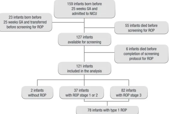
During the study period, a total of 182 infants born before 25 weeks gestation were enrolled (Fig. 1). Of these, 55 infants died before the ophthalmologic screening for ROP and additional 6 infants died before the completion of the ROP screening proto- col. Finally, 121 infants born before 25 weeks gestation were in- cluded in the analysis (Fig. 1). Demographic and systemic char- acteristics of included infants are shown in Table 1. The mean GA of included infants was 24.0 ± 0.6 weeks (range, 22.4-24.9).
The mean birth weight was 652.9 ± 109.3 g (range, 370-1,104 g).
Of the 121 infants, 119 (98.4%) infants developed ROP. Thirty- seven (30.6%) infants developed mild ROP (stages 1-2), and 82 (67.8%) infants developed severe ROP (stages 3-5). The inci- dences of maximum stages of ROP in relation to GA at birth are
23 infants born before 25 weeks GA and transferred
before screening for ROP
6 infants died before completion of screening
protocol for ROP 55 infants died before
screening for ROP 159 infants born before
25 weeks GA and admitted to NICU
127 infants available for screening
121 infants included in the analysis
78 infants with type 1 ROP 37 infants
with ROP stage 1 or 2 2 infants
without ROP
82 infants with ROP stage 3
Fig. 1. Flow-chart of the study pop- ulation. GA, gestational age; NICU, neonatal intensive care unit; ROP, retinopathy of prematurity.
shown in Table 1. Seventy-eight infants (64.5%) showed type 1 ROP, and laser treatment was performed in both eyes. Of the
121 infants, 17 (14.0%) infants with AP-ROP defined by revised IC-ROP were identified (Table 1) (12). Fourteen of 17 infants with AP-ROP showed vascularization in zone I, and others showed posterior zone II. Of the 17 infants with AP-ROP, 15 in- fants showed stage 3 ROP or less, but 2 infants developed bilat- eral stage 5 ROP.
In 6 (5.0%) of 121 infants, ROP was shown in at least one eye at the first screening examination performed between 29 to 31 weeks of PMA. Three infants had ROP stage 1, one had stage 2, and two had stage 3 at the first examination. The mean postna- tal ages (PNAs) and PMAs at the onset of any stage of ROP and type 1 ROP in the worse eye are shown in Table 2. The mean PMAs at the onset of type 1 ROP in the worse eyes of infants with Table 1. Incidence, maximal stage of retinopathy of prematurity (ROP), and clinical characteristics of included infants born before 25 weeks gestation
Parameters Gestation age
22 weeks (n = 5) 23 weeks (n = 42) 24 weeks (n = 74) Total (n = 121) Maximal stage of ROP
No ROP ROP Stage 1 Stage 2 Stage 3 Stage 4 Stage 5 Type 1 ROP AP-ROP
0 (0) 5 (100)
0 (0) 0 (0) 3 (60)
0 (0) 2 (40) 5 (100)
2 (40)
0 (0) 42 (100) 5 (11.9) 11 (26.2) 23 (54.8) 3 (7.1)
0 (0) 28 (66.7) 5 (11.9)
2 (2.7) 72 (97.3)
6 (8.1) 15 (20.3) 50 (67.6)
0 (0) 1 (1.4) 45 (60.8) 10 (13.5)
2 (1.7) 119 (98.4)
11 (9.1) 26 (21.5) 76 (62.8) 3 (2.5) 3 (2.5) 78 (64.5) 17 (14.0) Clinical characteristics
Gender, No. of male (%)
Birth weight (g, Mean ± SD) (range) VSGA No. (%)
SGA No. (%) AGA No. (%) Multiple births No. (%) Single
Twin Triplet
Apgar score (Mean ± SD) 1 min
5 min PDA ligation No. (%) IVH grade No. (%) None Grade I Grade II Grade III Grade IV
BPD ≥ moderate No. (%) NEC operation No. (%) Culture-proven sepsis No. (%) Early sepsis No. (%)
Surfactant (120 mg) use (No., Mean ± SD) TPN duration (days, Mean ± SD) Transfusion amount (unit, Mean ± SD) O2 therapy (days, Mean ± SD) Total duration
Mechanical ventilation CPAP
3 (60.0) 511.4 ± 108.7
(370-647) 0 (0) 1 (20.0) 4 (80.0) 3 (60) 2 (40) 0 (0) 3.3 ± 1.3 6.3 ± 1.3 4 (80) 1 (20) 1 (20) 0 (0) 3 (60)
0 (0) 4 (80) 2 (40) 5 (100)
1 (20) 2.0 ± 0.7 78.6 ± 67.1 21.4 ± 20.3 129.3 ± 35.2 52.4 ± 25.2 51.4 ± 21.3
19 (45.2) 588.3 ± 68.3
(441-720) 0 (0) 3 (7.1) 39 (92.9) 23 (54.8) 16 (38.1) 3 (7.1) 3.9 ± 1.4 6.7 ± 1.5 36 (85.7) 3 (7.1) 6 (14.3) 14 (33.3) 13 (30.9) 6 (14.3) 34 (80.9) 9 (21.4) 11 (26.2) 5 (11.9) 1.4 ± 0.5 56.3 ± 37.2
7.5 ± 4.5 114.1 ± 42.2 45.0 ± 19.3 33.5 ± 31.5
37 (50.0) 699.1 ± 102.1
(440-1,104) 1 (1.4) 2 (2.7) 71 (95.9) 48 (64.8) 25 (33.8) 1 (1.4) 4.4 ± 1.5 6.9 ± 1.4 46 (62.2) 30 (40.5) 14 (18.9) 9 (12.2) 15 (20.3) 6 (8.1) 40 (54.1) 9 (12.2) 17 (22.9) 5 (6.8) 1.3 ± 0.5 47.8 ± 42.3
6.1 ± 3.8 105.9 ± 61.4 43.4 ± 47.8 41.0 ± 32.7
59 (48.8) 652.9 ± 109.3
(370-1,104) 1 (0.8) 6 (5.0) 114 (94.2) 74 (61.2) 43 (35.5) 4 (3.3) 4.2 ± 1.5 6.8 ± 1.4 86 (71.1) 34 (28.1) 21 (17.4) 23 (19.0) 31 (25.6) 12 (9.9) 78 (64.4) 20 (16.5) 33 (27.3) 11 (9.1) 1.4 ± 0.5 52.1 ± 41.9
7.2 ± 6.2 109.6 ± 54.6 44.3 ± 39.3 38.8 ± 32.0
AP-ROP, aggressive posterior retinopathy of prematurity; VSGA, very small for gestational age; SGA, small for gestational age; AGA, appropriate for gestational age; PDA, patent ductus arteriosus; IVH, intraventricular hemorrhage; BPD, bronchopulmonary dysplasia; NEC, necrotizing enterocolitis; TPN, total parenteral nutrition; CPAP, continuous positive airway pressure.
Table 2. Onset and progression of retinopathy of prematurity (ROP) in worse eyes
Age Onset of any ROP
(n = 119) Onset of type 1 ROP (n = 78) Postmenstrual age (weeks)
Mean Median Range
33.5 ± 1.9 33.3 30.1-41.4
36.1 ± 2.5 35.9 30.3-41.9 Postnatal age (weeks)
Mean Median Range
9.5 ± 2.1 9.3 5.7-18.3
12.1 ± 2.6 12.1 6.1-17.1
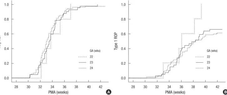
a GA of 22, 23, and 24 weeks were 36.4 ± 2.4 (range, 33+0-39+2), 36.2 ± 2.3 (range, 31+5-40+5), and 36.0 ± 2.6 weeks (range, 30+2 -41+6), respectively. The onset of any ROP and type 1 ROP in the worse eye relative to GA at birth is summarized in Fig. 2. In Ka-
plan-Meier survival plots, the onset of any ROP and type 1 ROP were not significantly different between GA groups (Fig. 2). All but one infant developed type 1 ROP after 31 weeks PMA. One infant born at 24+1 weeks gestation with an unstable clinical course showed zone I plus ROP at the first examination at 30+2 weeks PMA.
Systemic factors and ROP progression in relation to type 1 ROP are shown in Table 3. The duration of TPN was significant- ly associated with the development of type 1 ROP (P = 0.002, t- test). The mean PMA at the onset of any stage of ROP was signif- icantly shorter in patients with type 1 ROP than in others (P = 0.012, t-test). The 1-min Apgar score and mean PNAs at the on- set of any stage of ROP were lower in type 1 ROP with border- line significance (P = 0.052 and 0.057, respectively, t-test). How- ever, neither birth weight nor GA at birth was associated with the development of type 1 ROP in this cohort. Multiple logistic regression analysis was performed to identify systemic and oc- ular risk factors that had significant associations with the devel- opment of type 1 ROP (Table 4). The IVH ≥ grade 3 was associ- ated with the development of type 1 ROP with borderline signif- Table 3. Systemic and ocular risk factors for the development of type 1 retinopathy
of prematurity (ROP) among infants born before 25 weeks gestation
Parameters Type 1 ROP
(n = 78) No Type 1 ROP
(n = 43) P value
Gender, No. of male (%) 39 (50) 20 (46.5) 0.713
GA (mean ± SD) 24.0 ± 0.7 24.1 ± 5.7 0.282
Birth weight (mean ± SD)
SGA and VSGA, No. (%) 643.2 ± 111.5
6 (7.7) 670.5 ± 104.2
1 (2.3) 0.281
0.226 Multiple pregnancy, No. (%) 33 (42.3) 14 (32.6) 0.292 Apgar score (mean ± SD)
1 min
5 min 4.0 ± 1.4
6.7 ± 1.5 4.5 ± 1.5
7.0 ± 1.2 0.052 0.176 PDA ligation, No. (%) 51 (65.4) 35 (81.4) 0.063 IVH ≥ grade 3, No. (%) 32 (41.0) 11 (25.6) 0.089 BPD ≥ moderate, No. (%) 51 (65.4) 27 (62.8) 0.775 NEC operation, No. (%) 13 (16.7) 7 (16.3) 0.956 Culture-proven sepsis, No. (%) 17 (21.8) 16 (37.2) 0.068 Early sepsis, No. (%) 5 (6.4) 6 (14.0) 0.167 TPN duration (days) 44.1 ± 34.8 66.5 ± 49.7 0.002*
Transfusion amount (unit) 7.7 ± 7.2 6.4 ± 3.7 0.292 O2 therapy
Mechanical ventilation (days) CPAP (days)
Mechanical + CPAP (days) Total (days)
43.1 ± 39.6 37.6 ± 26.3 80.6 ± 45.0 108.5 ± 46.8
46.6 ± 39.0 41.1 ± 40.7 87.7 ± 70.1 111.9 ± 66.6
0.537 0.475 0.391 0.647 Onset of any ROP (PMA) 33.2 ± 1.5 34.1 ± 2.4 0.012*
Onset of any ROP (PNA) 9.2 ± 1.7 10.1 ± 2.6 0.057
*P < 0.05. GA, gestational age; VSGA, very small for gestational age; SGA, small for gestational age; PDA, patent ductus arteriosus; IVH, intraventricular hemorrhage; BPD, bronchopulmonary dysplasia; NEC, necrotizing enterocolitis; TPN, total parenteral nu- trition; CPAP, continuous positive airway pressure; PMA, postmenstrual age; PNA, postnatal age.
Table 4. Risk factors for the development of type 1 ROP among infants born before 25 weeks gestation using multiple logistic regression analysis
Risk factors P value OR (95% CI)
Apgar score 1 min 0.309 0.850 (0.621-1.163)
PDA ligation 0.399 0.629 (0.215-1.846)
IVH ≥ grade 3 0.064 2.415 (0.949-6.144)
Culture-proven sepsis 0.141 0.481 (0.182-1.274)
TPN duration 0.132 0.991 (0.980-1.003)
Onset of any ROP (PMA) 0.136 0.836 (0.660-1.058) OR, odds ratio; CI, confidence interval; PDA, patent ductus arteriosus; IVH, intraven- tricular hemorrhage; TPN, total parenteral nutrition.
Fig. 2. Onset of any retinopathy of prematurity (ROP) (A) and type 1 ROP (B) in worse eyes relative to gestational age (GA) at birth. P = 0.769, 0.511, 0.566, the onset of any ROP between GA of 22 and 23, 22 and 24, and 23 and 24, respectively, P = 0.122, 0.062, 0.654, the onset of type 1 ROP between GA of 22 and 23, 22 and 24, and 23 and 24, respectively, log-rank test. PMA, postmenstrual age.
Any ROP Type 1 ROP
PMA (weeks) PMA (weeks)
28 30 32 34 36 38 40 42 28 30 32 34 36 38 40 42
1.0
0.8
0.6
0.4
0.2
0.0
1.0
0.8
0.6
0.4
0.2
0.0
A B
GA (wks) 22 23 24
GA (wks) 22 23 24
icance (OR, 2.415; P = 0.064).
DISCUSSION
This study investigated the incidence and natural course of ROP in 121 extremely preterm infants born before 25 weeks gestation in a neonatal intensive care unit at a single medical center in Korea. The incidence of any ROP and type 1 ROP in this cohort was high, 98.4% and 64.5%, respectively. However, the incidence (5.0%) of stage 4 or 5 ROP was relatively low. Other studies on the incidence of ROP in extremely preterm infants also showed high incidences of any ROP, around 90% (Table 5) (13-17). How- ever, compared with recent studies, our study showed higher incidence of severe ROP (Table 5) (13-17). Differences in the study design, definition of severe ROP, comorbidity of infants, proportion of inborn infants, and disagreement on plus sign may explain the reason (18). As GA at birth decreases, incidence of severe ROP usually increases (10, 11, 13, 14). However, in this study, the incidence of type 1 ROP was not significantly differ- ent between 23 and 24 weeks GA groups.
The natural course of ROP in this cohort showed a relatively early onset and progression compared to the ET-ROP study. The mean PMAs at the onset of any ROP and type 1 ROP were simi- lar to those of a Swedish population-based study (19). Although only 5% of infants showed any ROP at ≤ 31 weeks PMA, one (0.8%) infant born at 24+1 weeks’ gestation developed type 1 ROP as early as 30+2 weeks PMA. The development of type 1 ROP be- fore 31 weeks PMA was not reported in two recent studies in- cluding more than 100 preterm infants born before 25 weeks gestation. In a German cohort study, no preterm infants required treatment before the 33rd postmenstrual week (14). In a Swed- ish study, ROP at stages 2 and 3 was seen as early as 29.9 and 31.6 weeks, respectively (19, 20). Thus, the authors recommend postponing the first examination until PMA of 31 weeks (20).
For the infants born before 27 weeks gestation, British guide-
lines recommend that ROP screening should start at 30 to 31 weeks PMA, while American guidelines recommend the first screening to be performed at PMA of 31 weeks (7, 8). Although these current screening protocols are feasible for most preterm infants born before 25 weeks gestation, the results of this study suggest that screening at 30 weeks PMA may be necessary in these infants with an unstable clinical course.
The present study investigated systemic and ocular risk fac- tors in relation to the development of type 1 ROP. In univariate analysis, a shorter duration of TPN was significantly associated with a higher incidence of type 1 ROP. A recent study about ag- gressive parenteral nutrition in preterm infants revealed the se- rum levels of insulin-like growth factor-1 (IGF-1) and insulin- like growth factor-binding protein 3 (IGFBP3) were higher in aggressive parenteral nutrition group than those of conventional parenteral nutrition group and lower levels of IGF-I and IGFBP3 in conventional parenteral nutrition group were negatively cor- related with development of ROP (21). Thus, shorter duration of TPN might affect the serum level of IGF-1 and IGFBP3, which might result in frequent development of type 1 ROP. However, the exact mechanism remains to be elucidated. In addition, an earlier onset of ROP was significantly related to a higher inci- dence of type 1 ROP. This finding is consistent with the Swedish population-based study, which revealed that PMA at the onset of ROP was significantly related to the severity of ROP (19). How- ever, this finding is inconsistent with results of the CRYO-ROP study (22, 23). Differences in clinical characteristics, especially GA at birth, among the enrolled infants, may explain the incon- sistency. In multiple logistic regression analysis, IVH ≥ grade 3 was associated with the development of type 1 ROP with bor- derline significance. This is consistent with the report that IVH was predictive of the development of ROP (24). In our study, previously reported risk factors including birth weight, multiple birth, and duration of oxygen therapy were not associated with type 1 ROP. In many studies including the ET-ROP and CRYO- Table 5. Incidence of retinopathy of prematurity (ROP) in infants before 25 weeks’ gestation compared with recent studies
Items Present study Sweden (13) Germany (14) Australia (15) Scotland (16) USA (17)
Study type Single center Population-based Single center Single center Population-based Single center
Study period 2004-2011 2004-2007 2001-2009 1992-2009 1990-2009 2003-2007
No. of infants GA 22-24 wk GA 22 wk GA 23 wk GA 24 wk Any ROP (%) GA 22 wk GA 23 wk GA 24 wk Severe ROP (%)*
GA 22 wk GA 23 wk GA 24 wk
121 5 42 74 98.4 100 100 97.3 67.8 100
61.9 68.9
157 5 53 99 87.9 100
90.6 85.9 54.8 80.0 62.3 49.5
125 13 57 55 NA NA NA NA 29.6† 61.5† 24.6† 27.3†
147 NA 21 126 89.8
NA 90.5 89.7 19.2 NA 18.6 18.3
72 NA NA NA 88.9
NA NA NA 68.1
NA NA NA
79 NA NA NA 87
NA NA NA 23† NA NA NA
*Stage 3 or more ROP; †laser-treated patients. NA, not available; GA, gestational age; wk, week.
ROP studies, low birth weight was significantly associated with the development of severe ROP (22-24). In a German cohort study, the odds ratio for treatment increased by 1.22 per 100 g of decrease in body weight (14). However, in this study, birth weight was not associated with the development of type 1 ROP.
This finding is consistent with a study by Teed and Saunders (17) study, which showed no association of birth weight with type 1 ROP in infants born before 25 weeks gestation. In addi- tion, multiple births, which was associated with an increased risk of reaching threshold ROP in the CRYO-ROP study, and the duration of oxygen therapy, which was also associated with ROP in several studies, were not associated with type 1 ROP in this study (22, 25, 26). This may imply that risk factors for ROP may be more complex in extremely preterm infants born before 25 weeks gestation than in more mature infants, and other neona- tal factors may contribute more to the development of severe ROP.
In conclusion, this study revealed the incidence, natural course, and risk factors of ROP in infants born before 25 weeks gestation in Korea. Although the incidence of ROP is very high, the proportion of infants with stage 4 or stage 5 ROP is relatively low when timely treatment is administered. Although rare, ear- ly onset of type 1 ROP before 31 weeks PMA is observed. Thus, earlier screening may be necessary in these extremely preterm infants with an unstable clinical course. Risk factors associated with an increased risk of type 1 ROP appear different in extreme- ly preterm infants from those of more mature infants.
REFERENCES
1. Gilbert C, Fielder A, Gordillo L, Quinn G, Semiglia R, Visintin P, Zin A;
International NO-ROP Group. Characteristics of infants with severe reti- nopathy of prematurity in countries with low, moderate, and high levels of development: implications for screening programs. Pediatrics 2005;
115: e518-25.
2. Gilbert C. Retinopathy of prematurity: a global perspective of the epi- demics, population of babies at risk and implications for control. Early Hum Dev 2008; 84: 77-82.
3. Good WV; Early Treatment for Retinopathy of Prematurity Cooperative Group. The Early Treatment for Retinopathy of Prematurity Study: struc- tural findings at age 2 years. Br J Ophthalmol 2006; 90: 1378-82.
4. Austeng D, Källen KB, Ewald UW, Wallin A, Holmström GE. Treatment for retinopathy of prematurity in infants born before 27 weeks of gesta- tion in Sweden. Br J Ophthalmol 2010; 94: 1136-9.
5. Wilson-Costello D, Friedman H, Minich N, Fanaroff AA, Hack M. Im- proved survival rates with increased neurodevelopmental disability for extremely low birth weight infants in the 1990s. Pediatrics 2005; 115:
997-1003.
6. Stoll BJ, Hansen NI, Bell EF, Shankaran S, Laptook AR, Walsh MC, Hale EC, Newman NS, Schibler K, Carlo WA, et al. Neonatal outcomes of ex- tremely preterm infants from the NICHD Neonatal Research Network.
Pediatrics 2010; 126: 443-56.
7. Section on Ophthalmology American Academy of Pediatrics; Ameri- can Academy of Ophthalmology; American Association for Pediatric Ophthalmology and Strabismus. Screening examination of premature infants for retinopathy of prematurity. Pediatrics 2006; 117: 572-6.
8. Wilkinson AR, Haines L, Head K, Fielder AR; Guideline Development Group of the Royal College of Paediatrics and Child Health; Royal Col- lege of Ophthalmologists; British Association of Perinatal Medicine.
UK retinopathy of prematurity guideline. Eye (Lond) 2009; 23: 2137-9.
9. Early Treatment For Retinopathy Of Prematurity Cooperative Group.
Revised indications for the treatment of retinopathy of prematurity: re- sults of the early treatment for retinopathy of prematurity randomized trial. Arch Ophthalmol 2003; 121: 1684-94.
10. Good WV; Early Treatment for Retinopathy of Prematurity Cooperative Group. Final results of the early treatment for retinopathy of prematu- rity (ETROP) randomized trial. Trans Am Ophthalmol Soc 2004; 102:
233-48.
11. Palmer EA, Flynn JT, Hardy RJ, Phelps DL, Phillips CL, Schaffer DB, Tung B. Incidence and early course of retinopathy of prematurity. The Cryotherapy for Retinopathy of Prematurity Cooperative Group. Oph- thalmology 1991; 98: 1628-40.
12. International Committee for the Classification of Retinopathy of Pre- maturity. The international classification of retinopathy of prematurity revisited. Arch Ophthalmol 2005; 123: 991-9.
13. Austeng D, Källen KB, Ewald UW, Jakobsson PG, Holmström GE. Inci- dence of retinopathy of prematurity in infants born before 27 weeks’ ges- tation in Sweden. Arch Ophthalmol 2009; 127: 1315-9.
14. Muether PS, Kribs A, Hahn M, Schumacher J, Eifinger F, Kirchhof B, Roth B, Fauser S. No advanced retinopathy of prematurity stages 4 or 5 in a large high-risk German cohort. Br J Ophthalmol 2012; 96: 400-4.
15. Gunn DJ, Cartwright DW, Gole GA. Incidence of retinopathy of prema- turity in extremely premature infants over an 18-year period. Clin Ex- periment Ophthalmol 2012; 40: 93-9.
16. Tan SZ, Dhaliwal C, Becher JC, Fleck B. Trends in the incidence of reti- nopathy of prematurity in Lothian, south-east Scotland, from 1990 to 2009. Arch Dis Child Fetal Neonatal Ed 2012; 97: F310-1.
17. Teed RG, Saunders RA. Retinopathy of prematurity in extremely prema- ture infants. J AAPOS 2009; 13: 370-3.
18. Slidsborg C, Forman JL, Fielder AR, Crafoord S, Baggesen K, Bangsgaard R, Fledelius HC, Greisen G, la Cour M. Experts do not agree when to treat retinopathy of prematurity based on plus disease. Br J Ophthalmol 2012; 96: 549-53.
19. Austeng D, Källen KB, Hellström A, Tornqvist K, Holmström GE. Natu- ral history of retinopathy of prematurity in infants born before 27 weeks’
gestation in Sweden. Arch Ophthalmol 2010; 128: 1289-94.
20. Austeng D, Källen KB, Hellström A, Jakobsson PG, Johansson K, Torn- qvist K, Wallin A, Holmström GE. Screening for retinopathy of prematu- rity in infants born before 27 weeks’ gestation in Sweden. Arch Ophthal- mol 2011; 129: 167-72.
21. Can E, Bülül A, Uslu S, Cömert S, Bolat F, Nuhoğlu A. Effects of aggres- sive parenteral nutrition on growth and clinical outcome in preterm in- fants. Pediatr Int 2012. doi: 10.1111/j.1442-200X.2012.03713.X.
22. Schaffer DB, Palmer EA, Plotsky DF, Metz HS, Flynn JT, Tung B, Hardy RJ. Prognostic factors in the natural course of retinopathy of prematurity.
The Cryotherapy for Retinopathy of Prematurity Cooperative Group.
Ophthalmology 1993; 100: 230-7.
23. Good WV, Hardy RJ, Dobson V, Pamer EA, Phelps DL, Quintos M, Tung B; Early Treatment for Retinopathy of Prematurity Cooperative Group.
The incidence and course of retinopathy of prematurity: findings from the early treatment for retinopathy of prematurity study. Pediatrics 2005;
116: 15-23.
24. Lad EM, Nguyen TC, Morton JM, Moshfeghi DM. Retinopathy of pre-
maturity in the United States. Br J Ophthalmol 2008; 92: 320-5.
25. Hussain N, Clive J, Bhandari V. Current incidence of retinopathy of pre- maturity, 1989-1997. Pediatrics 1999; 104: e26.
26. Shah VA, Yeo CL, Ling YL, Ho LY. Incidence, risk factors of retinopathy of prematurity among very low birth weight infants in Singapore. Ann Acad Med Singapore 2005; 34: 169-78.