광범위한 뇌경색증이 합병된 지발형 B군 사슬알균에 의한 수막염 증례
조민수1ᆞ김용민1ᆞ조혜경2ᆞ최수한1
한림대학교 동탄성심병원 소아청소년과1, 가천대학교 길병원 소아청소년과2
Late-Onset Group B Streptococcal Meningitis Complicated with Extensive Cerebral Infarction
Min Su Cho1, Yongmin Kim1, Hye-Kyung Cho2, Soo-Han Choi1
1Department of Pediatrics, Hallym University Dongtan Sacred Heart Hospital, Hallym University College of Medicine, Hwaseong; 2Department of Pediatrics, Gil Medical Center, Gachon University College of Medicine, Incheon, the Republic of Korea
Group B streptococcus (GBS) is the leading cause of neonatal morbidity and mortality. Late-onset GBS disease commonly manifests as occult bacteremia or meningitis. Approximately 50% of survivors of late-onset meningitis have long-term neurologic sequelae. Cerebrovascular complications are often associated with unfavorable clinical outcomes of GBS meningitis. There have been a few reports of cerebral infarction accompanied by GBS meningitis. We report a 29-day-old girl with severe, widespread cerebral infarction due to late-onset GBS meningitis.
Isolated GBS strain from this patient was serotype III, ST-19. Currently, she has cortical blindness and significant developmental delay.
Key Words: Streptococcus agalactiae; Neonatal sepsis; Meningitis; Cerebral infarction
접수: 2017년 9월 6일 수정: 2017년 9월 26일 승인: 2017년 9월 28일
책임저자: 최수한
한림대학교 의과대학 동탄성심병원 소아청소년과 Tel: 031)8086-8560, Fax: 031)8086-3000 E-mail: [email protected]
REPORT
이 발생하는 것으로 알려져 있다1,2).
세균성 수막염은 뇌경색증의 위험인자 중 하나이며, 소 아 뇌경색증 원인의 3%–5% 정도로 보고되었다3,4). 뇌경 색증 발생과 관련된 수막염의 원인균으로 폐렴사슬알균과 결핵균이 잘 알려져 있다5,6).
저자들은 지발형 GBS 침습성 감염이 발생한 생후 29일 된 신생아에서 다발성 뇌경색증이 합병된 수막염 사례를 보고하는 바이다.
증례
생후 29일 된 여아가 내원 당일 발생한 발열을 주소로 응급실을 통해 입원하였다. 발열 이외 동반된 다른 증상은 없었다. 환아는 임신 나이 39주 1일, 체중 3.8 kg, 정상 질 식분만으로 출생하였다. 산전 진찰 및 입원 시까지 특이
서론
B군 사슬알균(Streptococcus agalactiae, group B strep
tococcus [GBS])은 3개월 미만 영아에서 발생하는 침습 성 감염증의 주요한 원인균이다. 국소 병변이 없는 균혈증 과 수막염은 지발형 GBS 질환의 가장 흔한 임상 양상이 다. 20%–30%의 GBS 수막염 환자들에서 발달 지연, 인 지 장애, 경직성 사지마비, 피질맹, 청력 손실 등의 후유증
질환의 병력 없었으며 2자매 중 둘째로 가족력에서 특이 소견 없었다.
입원 당시 환아의 생체 활력징후는 혈압 94/58 mm Hg, 심박수 160회/분, 호흡수 42회/분, 체온 38.9℃이었다. 신 체 계측은 체중 4.2 kg (25–50 백분위수 범위), 신장 53 cm (25–50 백분위수 범위), 두위 38 cm (50–75 백분위수 범위)이었다. 진찰에서 환아는 급성 병색에 끙끙거림을 보 였으나 자극에 대한 반응은 적절하였다. 두경부, 흉부 및 복부 진찰에서 특이 소견 없었고 피부 병변 관찰되지 않 았다. 입원 당시 혈액검사에서 백혈구 5,300/mm3 (호중 구 46.6%), 혈색소 13.9 g/dL, 혈소판 372,000/mm3, C
반응단백 1.1 mg/dL (범위, 0–5 mg/dL)이었고 소변검사 에서 특이 소견 없었다. 하지만 뇌척수액 검사에서 백혈구 3,345/mm3 (다형핵세포 90%), 적혈구 370/mm3, 단백 307 mg/dL, 당 11 mg/dL 소견을 보여 세균성 수막염으 로 추정 진단하고 vancomycin (60 mg/kg/day), cefota
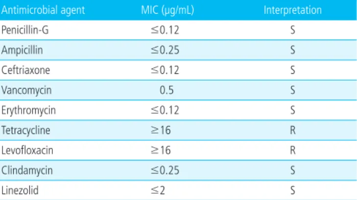
xime (200 mg/kg/day) 및 ampicillin (300 mg/kg/day) 으로 경험적 항생제 치료를 시작하였다. 입원 2일째부터 발열은 호전 추세를 보였으나 환아는 기면 상태로 자극에 대한 반응이 떨어지는 양상을 보였다. 입원 2일째 다시 시 행한 혈액검사에서 백혈구 3,900/mm3 (호중구 57.4%), 혈색소 13.6 g/dL, 혈소판 273,000/mm3이었고 C반응단 백이 172.5 mg/dL로 상승을 보였다. 같은 날 시행한 뇌 컴퓨터단층촬영에서 좌측 전두두정 부위 경막하삼출 소 견이 경미하게 관찰되었으나 뇌 실질 내 병변은 뚜렷하지 않았다. 입원 3일째 우측 사지에 국한된 부분 발작이 발생 하여 간헐적으로 반복되어 항경련제를 투여하였다. 입원 4일째에 시행한 뇌 자기공명영상에서 다발성의 광범위한 대뇌피질(양측 전두엽, 측두엽 및 후두엽)과 뇌량 및 좌측 후두엽에서 급성 뇌경색증 소견이 보였다(Fig. 1). 혈액응고 검사 및 심장초음파검사에서 이상 소견은 없었다. 뇌경색 증에 대해서 혈전용해 또는 항응고제 치료는 시행하지 않 았다. 경련은 입원 6일째부터 소실되어 더 이상 발생하지 않았고 뚜렷한 신경학적 이상 징후는 보이지 않았다.
입원 당시 시행한 혈액과 뇌척수액 배양검사에서 S. aga
lactiae가 확인되었다. 항생제 감수성 검사에서 erythro
mycin과 clindamycin에는 감수성을 보였고 levofloxacin 에는 내성을 보였다(Table 1). 환아에서 동정된 S. agalac
tiae에 대해서 추가적인 분자유전검사를 외부 의뢰하여 시 행하였다. 혈청형 분석은 serotype Ia, Ib, II, III, IV, V에 대한 특이 항혈청(Denka Seiken, Tokyo, Japan)을 이용 한 슬라이드 응집검사(slide agglutination test)를 이용하 였다7). 기존의 알려진 연구를 참조하여 다좌위 서열 형별
분석(multilocus sequence typing)을 시행하였다8). 본 증례 환자에서 분리된 S. agalactiae 균주는 serotype III, ST19 으로 확인되었다.
배양검사 결과 확인 후 입원 4일째 penicillin G (500,000 unit/kg/day)로 항생제를 변경하였다. 추적 검사로 시행한 뇌척수액 검사에서 백혈구 108/mm3 (다형핵세포 10%), 단백 302.4 mg/dL, 당 39 mg/dL이었고 배양검사에서 균
A B
C D
Fig. 1. Magnetic resonance image and angiography performed on the 4th hospital day showed (A, B) multifocal diffusion restriction at cortex of cerebral hemisphere and corpus cal- losum, (C) suspicious multifocal stenosis, and (D) extra-axial fluid collection at bilateral cerebral convexity.
Table 1. The Result of Antimicrobial Susceptibility Test of Strep- tococcus agalactiae Isolated from the Patient
Antimicrobial agent MIC (µg/mL) Interpretation
Penicillin-G ≤0.12 S
Ampicillin ≤0.25 S
Ceftriaxone ≤0.12 S
Vancomycin 0.5 S
Erythromycin ≤0.12 S
Tetracycline ≥16 R
Levofloxacin ≥16 R
Clindamycin ≤0.25 S
Linezolid ≤2 S
Abbreviations: S, sensitive; R, resistant.
은 동정 되지 않았다. 항생제 치료는 총 4주간 시행하였다.
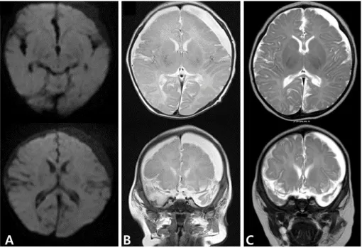
입원 24일째 추적검사로 시행한 뇌 자기공명영상에서 급 성 뇌경색증 병변은 호전을 보이나(Fig. 2A) 좌측 전두두정 부위의 경막하삼출이 증가된 소견을 보였다(Fig. 2B). 이 후 1개월 뒤 다시 시행한 검사에서는 경막하삼출 소견은 호전을 보였다(Fig. 2C).
항생제 치료 종결 전 시행한 청력검사는 정상이었다. 하 지만 치료 종결 1개월 후 시행한 안과 검진에서 피질맹이 의심되었다. 만 1세 때 시행한 Bayley 영유아 발달검사에 서 발달 연령은 인지척도 5개월, 수용언어척도 7개월, 표 현언어척도 10개월, 소근육운동척도 6개월, 대근육운동척 도 7개월 수준으로 발달 지연을 보였다.
고찰
GBS는 신생아 및 영아의 침습성 감염의 중요한 원인균
이다1,2,9). 1996–2005년 동안 8세 이하 소아 세균성 수막
염의 원인균을 조사한 국내 다기관 후향적 연구에서는 GBS 가 가장 흔한 원인균으로 24.6%였고, 3개월 미만 환자에 서는 47.6%로 보고되었다10).
생후 첫 6일 이내에 발현하는 조발형 GBS 감염은 생식 기나 직장에 GBS가 집락화된 모체로부터 출산 직전이나
출산하는 동안 수직 감염으로 주로 발생한다. 지발형 GBS 감염은 생후 7일에서 90일 사이에 발현하며 모체로부터 수직 전파되거나, 원내 감염, 지역사회 감염원으로부터 획 득될 수 있다1). 미국에서는 신생아 침습성 GBS 질환 예 방을 위해 산모의 일괄적인 검사 및 분만 중 항생제 예방 요법 시행으로 출생아 1,000명당 침습성 GBS 감염의 발 생 빈도가 1990년대 초반 1.7건에서 2011년 0.26건으로 크게 감소하였다. 하지만 지발형 감염의 발생률은 출생아 1,000명당 0.3건 정도로 유지되어 큰 변화가 없었다1,9). 국 내 연구에서 임산부의 GBS 집락화 유병률은 8%–10%까 지 보고되고 있으며 이전의 연구에서 보다 증가한 양상을 보이고 있다1114). 1996–2005년 국내 신생아 GBS 질환을 조사한 다기관 연구에서 조발형(20%)은 지발형(80%) 보 다 낮은 비율을 보였으나 연도별 환자 수는 증가 추이를 보였다15).
세균성 수막염에서 뇌혈관 합병증이 발생하는 기전은 명확히 확립되지 않았으나 여러 요인이 작용할 것으로 보 인다. 세균성 수막염에서는 이차적인 혈관 염증으로 뇌혈 관 내피 손상, 과다 응고 상태, 혈류 정체가 발생할 수 있 으며 이런 요인들이 뇌경색증 발생에 기인하는 것으로 알 려져 있다6,16).
뇌경색증이 세균성 수막염에서 중요한 합병증으로 알려 져 있으나 소아 환자에서 역학이나 예후 등에 대한 연구
A B
C
Fig. 2. (A) Magnetic resonance image (MRI) on the 24th hospital day showed no more diffusion restriction, (B) but increased in amount of extra-axial fluid collection at left cerebral convexity. (C) One month later, follow-up MRI showed decreased in amount of extra-axial fluid collection.
는 제한적이다. 캐나다의 국가 인구 기반 연구에서 1992 년부터 2001년까지 소아 허혈성 뇌졸중의 발생률은 1.72/
100,000/년이었고 이 중 수막염이 원인인 경우는 5%이 었다4). 캐나다 어린이병원 2곳에서 조사된 연구에서는 1992–2010년 동안 뇌경색증으로 진단받았던 1개월 이 상 소아 환자의 4.6% (24/515)가 세균성 수막염을 동반 하였다. 폐렴사슬알균이 원인균이었던 경우가 10건으로 가장 많았고 4건이 GBS가 원인균이었다6). 대만에서 시행 된 단일기관 후향적 연구에서 1986년부터 2001년 동안 배 양 양성 세균성 수막염으로 진단되었던 소아 환자의 8.4%
(14/166)에서 입원 당시 뇌경색증 소견이 있었고 원인균 으로 살모넬라균과 폐렴사슬알균이 각각 4건으로 가장 많 았다17).
GBS는 피막 다당질의 종류에 따라 10개의 혈청형(Ia, Ib, II–IX)으로 구분되며 혈청형 특이 피막 다당질이 병 리기전에 중요한 역할을 한다. 조발형에서는 serotype Ia, Ib, II, III, V이 주된 원인이 되며, 지발형 GBS 감염에서 serotype III가 우세하다2). Serotype III GBS는 다른 혈청 형보다 더 침습적인 감염을 유발하는 것으로 알려져 있다
16,18)
. Serotype III GBS 피막 다당질의 sialic acid 성분이 보체계의 대체경로 활성화를 방지하여 GBS의 포식을 억 제한다. Hypervirulent GBS adhesion (HvgA)은 표면 정 박 단백질(surfaceanchored protein)로 주로 serotype III, ST17 균주에서 생성되며 초고독성과 연관된 것으로 알려져 있다2,18,19).
Tibussek 등16)은 2009–2014년 동안 지발형 GBS 수막 염 환자에서 뇌혈관 합병증이 발생했던 14건에 대해 분석 발표하였다. 10명의 환자에서 허혈성 뇌졸중이 발생하였 고 2명에서 뇌정맥굴 혈전증이 발생하였다. GBS 혈청형 분석이 가능했던 9명 중에서 6명의 GBS 균주가 serotype III이었고, 이 중 5개가 ST17으로 HvgA 유전자 양성이었 다. 혈청형이 분석된 나머지 3개의 균주는 serotype Ia (2 개), Ib (1개)이었다.
본 증례의 GBS 균주는 serotype III, ST19 이었고 HvgA 유전자 검사는 시행하지 않았다. 최근 국내 연구에서 Kang 등19)은 1995–2015년 동안 침습성 GBS 질환이 발생했던 영아에서 분리된 98개의 GBS 균주를 분석하였고, sero
type III가 50% 이상을 차지하였다. HvgA 유전자 양성은 19.4%로 모두 serotype III, ST17이었다. 이 연구에서도 본 증례의 GBS 균주가 속해 있는 ST19의 비율이 18.4%
(18/98)로 ST1 (20/98) 및 ST17 (19/98) 다음으로 많 았다.
세균성 수막염에 의해 발생한 뇌경색증에서 항응고제나
항혈전제 치료에 대한 효과와 안전성에 대한 연구는 제한
적이다6,16,20). 따라서 세균성 수막염에 의한 뇌혈관 합병증
발생 여부에 대한 모니터링과 예방이 중요하다.
지발형 GBS 수막염은 심각한 뇌혈관 합병증을 유발할 수 있다. 본 저자들은 균혈증이 동반된 지발형 GBS 수막 염 환자에서 광범위한 다발성 뇌경색증이 합병된 예를 경 험하였기에 본 증례를 보고하는 바이다.
References
1. Pannaraj PS, Baker CJ. Group B streptococcal infections.
In: Cherry JD, Demmler-Harrison GJ, Kaplan SL, Stein- bach WJ, Hotez PJ, editors. Feigin and Cherry’s Textbook of pediatric infectious diseases. 7th ed. Philadelphia:
Saunders Elsevier, 2014:1153-69.
2. Lachenauer CS, Wessels MR. Group B streptococcus. In:
Kliegman RM, Nelson WE, editors. Nelson textbook of pediatrics. 20th ed. Philadelphia: Elsevier, 2016:1337-41.
3. Hernandez MI, Sandoval CC, Tapia JL, Mesa T, Escobar R, Huete I, et al. Stroke patterns in neonatal group B strepto- coccal meningitis. Pediatr Neurol 2011;44:282-8.
4. deVeber GA, Kirton A, Booth FA, Yager JY, Wirrell EC, Wood E, et al. Epidemiology and outcomes of arterial ischemic stroke in children: the Canadian Pediatric Is- chemic Stroke Registry. Pediatr Neurol 2017;69:58-70.
5. Pryde K, Walker WT, Hollingsworth C, Haywood P, Baird J, Hussey M, et al. Stroke in paediatric pneumococcal meningitis: a cross-sectional population-based study.
Arch Dis Child 2013;98:647-9.
6. Lehman LL, Rivkin MJ. Perinatal arterial ischemic stroke:
presentation, risk factors, evaluation, and outcome. Pe- diatr Neurol 2014;51:760-8.
7. Cho HK, Nam HN, Cho HJ, Son DW, Cho YK, Seo YH, et al. Serotype distribution of invasive group B streptococcal diseases in infants at two university hospitals in Korea.
Pediatr Infect Vaccine 2017;24:79-86.
8. Jones N, Bohnsack JF, Takahashi S, Oliver KA, Chan MS, Kunst F, et al. Multilocus sequence typing system for group B streptococcus. J Clin Microbiol 2003;41:2530-6.
9. Pintye J, Saltzman B, Wolf E, Crowell CS. Risk factors for late-onset group B streptococcal disease before and after implementation of universal screening and intrapartum
antibiotic prophylaxis. J Pediatric Infect Dis Soc 2016;5:
431-8.
10. Cho HK, Lee H, Kang JH, Kim KN, Kim DS, Kim YK, et al. The causative organisms of bacterial meningitis in Korean children in 1996-2005. J Korean Med Sci 2010;25:
895-9.
11. Chong Y, Lee K, Kwon OH, Nahm CH, Murai T, Inazumi Y.
Trend of isolation and serotypes of group B streptococci in Korea. Yonsei Med J 1993;34:78-83.
12. Uh Y, Jang IH, Yoon KJ, Lee CH, Kwon JY, Kim MC. Co- lonization rates and serotypes of group B streptococci isolated from pregnant women in a Korean tertiary hos- pital. Eur J Clin Microbiol Infect Dis 1997;16:753-6.
13. Hong JS, Choi CW, Park KU, Kim SN, Lee HJ, Lee HR, et al. Genital group B Streptococcus carrier rate and serotype distribution in Korean pregnant women: implications for group B streptococcal disease in Korean neonates. J Pe- rinat Med 2010;38:373-7.
14. Lee BK, Song YR, Kim MY, Yang JH, Shin JH, Seo YS, et al. Epidemiology of group B streptococcus in Korean pre- gnant women. Epidemiol Infect 2010;138:292-8.
15. Park KH, Kim KH, Kang JH, Kim KN, Kim DS, Kim YK,
et al. Current status and clinical presentations of invasive neonatal Group B streptococcal infections in Korea.
Pediatr Int 2011;53:236-9.
16. Tibussek D, Sinclair A, Yau I, Teatero S, Fittipaldi N, Ri- chardson SE, et al. Late-onset group B streptococcal me- ningitis has cerebrovascular complications. J Pediatr 2015;
166:1187-92.e1.
17. Chang CJ, Chang WN, Huang LT, Chang YC, Huang SC, Hung PL, et al. Cerebral infarction in perinatal and child- hood bacterial meningitis. QJM 2003;96:755-62.
18. Tazi A, Disson O, Bellais S, Bouaboud A, Dmytruk N, Dramsi S, et al. The surface protein HvgA mediates group B streptococcus hypervirulence and meningeal tropism in neonates. J Exp Med 2010;207:2313-22.
19. Kang HM, Lee HJ, Lee H, Jo DS, Lee HS, Kim TS, et al.
Genotype characterization of group B streptococcus iso- lated from infants with invasive diseases in South Korea.
Pediatr Infect Dis J 2017;36:e242-7.
20. Boelman C, Shroff M, Yau I, Bjornson B, Richrdson S, deVeber G, et al. Antithrombotic therapy for secondary stroke prevention in bacterial meningitis in children. J Pediatr 2014;165:799-806.
요약
B군 사슬알균은 3개월 미만 영아에서 발생하는 침습성 감염증의 주요한 원인균이다. 신생아 침습성 B 군 사슬알균 수막염에 의한 뇌혈관 합병증은 드물게 보고되고 있다. 발열을 주소로 내원한 생후 29일 신생아에서 세균성 수막염이 진단되었다. 입원 3일째 경련이 발생하였고 뇌 자기공명영상에서 다발성 의 광범위한 대뇌피질(양측 전두엽, 측두엽 및 후두엽)과 뇌량 및 좌측 후두엽에서 급성 뇌경색증 합병 된 소견을 보였다. 환아의 혈액과 뇌척수액에서 B군 사슬알균이 분리되었고 serotype III, ST-19으로 확인되었다. 현재 환아는 피질맹과 발달 지연을 보이고 있다.