Copyright © 2021. Korean Society for Neurorehabilitation i
HIGHLIGHT
• Voxel-based lesion-symptom analysis could reveal the typical brain lesion related to language processing in acute stroke.
Brain Neurorehabil. 2021 Jul;14(2):e14 https://doi.org/10.12786/bn.2021.14.e14 pISSN 1976-8753·eISSN 2383-9910
Original Article
Received: Mar 24, 2021 Revised: Jun 11, 2021 Accepted: Jun 29, 2021 Correspondence to Deog Young Kim
Department and Research Institute of Rehabilitation Medicine, Yonsei University College of Medicine, 50 Yonsei-ro, Seodaemun-gu, Seoul 03722, Korea.
E-mail: [email protected]
Eun Ji Park, Yong Wook Kim, Hyo Suk Nam, Hyo Seon Choi, Deog Young Kim
Neural Substrates of Aphasia in Acute Left Hemispheric Stroke Using Voxel- Based Lesion-symptom Brain Mapping
Brain & NeuroRehabilitation
ABSTRACT
It is unclear how these brain lesions fit into the language processing in acute stroke. In this study, we aimed to investigate the neuroanatomical lesion related to language processing in acute stage of stroke patients using voxel-based lesion-symptom mapping (VLSM). 73 acute first-ever post-stroke patients were enrolled in this retrospective study, who had undertaken brain magnetic resonance imaging (MRI) and Korean version of the Western Aphasia Test within 1 month from onset. Each voxel was compared with aphasia quotient and subtest scores as dependent variables using VLSM. The aphasia group showed significantly much more involvement of extra-nuclear area, insula, inferior frontal gyrus and superior temporal gyrus compared to non-aphasia group. The deficit of spontaneous speech domain was associated with the inferior parietal lobule, inferior and middle frontal gyrus and insula.
The insular cortex, inferior parietal lobule, inferior frontal gyrus, middle frontal gyrus and superior temporal gyrus were related to deficit of comprehension. The inferior parietal lobule, insula, precentral gyrus, inferior frontal gyrus were related to the deficit of repetition.
The deficit of naming was related to inferior parietal lobule, insula and inferior frontal gyrus.
In conclusion, VLSM from early MRI imaging study after stroke may be useful to understand the language process network and establish early rehabilitation strategies after stroke.
Keywords: Stroke; Aphasia; Brain Mapping; Topographic Brain Mapping
INTRODUCTION
Aphasia, acquired communication disorders caused by injury in central nervous system, is most common as a result of stroke and has been reported to occur in 15 to 38% of ischemic stroke patients [1]. Aphasia is distinct from cognitive impairments or speech disorders which refer to the motor mechanism dysfunction involved in spoken words. It is well known that communication disorders could severely impact loss of social function and the quality of life of patients and their families as well as morbidity and mortality.
The location and size of brain lesions determine the type and severity of aphasia and moreover prognosis. A cortical regions of left cerebral hemisphere was traditionally well known to cause aphasia such as Wernicke's area, Broca's area, lateral frontal lobule,
Original Article
Received: Mar 24, 2021 Revised: Jun 11, 2021 Accepted: Jun 29, 2021 Correspondence to Deog Young Kim
Department and Research Institute of Rehabilitation Medicine, Yonsei University College of Medicine, 50 Yonsei-ro, Seodaemun-gu, Seoul 03722, Korea.
E-mail: [email protected]
Copyright © 2021. Korean Society for Neurorehabilitation
This is an Open Access article distributed under the terms of the Creative Commons Attribution Non-Commercial License (https://
creativecommons.org/licenses/by-nc/4.0) which permits unrestricted non-commercial use, distribution, and reproduction in any medium, provided the original work is properly cited.
ORCID iDs Eun Ji Park
https://orcid.org/0000-0002-4679-5553 Yong Wook Kim
https://orcid.org/0000-0002-5234-2454 Hyo Suk Nam
https://orcid.org/0000-0002-4415-3995 Hyo Seon Choi
https://orcid.org/0000-0002-2781-2557 Deog Young Kim
https://orcid.org/0000-0001-7622-6311 Conflict of Interest
The authors have no potential conflicts of interest to disclose.
Eun Ji Park ,1,2 Yong Wook Kim ,1,3 Hyo Suk Nam ,4 Hyo Seon Choi ,5 Deog Young Kim 1,3
1Research Institute of Rehabilitation Medicine, Yonsei University College of Medicine, Seoul, Korea
2Department of Rehabilitation Medicine, National Police Hospital, Seoul, Korea
3Department of Rehabilitation Medicine, Yonsei University College of Medicine, Seoul, Korea
4Department of Neurology, Yonsei University College of Medicine, Seoul, Korea
5Department of Rehabilitation Medicine, Nowon Eulji Medical Center, Eulji University, Seoul, Korea
Neural Substrates of Aphasia in Acute
Left Hemispheric Stroke Using Voxel-
Based Lesion-symptom Brain Mapping
superior temporal gyrus, angular and supramarginal gyrus. Recently, it has been argued that more complex processes may affect language formation. Some studies also reported that subcortical structures, such as thalamus or putamen was associated with aphasia [2].
The voxel-based lesion-symptomatic brain mapping (VLSM) study [3] has been suggested as a useful method of offering the statistical significant brain lesions related to certain performance ability. Also, VLSM analyses allow whether a certain performance could be predicted by the spatial location of brain lesions [4]. Many studies reported the relationship between language impairment and lesion site in chronic stroke patients [5,6]. However, the deficit of language in acute stroke were not fully explained by these study's findings from chronic stroke clinically because the acute local lesion may influence the wide complex brain networks. Also, there has been paucity of VLSM studies with comprehensive assessment of language including severity of aphasia and four language domains.
In line with the aforementioned studies, the aim of this study was to identify brain lesion locations of aphasia in acute stroke patients using voxel-based lesion-symptom mapping (VLSM). We evaluated not only the presence of aphasia, also subtests of aphasia using the Korean version of the Western Aphasia Test (K-WAB) in order to help establish the plan for early rehabilitation treatment.
MATERIALS AND METHODS
Participants
This retrospective study was conducted at the single tertiary university-based rehabilitation hospital. 433 in-patient adult patients with stroke from 2011 to 2016 were screened by review of medical records. Seventy-three patients (40 men and 37 women), who met all of the following criteria were included into this study: 1) age 18 years or older, 2) right handedness, 3) first-ever stroke restricted to the left supratentorial hemisphere, either ischemic or hemorrhagic, confirmed by magnetic resonance imaging (MRI) within 1 month or less from stroke onset, 4) language assessment using K-WAB within 1 month after onset of stroke 5) no history of neurological or psychiatric disorders, communication disorder including hearing loss. The exclusion criteria was as follows: 1) bilateral hemispheric lesion, 2) right hemispheric lesion, 3) combined infratentorial lesion, 4) previous stroke or traumatic brain lesion, 5) over 1 month after stroke, and 6) inadequate or missed data.
The age of patients was 63.3 ± 16.2 years and the elapsed time after stroke onset was 18.1 ± 5.7 days. Majority of subjects (94.5%) were ischemic stroke. The study protocol was approved by the Institutional Review Board of Yonsei University.
Evaluation of language performance
All the results of K-WAB performed by the experienced speech therapists within 1 months from onset of stroke were obtained from medical records [7]. The K-WAB assessment is well validated language evaluation tool for aphasia consisted of four language subtests including spontaneous speech, comprehension, repetition, and naming. Aphasia quotient (AQ) was obtained to quantify the severity of aphasia by summating the score of above four domains, and the score of four domains were also obtained. Subjects were classified into two groups by AQ scores; less than 92.8 as aphasia group and the rest as non-aphasia group based on previous studies, and severe aphasia was also defined as below 20 in AQ score [8-10].
2/8 https://doi.org/10.12786/bn.2021.14.e14
Neural Substrates of Aphasia in Acute Stroke Brain & NeuroRehabilitation
https://e-bnr.org
Acquisition of brain MRI
All brain MRI imaging study underwent with the same protocol in a single medical center were obtained. Brain MRI was performed using 3.0-Tesla scanners (Philips Gyroscan Intera, Netherlands). MRI images including 3-dimensional T1-weighted (axial plane, repetition time
= 9.9 ms, echo time = 4.6 ms, field of review = 220 mm, 160 slices), T2-weighted (axial plane, repetition time = 4,553 ms, echo time = 80 ms, field of review = 230 mm, 48 slices) and a fluid attenuation inversion recovery (FLAIR) scan (axial plane, repetition time: 11.000 ms, echo time: 125 ms, field of view: 230 mm, 20 slices) were obtained.
Lesion mapping and analysis
With brain MR images, regions of interest (ROI) were aligned manually at each affected slice on T1-weighted magnetic resonance images using MRIcron software (University of South Carolina, USA). Each slice was inspected with comparing corresponding FLAIR image to ensure the spatial location of lesion delineation and plausibility of ROI. And then, data was normalized to a standard brain template in order to examine the neural correlates of aphasia using VLSM analysis. Normalization was performed using Statistical Parametic Mapping 12 software (Wellcome Department of Cognitive Neurology, UK) running under MATLAB (MathWorks, USA).
Using normalized lesion images, overlay map was created of all and each group of subjects.
Also, voxel-by-voxel chi square analysis of each voxel was done for group comparison with statistical significance as p<0.01.
A VLSM analysis was executed to ascertain correlations between brain lesions and the severity and each score of four subtests of aphasia. For statistical analysis, only voxels which were lesion at least 20% of the patients were included. This test provides a Z score map which higher, the greater impact on lower score. Significant statistical value was defined as p <
0.001, corrected for multiple comparisons using the false discovery rate.
Statistical analysis
Descriptive statistics were used to characterize patients using SPSS Statistics software, Version 20.0 (IBM, USA). Statistical significance was set at a p value of < 0.05.
RESULTS
Baseline characteristics
Fifty-eight patients (79.5%) were classified into aphasia group, and 33 patients (45.2%) had severe aphasia. In aphasia group, the types of aphasia were as follows; global 31 (53.4%), Broca's (10.3%), transcortical motor 1 (1.7%), Wernicke's 8 (13.8%), transcortical sensory 2 (3.4%), conduction 1 (1.7%) and anomic 9 (15.5%). The age, sex of patients, type and onset duration of stroke were not significantly different between both groups.
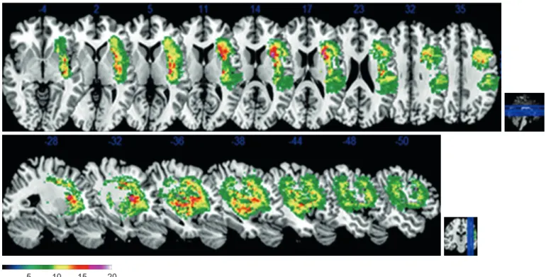
Group comparison analysis between aphasia and non-aphasia group Peak intensity was showed in left extra-nuclear area (χ2 = 20.3, MNI: −28, 14, 14) and high- powered regions were also presented in insular cortex (χ2 = 17.4, MNI: −32, 8, 14), claustrum (χ2 = 15.7, MNI: −32, 8, 4), inferior frontal gyrus (χ2 = 12.7, MNI: −38, 4, 34) and superior temporal gyrus (χ2 = 9.5, MNI: −50, 2, 4) in aphasia group compared to non-aphasia group (p
< 0.01), (Fig. 1).
Lesions relating with language performance
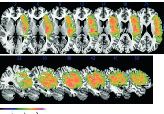
The regions of peak intensity associated with AQ were left inferior parietal lobule (Z = 7.2, MNI: −42, −24, 24), insula/BA 13(Z=6.9, MNI: −38, −8, 2; −36, −20, 12), middle frontal gyrus (Z = 6.0, MNI: −38, 10, 34), precentral gyrus (Z = 5.6, MNI: −38, −4, 38), inferior frontal gyrus (Z = 5.4, MNI: −38, 26, 12), superior temporal gyrus (Z = 4.12, MNI: −46, −28, 7) in order of Z score (p < 0.001) (Fig. 2).
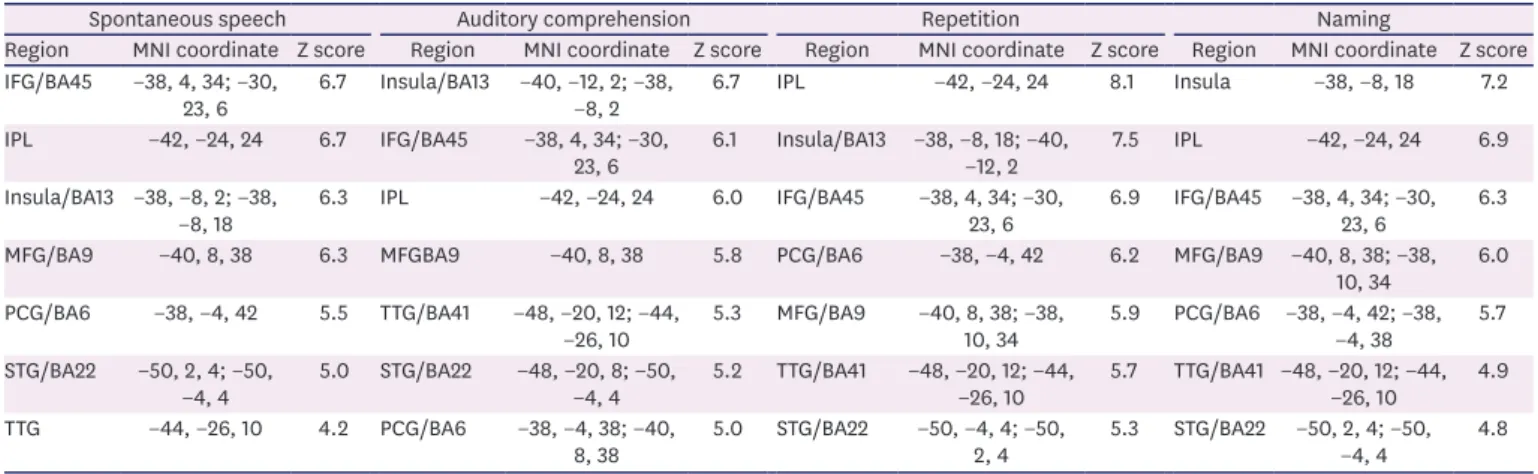
Most highly powered regions as highest Z score related to spontaneous speech were inferior frontal gyrus and inferior parietal lobule (p < 0.001). Left insula/Brodmann area 13 was determined to most highly associated area in auditory comprehension and naming (p <
0.001), Repetition was highest relationship with left inferior parietal lobule (p < 0.001). The detail information of correlation analysis of four subtests of K-WAB and brain lesions were shown in Fig. 3 and Table 1.
DISCUSSION
The remarkable findings of this study showed that an involvement of the inferior parietal lobule and insula were most strongly associated with severity of aphasia, and also subtests of K-WAB.
Traditionally the cortical areas such as Broca's area and Wernicke's area of brain were regarded as critical regions of aphasia [7,11], however, more recently, more complex processes have been reported to be involved in language formation. Insula, deep folded cerebral cortex, was known to most common region affected by middle cerebral artery territory infarction and greatest overlap among large strokes reflecting vulnerability to ischemia [12]. Also, complex process of speech probably depends on a network of brain regions including anterior insula [13]. Inferior parietal lobule, part of arcuate fasciculus, was known to important neural tract in language function.
4/8 https://doi.org/10.12786/bn.2021.14.e14
Neural Substrates of Aphasia in Acute Stroke Brain & NeuroRehabilitation
https://e-bnr.org
5 10 15 20
Fig. 1. Group comparison analysis between aphasia and non-aphasia group. The color bar indicates chi square (χ2) by p < 0.01. Higher χ2 indicates anatomical regions related to aphasia group.
2 4 6
Fig. 2. Voxel-based lesion-symptom maps of significant correlation between lesion and aphasia quotient (p< 0.001 with corrected false discovery rate).
Anatomical correlation was shown as axial and sagittal view. The color range indicates Z score.
2 4 6
A
B
E
F
C
D
G
H
Fig. 3. Anatomical correlation between brain lesions and four subtests of K-WAB shown as axial and sagittal view. (A, E) spontaneous speech, (B, F) auditory comprehension, (C, G) repetition, (D, H) naming. The color range indicates Z score by statistical significance p< 0.001, false discovery rate corrected. Higher Z score indicates areas associated with lower score of K-WAB subtests.
K-WAB, Korean version of the Western Aphasia Test.
Neural connectivity and preservation of arcuate fasciculus was related to better aphasia outcome in previous study [14]. In this context, neural correlates of language functions in acute phase of stroke may be extended and overlapped in spatial regions and then this will happen highest powered lesion correlation in insula and inferior parietal lobule such as arcuate fasciculus.
In this study, spontaneous speech was strongly associated with inferior frontal gyrus including BA 45, middle frontal gyrus including BA 9, inferior parietal lobule, insular area and followed by precentral gyrus. This is consistent with the previous study that showed substantial evidence of the association between verbal fluency and left inferior frontal gyrus, especially Broca's area known as BA 44/45 and premotor and precentral gyrus known as motor area [15]. Some neuroimaging studies also revealed that damage in anterior insula had shown predictive role in speech production [16].
Interestingly, auditory word comprehension was strongly associated with insular cortex, inferior frontal gyrus, inferior parietal lobule in order in this study opposite of traditional concept that comprehension engaged in Wernicke area. But, it is line with the study that major insula involvement is associated with large MCA territory infarcts, proximal MCA occlusions, and greater stroke severity, and neurologic abnormalities, including somatosensory, gustatory, neuropsychological, language, auditory processing, vestibular, and swallowing disorders [17], and that left inferior parietal lobe were associated with perisylvian cortex, were reported as brain regions involved in language comprehension [18], and the processing of syntactic information [19], selection and integration of semantic information for comprehension [20]. However, VLSM studies have a limitation that do not account for lesion size may mistakenly attribute disproportionate significance of injury to regions that tend to be associated with large lesion volumes (e.g., insular cortex) [21].
Auditory word repetition has been known to activation of auditory-motor pathway. Arcuate fasciculus, connecting white matter tracts, damage and also left posterior temporal cortex, angular gyrus and inferior frontal gyrus were associated to repetition [22] corresponding to this study.
In this study, naming was strongly associated with insular cortex, inferior posterior lobule, inferior frontal gyrus in order. The ability of naming is considered to multi-stages of complex processing, such as recognition of item, representation and articulation [5]. It is in line
6/8 https://doi.org/10.12786/bn.2021.14.e14
Neural Substrates of Aphasia in Acute Stroke Brain & NeuroRehabilitation
https://e-bnr.org
Table 1. Anatomic coordinates of four subtests of Korean version of the Western Aphasia Test (p < 0.001, false discovery rate corrected)
Spontaneous speech Auditory comprehension Repetition Naming
Region MNI coordinate Z score Region MNI coordinate Z score Region MNI coordinate Z score Region MNI coordinate Z score IFG/BA45 −38, 4, 34; −30,
23, 6 6.7 Insula/BA13 −40, −12, 2; −38,
−8, 2 6.7 IPL −42, −24, 24 8.1 Insula −38, −8, 18 7.2
IPL −42, −24, 24 6.7 IFG/BA45 −38, 4, 34; −30,
23, 6 6.1 Insula/BA13 −38, −8, 18; −40,
−12, 2 7.5 IPL −42, −24, 24 6.9
Insula/BA13 −38, −8, 2; −38,
−8, 18 6.3 IPL −42, −24, 24 6.0 IFG/BA45 −38, 4, 34; −30,
23, 6 6.9 IFG/BA45 −38, 4, 34; −30,
23, 6 6.3
MFG/BA9 −40, 8, 38 6.3 MFGBA9 −40, 8, 38 5.8 PCG/BA6 −38, −4, 42 6.2 MFG/BA9 −40, 8, 38; −38,
10, 34 6.0
PCG/BA6 −38, −4, 42 5.5 TTG/BA41 −48, −20, 12; −44,
−26, 10 5.3 MFG/BA9 −40, 8, 38; −38,
10, 34 5.9 PCG/BA6 −38, −4, 42; −38,
−4, 38 5.7
STG/BA22 −50, 2, 4; −50,
−4, 4 5.0 STG/BA22 −48, −20, 8; −50,
−4, 4 5.2 TTG/BA41 −48, −20, 12; −44,
−26, 10 5.7 TTG/BA41 −48, −20, 12; −44,
−26, 10 4.9
TTG −44, −26, 10 4.2 PCG/BA6 −38, −4, 38; −40,
8, 38 5.0 STG/BA22 −50, −4, 4; −50,
2, 4 5.3 STG/BA22 −50, 2, 4; −50,
−4, 4 4.8
All brain regions in left hemisphere.
IFG, inferior frontal gyrus; IPL, inferior parietal lobule; MFG, middle frontal gyrus; PCG, precentral gyrus; STG, superior temporal gyrus; TTG, transverse temporal gyrus.
with the previous studies that insula is associated with greater stroke severity and language, auditory processing [17], and that left middle and superior temporal gyrus, inferior parietal cortex and inferior frontal cortex were revealed to relationship with naming in [5,23], and that revealed the role of left middle and inferior temporal gyrus as word production or naming [5,6]. Also, these brain lesions can attribute to prognosis of aphasia on the basis of recent study of post-stroke aphasia using language subsets in chronic patients [24].
There are several limitations of this retrospective study. First, selection bias may affect our findings. The number of subjects without aphasia was relatively small and the location of brain in patients did not cover the whole hemisphere. Second, measurement bias may have been present, despite lesion mapping were conducted by a skilled clinician who was blinded to all clinical information, with same equipment and protocol, and K-WAB was assessed by experienced speech therapist. Third, the confounding factors such as age, education level, cognitive function, medication, which may affect K-WAB, could not be controlled because of the limitation of retrospective study. Forth, more advanced language functions including reading and writing were not evaluated because majority of patients were severe aphasia.
This study showed that the brain lesion related to language processing could be localized using voxel-based lesion-symptom analysis from early MRI imaging study in acute stroke. It may be useful to understand the language process and brain lesion in acute phase of stroke.
Further prospective studies will be required to verify these findings.
REFERENCES
1. Inatomi Y, Yonehara T, Omiya S, Hashimoto Y, Hirano T, Uchino M, Inatomi Y. Aphasia during the acute phase in ischemic stroke. Cerebrovasc Dis 2008;25:316-323.
PUBMED | CROSSREF
2. Alexander MP, Naeser MA, Palumbo CL. Correlations of subcortical CT lesion sites and aphasia profiles.
Brain 1987;110:961-991.
PUBMED | CROSSREF
3. Bates E, Wilson SM, Saygin AP, Dick F, Sereno MI, Knight RT, Dronkers NF. Voxel-based lesion-symptom mapping. Nat Neurosci 2003;6:448-450.
PUBMED | CROSSREF
4. Rorden C, Karnath HO, Bonilha L. Improving lesion-symptom mapping. J Cogn Neurosci 2007;19:1081-1088.
PUBMED | CROSSREF
5. Baldo JV, Arévalo A, Patterson JP, Dronkers NF. Grey and white matter correlates of picture naming:
evidence from a voxel-based lesion analysis of the Boston Naming Test. Cortex 2013;49:658-667.
PUBMED | CROSSREF
6. Schwartz MF, Kimberg DY, Walker GM, Faseyitan O, Brecher A, Dell GS, Coslett HB. Anterior temporal involvement in semantic word retrieval: voxel-based lesion-symptom mapping evidence from aphasia.
Brain 2009;132:3411-3427.
PUBMED | CROSSREF
7. Albert ML, Helm-Estabrooks N. Diagnosis and treatment of aphasia. Part II. JAMA 1988;259:1205-1210.
PUBMED | CROSSREF
8. Kim H, Na DL. Normative data on the Korean version of the Western Aphasia Battery. J Clin Exp Neuropsychol 2004;26:1011-1020.
PUBMED | CROSSREF
9. Choi JY, Lee KH, Na DL, Byun HS, Lee SJ, Kim H, Kwon M, Lee KH, Kim BT. Subcortical aphasia after striatocapsular infarction: quantitative analysis of brain perfusion SPECT using statistical parametric mapping and a statistical probabilistic anatomic map. J Nucl Med 2007;48:194-200.
PUBMED
10. Tak HJ, Jang SH. Relation between aphasia and arcuate fasciculus in chronic stroke patients. BMC Neurol 2014;14:46.
PUBMED | CROSSREF
11. Albert ML, Helm-Estabrooks N. Diagnosis and treatment of aphasia. Part I. JAMA 1988;259:1043-1047.
PUBMED | CROSSREF
12. Dronkers NF. A new brain region for coordinating speech articulation. Nature 1996;384:159-161.
PUBMED | CROSSREF
13. Hillis AE, Work M, Barker PB, Jacobs MA, Breese EL, Maurer K. Re-examining the brain regions crucial for orchestrating speech articulation. Brain 2004;127:1479-1487.
PUBMED | CROSSREF
14. Kim SH, Jang SH. Prediction of aphasia outcome using diffusion tensor tractography for arcuate fasciculus in stroke. AJNR Am J Neuroradiol 2013;34:785-790.
PUBMED | CROSSREF
15. Costafreda SG, Fu CH, Lee L, Everitt B, Brammer MJ, David AS. A systematic review and quantitative appraisal of fMRI studies of verbal fluency: role of the left inferior frontal gyrus. Hum Brain Mapp 2006;27:799-810.
PUBMED | CROSSREF
16. Borovsky A, Saygin AP, Bates E, Dronkers N. Lesion correlates of conversational speech production deficits. Neuropsychologia 2007;45:2525-2533.
PUBMED | CROSSREF
17. Fink JN, Selim MH, Kumar S, Voetsch B, Fong WC, Caplan LR. Insular cortex infarction in acute middle cerebral artery territory stroke: predictor of stroke severity and vascular lesion. Arch Neurol 2005;62:1081-1085.
PUBMED | CROSSREF
18. Dronkers NF, Wilkins DP, Van Valin RD Jr, Redfern BB, Jaeger JJ. Lesion analysis of the brain areas involved in language comprehension. Cognition 2004;92:145-177.
PUBMED | CROSSREF
19. Friederici AD, Rüschemeyer SA, Hahne A, Fiebach CJ. The role of left inferior frontal and superior temporal cortex in sentence comprehension: localizing syntactic and semantic processes. Cereb Cortex 2003;13:170-177.
PUBMED | CROSSREF
20. Homae F, Hashimoto R, Nakajima K, Miyashita Y, Sakai KL. From perception to sentence
comprehension: the convergence of auditory and visual information of language in the left inferior frontal cortex. Neuroimage 2002;16:883-900.
PUBMED | CROSSREF
21. Wu O, Cloonan L, Mocking SJ, Bouts MJ, Copen WA, Cougo-Pinto PT, Fitzpatrick K, Kanakis A, Schaefer PW, Rosand J, Furie KL, Rost NS. Role of acute lesion topography in initial ischemic stroke severity and long-term functional outcomes. Stroke 2015;46:2438-2444.
PUBMED | CROSSREF
22. Price CJ. The anatomy of language: contributions from functional neuroimaging. J Anat 2000;197:335-359.
PUBMED | CROSSREF
23. Walker GM, Schwartz MF, Kimberg DY, Faseyitan O, Brecher A, Dell GS, Coslett HB. Support for anterior temporal involvement in semantic error production in aphasia: new evidence from VLSM. Brain Lang 2011;117:110-122.
PUBMED | CROSSREF
24. Sul B, Lee KB, Hong BY, Kim JS, Kim J, Hwang WS, Lim SH. Association of lesion location with long-term recovery in post-stroke aphasia and language deficit. Front Neurol 2019;10:776.
PUBMED | CROSSREF
8/8 https://doi.org/10.12786/bn.2021.14.e14
Neural Substrates of Aphasia in Acute Stroke Brain & NeuroRehabilitation
https://e-bnr.org