ABSTRACT
Purpose: The aims of this study were to examine the salivary microbiota in conditions of periodontal health and disease and to explore microbial changes following nonsurgical periodontal treatment.
Methods: Non-stimulated saliva samples were collected from 4 periodontally healthy participants at baseline and from 8 patients with chronic periodontitis at baseline and 3 months following nonsurgical periodontal therapy. The V3 and V4 regions of the 16S rRNA gene from the DNA of saliva samples were amplified and sequenced. The salivary microbial compositions of the healthy participants and patients with periodontitis prior to and following nonsurgical treatment of periodontitis were compared based on the relative abundance of various taxa.
Results: On average, 299 operational taxonomic units were identified in each sample.
The phylogenetic diversity in patients with periodontitis was higher than that in healthy participants and decreased following treatment. The abundance of the phylum Spirochaetes and the genus Treponema in patients with periodontitis was 143- and 134-fold higher than in the healthy control group, respectively, but decreased significantly following treatment.
The species that were overabundant in the saliva of patients with periodontitis included the Peptostreptococcus stomatis group, Porphyromonas gingivalis, the Fusobacterium nucleatum group, Parvimonas micra, Porphyromonas endodontalis, Filifactor alocis, and Tannerella forsythia. The phylum Actinobacteria, the genus Streptococcaceae_uc, and the species Streptococcus salivarius group were more abundant in healthy participants than in those with periodontitis. There was a trend toward a decrease in disease-associated taxa and an increase in health-associated taxa following treatment.
Conclusions: Our results revealed differences in the taxa of salivary microbiota between conditions of periodontal health and disease. The taxa found to be associated with health or disease have potential for use as salivary biomarkers for periodontal health or disease.
Keywords: Microbiota; Periodontitis; Porphyromonas gingivalis; Saliva; Treponema
Research Article
Received: Sep 3, 2019 Revised: Apr 21, 2020 Accepted: Apr 27, 2020
*Correspondence:
Suk Ji
Department of Periodontology, Institute of Oral Health Science, Ajou University Hospital, Ajou University School of Medicine, 164 World cup-ro, Yeongtong-gu, Suwon 16499, Korea.
E-mail: sukji@ajou.ac.kr Tel: +82-31-219-5331 Fax: +82-31-219-4112
Copyright © 2020. Korean Academy of Periodontology
This is an Open Access article distributed under the terms of the Creative Commons Attribution Non-Commercial License (https://
creativecommons.org/licenses/by-nc/4.0/).
ORCID iDs Youngkyung Ko
https://orcid.org/0000-0002-6564-9156 Eun-Mi Lee
https://orcid.org/0000-0002-1995-2867 Joo Cheol Park
https://orcid.org/0000-0002-3162-7557 Man Bock Gu
https://orcid.org/0000-0002-0848-5394 Seongmin Bak
https://orcid.org/0000-0003-3159-5599 Suk Ji
https://orcid.org/0000-0001-9720-6731 Funding
This research was supported by a grant of the Korea Health Technology R&D Project through the Korea Health Industry Development Institute (KHIDI) funded by the Ministry of Health & Welfare, Republic of Korea (grant number: HI16C0220).
Youngkyung Ko 1, Eun-Mi Lee 1, Joo Cheol Park 2, Man Bock Gu 3, Seongmin Bak 4, Suk Ji 4,*
1 Department of Periodontics, Seoul St. Mary's Hospital, The Catholic University of Korea College of Medicine, Seoul, Korea
2Department of Oral Histology, Seoul National University School of Dentistry, Seoul, Korea
3Department of Biotechnology, Korea University College of Life Sciences and Biotechnology, Seoul, Korea
4 Department of Periodontology, Institute of Oral Health Science, Ajou University School of Medicine, Suwon, Korea
Salivary microbiota in periodontal health and disease and their changes following nonsurgical periodontal treatment
Periodontal Science
Author Contributions
Conceptualization: Youngkyung Ko, Suk Ji; Data curation: Youngkyung Ko, Eun-Mi Lee, Seongmin Bak, Suk Ji; Formal analysis:
Youngkyung Ko, Suk Ji; Funding acquisition:
Youngkyung Ko, Joo Cheol Park, Man Bock Gu; Investigation: Youngkyung Ko, Suk Ji;
Methodology: Youngkyung Ko, Eun-Mi Lee, Seongmin Bak, Suk Ji; Project administration:
Youngkyung Ko, Suk Ji; Resources: Joo Cheol Park, Man Bock Gu; Software: Youngkyung Ko, Suk Ji; Supervision: Youngkyung Ko, Joo Cheol Park, Man Bock Gu, Suk Ji; Validation:
Youngkyung Ko, Joo Cheol Park, Man Bock Gu, Suk Ji; Visualization: Suk Ji; Writing - original draft: Suk Ji; Writing - review & editing:
Youngkyung Ko, Joo Cheol Park, Man Bock Gu, Suk Ji.
Conflict of interest
No potential conflict of interest relevant to this article was reported.
INTRODUCTION
Periodontal disease is common among adults and can lead to tooth loss. It is accompanied by destruction of tissues surrounding the teeth following an inflammatory reaction that results from the host immune response to oral bacteria [1]. A homeostatic balance with symbiotic microbiota is maintained in the oral cavity in healthy conditions, whereas the dysbiosis of oral microbiota causes inflammatory destruction of periodontal tissue [2]. It was recently suggested that dysbiotic microbiota have distinct functions and act as accessory, pathobiont, or keystone pathogens, and that all these bacterial types can affect the pathogenesis of periodontitis [3]. Therefore, the entire dysbiotic subgingival microbiota, rather than a single bacterium, influences the progression of periodontitis [2-4]. Understanding dysbiotic microbiota can provide useful insights into the pathogenesis, diagnosis, and treatment of periodontitis.
Even before the advent of next-generation sequencing (NGS) as a method of analyzing oral microbiota, substantial evidence emerged regarding differences in the microbial composition of subgingival plaque between healthy periodontal conditions and periodontitis. Research by Socransky et al. [5] was essential in understanding the association between the oral bacterial community and periodontal disease. In their study, the distribution of 40 species was analyzed in subgingival plaque, and they reported that the subgingival bacteria in patients with periodontitis were characterized by a relative decrease of Actinomyces and a relative increase of the red complex, whereas Streptococci and Actinomyces were predominant in the healthy gingiva, with a relatively low proportion of the red complex [5]. Several studies on the effects of periodontal treatment on the bacterial composition of subgingival plaque have shown that improved clinical outcomes may be obtained when the levels of periodontal pathogens are reduced and the proportions of health-associated bacteria are increased [6,7].
Targeted approaches, including DNA hybridization and polymerase chain reaction (PCR)- based analyses, have provided information with regard to changes at the individual species level, but have not been able to provide a comprehensive view of the microbial community.
The analysis of oral microbiota using open-ended molecular analytic tests, such as NGS, can provide additional information to enhance our understanding of the role of microorganisms in periodontal health and disease.
There is an unmet need for simple, immediate forms of near-patient testing to diagnose periodontitis [8]. Saliva has become a focus as a near-patient diagnostic tool for monitoring periodontal health, as repeated sampling is relatively easy and noninvasive. However, as the saliva combines bacteria from all niches of the oral cavity, it is questionable whether differences in the salivary microbiota reflect periodontal disease status or changes following periodontal treatment.
Research into the oral microbiota using NGS has been performed predominantly via the analysis of subgingival plaque, whereas very few studies have analyzed the salivary microbiota. The present study examined the characteristics of the salivary microbiota in conditions of periodontal health and disease following nonsurgical periodontal treatment and analyzed which bacterial taxa were present at a relatively high rate in conditions of periodontal health or periodontitis.
MATERIALS AND METHODS
Participants and microbial sampling
Salivary samples were obtained from 4 periodontally healthy participants and 8 patients diagnosed with chronic periodontitis recruited from Ajou University Dental Hospital and Seoul St. Mary's Hospital. This study was approved by the Institutional Review Board for Human Subjects of Ajou University Dental Hospital (AJIRB-BMR-SMP-16) and Seoul St.
Mary's Hospital (KC16TIMI0755). Informed consent was obtained from all participants. The 12 participants were included in an ongoing investigation of the diagnosis and treatment of periodontal disease. The participants had no history of systemic disease that could influence the prognosis of periodontitis, untreated caries, or orthodontic appliances, and they did not smoke. None of the participants were pregnant/breastfeeding or treated with antibiotic, antimicrobial, and/or anti-inflammatory drugs during the 3 months before the examinations and sampling. The inclusion criteria for the chronic periodontitis group were as follows: with all teeth divided into 4 quadrants, each quadrant had at least 2 teeth with a probing depth (PD) ≥5 mm and attachment loss of ≥3 mm, and participants must not have had received periodontal treatment in the last 2 years [9]. Participants in the healthy control group were required to have an average PD of <3 mm and <20% of sites presenting with bleeding on probing (BOP). Two examiners (Suk Ji and Youngkyung Ko) carried out the periodontal examinations and microbial sampling. All participants were instructed to avoid ingestion, rinsing, and oral hygiene measures for 1 hour prior to sampling. From each participant, approximately 3 mL of a non-stimulated whole saliva sample was obtained and stored at
−80°C until DNA extraction. Clinical parameters were measured, including the plaque index (PI), PD, gingival recession, and BOP. Patients with periodontitis received nonsurgical periodontal treatment, and saliva sampling and measurements of clinical parameters were repeated 3 months after treatment.
DNA extraction, amplification of 16S rDNA, and Illumina sequencing Of the saliva sample, 1 mL was centrifuged at 15,928×g for 5 min, and the pellet was transferred to ChunLab, Inc. (Seoul, Korea) for DNA extraction and sequencing analysis.
DNA was extracted using a FastDNA SPIN Kit for Soil (MP Biomedicals, Santa Ana, CA, USA), and PCR amplification was performed using primers targeting the V3 and V4 regions of the 16S rRNA gene, as previously described [10]. The PCR products were sequenced with an Illumina MiSeq sequencing system at ChunLab, Inc.
Sequencing analysis and statistical analysis
UCHIME [11] and the non-chimeric 16S rRNA database from EzBioCloud were used to detect chimera on reads that contained a <97% best hit similarity rate. The taxonomic profile of the microbiome was analyzed and compared between the healthy and periodontitis groups and between pre-treatment and post-treatment in the periodontitis group using BIOiPLUG (https://www.bioiplug.com/), a web-based life information analysis cloud platform provided by ChunLab, Inc. The analysis was based on the relative abundance of each taxonomic group, and the results were reconstructed for this study. The comparison of the number of operational taxonomic units (OTUs), the Chao 1 index, the Shannon index, and the phylogenetic diversity among the healthy, pre-treatment, and post-treatment groups were performed using the Wilcoxon rank-sum test. Kruskal-Wallis H statistical analysis was conducted to evaluate the differences in the dominant OTUs between the healthy and periodontitis groups and between pre-treatment and post-treatment in the periodontitis group. The sum of the whole-mouth PI, PD, clinical attachment level (CAL), the count of sites
with PD ≥5 mm, and the percentage of BOP of the full mouth were compared between healthy participants and those diagnosed with periodontitis using the Mann-Whitney U test, and those clinical parameters were compared in pre-treatment and post-treatment samples using the Wilcoxon test. Analyses were performed using SPSS version 20 (IBM Corp., Armonk, NY, USA). P values <0.05 were considered to indicate statistical significance.
RESULTS
Sample groups and clinical responses to treatment
Four periodontally healthy participants (1 man and 3 women) with a mean age of 41 years (range, 26–52 years) and 8 patients with chronic periodontitis (5 men and 3 women) with a mean age of 52 years (range, 38–58 years) were selected for microbiome analysis. In total, the 20 samples were divided into 3 groups (healthy, pre-treatment of periodontitis, and post- treatment of periodontitis). The clinical parameters of the 3 groups are detailed in Table 1.
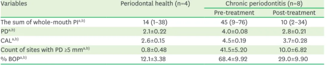
There were statistically significant differences in PI, PD, CAL, count of sites with PD ≥5 mm, and percentage of BOP of the full mouth between the healthy participants and the pre-treatment periodontitis participants and between the pre- and post-treatment samples (Table 1). Clinical improvements in all parameters were observed in the periodontitis group after 3 months of nonsurgical treatment.
Taxa diversity of the samples
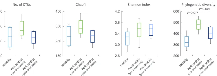
From the 4 periodontally healthy, 8 pre-treatment, and 8 post-treatment bacterial communities, the average number of reads used for data analysis was 37,101 (range, 21,234–51,052 per sample) with an average length of 422 bp and an average number of OTUs per sample of 299. The number of OTUs, Chao 1 index (species richness index), and Shannon index (species evenness index) did not differ among groups (Figure 1). However, the phylogenetic diversity of the pre-treatment group (median, 489.5) was significantly higher than that of the healthy group (median, 325.5) and the post-treatment group (median, 404.0) groups (Figure 1).
Differential abundance of taxa in health and disease
To investigate differences in the composition of the salivary microbiota and to evaluate microbial changes following periodontal treatment, the relative abundance of each taxon was compared between the healthy and pre-treatment periodontitis group and between the pre- and post-treatment samples at the phylum, genus, and species levels (Figures 2-4). In total, 18 OTUs at the phylum level were identified. The distribution pattern of the top 10 phyla (comprising 99.90%–99.98% of the total counts) in each sample group is shown in Figure 2A.
Table 1. Clinical characteristics of healthy participants and patients with periodontitis pre- and post-treatment
Variables Periodontal health (n=4) Chronic periodontitis (n=8)
Pre-treatment Post-treatment
The sum of whole-mouth PIa,b) 14 (1–38) 45 (9–76) 10 (2–34)
PDa,b) 2.1±0.22 4.0±0.08 2.8±0.21
CALa,b) 2.6±0.15 4.5±0.19 3.7±0.28
Count of sites with PD ≥5 mma,b) 0.8±0.48 41.5±5.20 10.0±6.82
% BOPa,b) 12.1±3.38 68.4±9.92 29.0±9.90
Values are presented as median (minimum–maximum) or mean±standard deviation.
PI: plaque index; PD: probing depth; CAL: clinical attachment level; BOP: bleeding on probing.
a)P<0.05 by the Mann-Whitney U test between the healthy group and pre-treatment in the periodontitis group;
b)P<0.05 by the Wilcoxon signed rank test between pre-treatment and post-treatment in the periodontitis group.
The 5 most abundant phyla in all 3 groups were Firmicutes, Bacteroidetes, Proteobacteria, Actinobacteria, and Fusobacteria, which comprised 97.7%–98.9% of the total count. The level of Actinobacteria was twofold higher in the saliva of the healthy controls than in the periodontitis group (16.5% vs. 8.7%) (Figure 2A), whereas the level of Spirochaetes was 143- No. of OTUs
150 350
250 450
Healthy Peridontitis (pre-treatment)Peridontitis
(post -treatment)
Chao 1
150 350
250 450
Healthy Peridontitis (pre-treatment)Peridontitis
(post -treatment)
Shannon index
2.6 3.8
3.0 3.4 4.2
Healthy Peridontitis (pre-treatment)Peridontitis
(post -treatment)
Phylogenetic diversity P=0.017
200 500
300 400 600
Healthy Peridontitis (pre-treatment)Peridontitis
(post -treatment) P=0.021
Figure 1. Comparison of the number of OTUs, Chao 1 index, Shannon index, and phylogenetic diversity. Saliva samples were collected from 4 periodontally healthy participants at baseline and from 8 subjects diagnosed with chronic periodontitis at baseline and 3 months following nonsurgical periodontal treatment.
Each value is presented as a box plot. The top, middle, and bottom lines of the boxes represent the 25th, 50th (median), and 75th percentiles, respectively. The significance of differences among the 3 groups was evaluated using the Wilcoxon rank-sum test, and P<0.05 was considered to indicate statistical significance.
Only P values of <0.05 are indicated. OTU: operational taxonomic unit.
0 20 40 60 80 100
%
% Healthy
Peridontitis (pre-treatment)
A
Peridontitis (post-treatment)
Actinobacteriaa) Bacteroidetes
SR1
Saccharibacteria_TM7 Tenericutesa,b) Firmicutes
Fusobacteria Spirochaetesa,b) Proteobacteria
Synergistetesa,b)
Healthy Peridontitis (pre-treatment) Peridontitis (post-treatment)
Spirochaetes B
Tenericutes
Healthy Peridontitis (pre-treatment) Peridontitis (post-treatment)
Synergistetes
Healthy Peridontitis (pre-treatment) Peridontitis (post-treatment)
0 0.2 0.4 0.6 0.8
a) b)
a) b)
a) b)
Figure 2. Differences between conditions of periodontal health and periodontitis at the phylum level. (A) Distribution pattern of the top 10 phyla in the healthy group and the pre-and post-treatment periodontitis groups.
(B) Three phyla dominant in the periodontitis group compared with the healthy control group.
a)P<0.05 by the Kruskal-Wallis H test between the healthy and periodontitis (pre-treatment) groups; b)P<0.05 by the Kruskal-Wallis H test between the pre- and post-treatment periodontitis groups.
fold higher in the periodontitis group than in the healthy control group (0.6% vs. 0.004%) (Figure 2B). The levels of Spirochaetes, Synergistetes, and Tenericutes, which were dominant in the periodontitis group, significantly decreased following nonsurgical periodontal treatment (Figure 2B).
The results revealed that 28 genera and 62 species were significantly more abundant in the periodontal saliva than in the healthy saliva. By contrast, 5 genera and 10 species were more abundant in the healthy saliva. Among the genera and species exhibiting relative differences in abundance between periodontal health and disease, only those genera or species with a
>0.05% relative abundance in disease compared with healthy conditions and those with a
>0.01% relative abundance in healthy conditions compared with disease are presented in Figures 3 and 4.
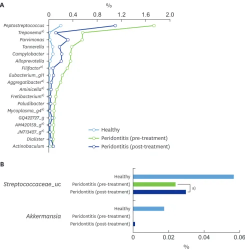
The top 7 genera showing a high abundance in the saliva from the pre-treatment periodontitis group were Peptostreptococcus, Treponema, Parvimonas, Tannerella, Campylobacter, Alloprevotella, and Filifactor (Figure 3A), whereas Streptococcaceae_uc and Akkermansia were present
A
0 0.4 0.8 1.2 1.6 2.0
%
Peptostreptococcus Treponemaa) Parvimonas Tannerella Campylobacter Alloprevotella Filifactora) Eubacterium_g11 Aggregatibactera) Aminicellaa) Fretibacteriuma) Paludibacter Mycoplasma_g4a) GQ422727_g AM420159_ga) JN713427_ga) Dialister Actinobaculum
Healthy
Peridontitis (post-treatment) Peridontitis (pre-treatment)
%
Healthy Peridontitis (pre-treatment) Peridontitis (post-treatment)
B
Akkermansia
Healthy Peridontitis (pre-treatment) Peridontitis (post-treatment)
Streptococcaceae_uc
0 0.02 0.04 0.06
a)
Figure 3. Differences between conditions of periodontal health and periodontitis at the genus level. (A) Genera significantly more abundant in saliva samples from the pre-treatment periodontitis group than in those from the healthy group among genera with a distribution of >0.05% in the untreated periodontitis group. (B) Genera significantly more abundant in saliva samples from the healthy group than in those from the pre-treatment periodontitis group among genera with a distribution >0.01% in the healthy group.
a)P<0.05 was considered to indicate statistical significance by the Kruskal-Wallis H test between the pre- and post-treatment periodontitis.
at higher levels in the saliva from the healthy group than in the saliva from the pre-treatment periodontitis group (Figure 3B). In particular, the relative abundance of Treponema in the pre- treatment periodontitis group was 134-fold higher than that of the healthy controls (0.6% vs.
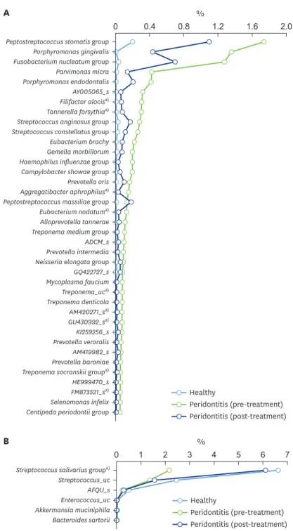
0.004%) (Figure 3A). The ratio of Filifactor in the periodontitis group was 0.3%, whereas that A
0 0.4 0.8 1.2 1.6 2.0
%
Peptostreptococcus stomatis group Porphyromonas gingivalis Fusobacterium nucleatum group Parvimonas micra Porphyromonas endodontalis AY005065_s Filifactor alocisa) Tannerella forsythiaa) Streptococcus anginosus group Streptococcus constellatus group Eubacterium brachy Gemella morbillorum Haemophilus influenzae group Campylobacter showae group Prevotella oris Aggregatibacter aphrophilusa) Peptostreptococcus massiliae group Eubacterium nodatuma) Alloprevotella tannerae Treponema medium group ADCM_s Prevotella intermedia Neisseria elongata group GQ422727_s Mycoplasma faucium Treponema_uca) AM420271_sa) GU430992_sa) KI259256_s Treponema denticola
Prevotella veroralis AM419982_s Prevotella baroniae Treponema socranskii groupa) HE999470_s FM873521_sa) Selenomonas infelix Centipeda periodontii group
Healthy
Peridontitis (post-treatment) Peridontitis (pre-treatment)
B
0 2 4 5 6 7
%
Streptococcus salivarius groupa) Streptococcus_uc AFQU_s Enterococcus_uc Akkermansia muciniphila Bacteroides sartorii
Healthy
Peridontitis (post-treatment) Peridontitis (pre-treatment)
1 3
Figure 4. Differences between conditions of periodontal health and periodontitis at the species level. (A) Species significantly more abundant in saliva samples from the pre-treatment periodontitis group than in those from the healthy group among genera with a distribution >0.05% in the periodontitis group. (B) Species significantly more abundant in saliva samples from the healthy group than in those from the pre-treatment periodontitis group among species with a distribution >0.01% in the healthy group.
a)P<0.05 was considered to indicate statistical significant by the Kruskal-Wallis H test between the pre- and post- treatment periodontitis groups.
in the healthy controls was 0%. There was a trend for the genera dominant in periodontitis to decrease and for the genera dominant in health to increase following treatment. Significant differences between pre-treatment and post-treatment were found in Treponema, Filifactor, Aggregatibacter, Aminicella, Fretibacterium, Mycoplasma_g4, and 2 unclassified genera (AM420159_g and JN713427_g) among the genera dominant in untreated periodontitis, and in this group, the Streptococcus anginosus group and the Streptococcus constellatus group were also found. The genera dominant in healthy conditions included Streptococcaceae_uc (Figure 3B).
The top 8 species that were more abundant in diseased saliva included the P. stomatis group, Porphyromonas gingivalis, the Fusobacterium nucleatum group, Parvimonas micra, Porphyromonas endodontalis, AY005065_s (Prevotella genus), Filifactor alocis, and Tannerella forsythia (Figure 4A). The abundance ratios of these species were 0.3%–1.73% in the periodontitis group and <0.04% in the healthy group, with the exception of the P. stomatis group (0.2% in the healthy group). Among these 7 species, the distributions of P. gingivalis and F. alocis in healthy controls were zero. There was a trend for species dominant in periodontitis to decrease following treatment. Nine species, including F. alocis and T. forsythia, decreased significantly following treatment (Figure 4A). Six species, including the Streptococcus salivarius group, were present at higher levels in healthy saliva than in diseased saliva, and the S. salivarius group increased in abundance following treatment (Figure 4B).
DISCUSSION
The present study examined the distribution and characteristics of the salivary microbiota in periodontal health and disease and their changes following nonsurgical periodontal treatment. Sequencing analysis of the 16S rRNA gene revealed that healthy and diseased saliva samples had compositionally distinct microbiota and that the samples of saliva from participants with periodontitis had more diverse microbiota than the samples from healthy participants. In patients with periodontitis, the phylum Spirochaetes and the genus Treponema were present at 143- and 134-fold higher levels than in healthy participants, respectively, and these taxa significantly decreased following periodontal treatment. The association between these 2 taxa and periodontitis has been well demonstrated [12-14]. Among the OTUs at the species level, the distributions of well-established periodontal pathogens of the P. stomatis group, P. gingivalis, the F. nucleatum group, and T. forsythia [2,3,5,13,15,16] were significantly higher than those in the healthy group. In addition, P. micra, P. endodontalis, and F. alocis, which have been reported as newly identified pathogenic bacteria in recent NGS studies [13,16,17], were also identified to be highly abundant in the periodontitis group. The phylum Actinobacteria, the genus Streptococcaceae_uc, and the species S. salivarius group were found to be markedly more abundant in the samples from healthy participants than in the periodontitis group. The association between Actinobacteria and periodontal health is consistent with previous studies [13,15], and S. salivarius is frequently described as a commensal bacterium [18], with a strain of S. salivarius under trial for use as a probiotic for the prevention of oral infections [19]. These results show that the periodontitis- or health-associated bacteria identified through the analysis of subgingival plaque bacteria correspond closely to the salivary bacteria associated with periodontitis or healthy periodontal conditions in this study.
Due to its simplicity and easy accessibility, saliva sampling has been suggested as a tool for monitoring periodontitis. Therefore, the taxa identified as health-associated or disease- associated may be used as candidate salivary biomarkers of periodontal health or disease.
The microbiota of the subgingival space has been the focus of research into the pathogenesis of periodontitis. However, the sampling of subgingival plaque is relatively complicated and requires more equipment and trained personnel. Our results show that the relative abundance of well-known health- or periodontitis-associated bacteria in the salivary microbiota closely parallels that of the subgingival microbiota in health or disease, as supported by recent NGS studies analyzing the association between subgingival and salivary microbiota. Periodontal pathogens observed at a high proportion in subgingival plaque were observed in saliva at levels correlated with their abundance in subgingival plaque [16,20]. Kageyama et al. [20] demonstrated that the relative abundance of subgingival plaque-specific bacteria in the salivary microbiota was closely correlated with the severity of periodontal disease. They concluded that the salivary microbiota may be a promising target for the evaluation of subgingival plaque-derived bacteria representing periodontal health. In addition, Belstrom et al. [16] compared the subgingival and salivary microbiota before and after periodontal treatment to determine whether changes in the subgingival microbiota were reflected in the salivary microbiota; it was concluded that the subgingival and salivary abundance of periodontal pathogens was correlated before and after treatment.
In research aiming to identify biomarkers for periodontitis using saliva, the levels of well- known pathogenic bacteria, including the 3 red complex species, are determined by targeting a specific region of the16S rRNA gene [21-24]. Multiple promising microbial biomarkers that correlate with the clinical parameters of periodontitis have been identified in saliva. Among them, P. gingivalis, Prevotella intermedia, and T. forsythia have been endorsed by multiple studies as potentially useful biomarkers of periodontitis [8]. In the present study, these 3 species were found to be more abundant in the saliva of patients with periodontitis than that of healthy participants.
An optimal biomarker can only be validated through large-scale analysis of the salivary microbiota of a large number of individuals. In the present study, the distribution of P.
gingivalis was zero in all healthy controls, whereas patients with periodontitis had an average distribution of 1.35%, which decreased to 0.43% following treatment. Therefore, P. gingivalis in saliva can be considered as a strong candidate biomarker of periodontitis. However, P. gingivalis was not detected in 1 of the 8 patients with periodontitis. In addition, in another participant, the relative abundance of P. gingivalis increased following periodontal treatment, although the clinical parameters showed improvement (Supplementary Figure 1). This result indicates that diagnosis based on a single species is not feasible, and it is instead suggested that diagnosing periodontitis based on the combined detection of multiple bacterial taxa in saliva is superior to diagnosing periodontitis based on the detection of a single bacterial species.
Relatively few reports have described changes in the salivary microbiota following periodontal treatment [14]. To date, only 1 paper has been published on changes in the salivary microbiome of periodontitis patients after periodontal therapy, and more studies are needed to uncover the whole picture of the salivary microbiome and periodontitis.
Differences in the oral microbiome have been reported from even closely neighboring countries [25]. This is the first study to describe salivary microbiome changes in Koreans.
Scaling and subgingival debridement disrupt the biofilm in the subgingival space and alter the number and composition of subgingival microbes [26]. This eventually attenuates inflammation in the periodontal tissues and reduces the depth of the periodontal pocket, resulting in ecological changes in the subgingival environment. These ecological changes are unfavorable to most periodontopathic bacteria and enable the change in composition
of subgingival microbiota to be maintained for a longer period [27]. In the present study, we examined changes in the salivary microbiota 3 months after nonsurgical periodontal treatment. Reductions in the periodontal pockets and BOP were maintained, and clinical changes were accompanied by alterations in the composition of the salivary microbiota, with a decrease in the relative abundance of P. gingivalis (3-fold, 1.35% vs. 0.43%), P. micra (3-fold, 0.425% vs. 0.14%), F. alocis (4.5-fold, 0.3% vs. 0.07%), and T. forsythia (3.9-fold, 0.3% vs. 0.08%) and an increase in the relative abundance of the S. salivarius group (2.8-fold, 6.13% vs. 2.16%). This suggests that ecological changes in the subgingival environment are associated with sustained changes in the salivary microbiota. We did not detect in the salivary microbiome Halomonas hamiltonii, which was the microorganism most often discovered in Koreans with a healthy periodontium in a report by Park et al. [15]. This discrepancy may have resulted from differences in the sampled groups. An important finding of the present study was that there were distinctions in the taxa of the salivary microbiota between periodontal health and disease. The presence of Streptococcus anginosus group and the Streptococcus constellatus group in the saliva of periodontitis patients has not been reported previously, and further research should explore possible correlations of these taxa with periodontitis in the wider population. Well-known pathogenic and health-associated bacteria were confirmed to be highly related to periodontitis and health, respectively, making them suitable as candidate salivary biomarkers for periodontal health and disease. However, this study has limitations in that the sample size was small and the functional activity of the microbial community was not identified. Another limitation is that quantitative changes in specific bacteria were not analyzed according to participants’ age or periodontitis progression. Large-scale, long-term follow-up studies are needed to observe the causal relationships between periodontopathic bacteria and disease progression.
SUPPLEMENTARY MATERIAL
Supplementary Figure 1
Differences in Porphyromonas gingivalis composition between pre- and post-treatment measurements of 8 patients with periodontitis. The relative abundance of P. gingivalis between the pre- and post-treatment periodontitis communities was compared.
Click here to view
REFERENCES
1. Ji S, Choi YS, Choi Y. Bacterial invasion and persistence: critical events in the pathogenesis of periodontitis? J Periodontal Res 2015;50:570-85.
PUBMED | CROSSREF
2. Darveau RP. Periodontitis: a polymicrobial disruption of host homeostasis. Nat Rev Microbiol 2010;8:481-90.
PUBMED | CROSSREF
3. Hajishengallis G, Lamont RJ. Dancing with the stars: how choreographed bacterial interactions dictate nososymbiocity and give rise to keystone pathogens, accessory pathogens, and pathobionts. Trends Microbiol 2016;24:477-89.
PUBMED | CROSSREF
4. Roberts FA, Darveau RP. Microbial protection and virulence in periodontal tissue as a function of polymicrobial communities: symbiosis and dysbiosis. Periodontol 2000 2015;69:18-27.
PUBMED | CROSSREF
5. Socransky SS, Haffajee AD, Cugini MA, Smith C, Kent RL Jr. Microbial complexes in subgingival plaque.
J Clin Periodontol 1998;25:134-44.
PUBMED | CROSSREF
6. Cugini MA, Haffajee AD, Smith C, Kent RL Jr, Socransky SS. The effect of scaling and root planing on the clinical and microbiological parameters of periodontal diseases: 12-month results. J Clin Periodontol 2000;27:30-6.
PUBMED | CROSSREF
7. De Soete M, Mongardini C, Peuwels M, Haffajee A, Socransky S, van Steenberghe D, et al. One-stage full-mouth disinfection. Long-term microbiological results analyzed by checkerboard DNA-DNA hybridization. J Periodontol 2001;72:374-82.
PUBMED | CROSSREF
8. Ji S, Choi Y. Point-of-care diagnosis of periodontitis using saliva: technically feasible but still a challenge.
Front Cell Infect Microbiol 2015;5:65.
PUBMED | CROSSREF
9. Shi M, Wei Y, Hu W, Nie Y, Wu X, Lu R. The subgingival microbiome of periodontal pockets with different probing depths in chronic and aggressive periodontitis: a pilot study. Front Cell Infect Microbiol 2018;8:124.
PUBMED | CROSSREF
10. Lee SY, Eom YB. Analysis of microbial composition associated with freshwater and seawater. Biomedical Science Letters. 2016;22:150-9.
CROSSREF
11. Edgar RC, Haas BJ, Clemente JC, Quince C, Knight R. UCHIME improves sensitivity and speed of chimera detection. Bioinformatics 2011;27:2194-200.
PUBMED | CROSSREF
12. Visser MB, Ellen RP. New insights into the emerging role of oral spirochaetes in periodontal disease. Clin Microbiol Infect 2011;17:502-12.
PUBMED | CROSSREF
13. Griffen AL, Beall CJ, Campbell JH, Firestone ND, Kumar PS, Yang ZK, et al. Distinct and complex bacterial profiles in human periodontitis and health revealed by 16S pyrosequencing. ISME J 2012;6:1176-85.
PUBMED | CROSSREF
14. Chen C, Hemme C, Beleno J, Shi ZJ, Ning D, Qin Y, et al. Oral microbiota of periodontal health and disease and their changes after nonsurgical periodontal therapy. ISME J 2018;12:1210-24.
PUBMED | CROSSREF
15. Park OJ, Yi H, Jeon JH, Kang SS, Koo KT, Kum KY, et al. Pyrosequencing analysis of subgingival microbiota in distinct periodontal conditions. J Dent Res 2015;94:921-7.
PUBMED | CROSSREF
16. Belstrøm D, Grande MA, Sembler-Møller ML, Kirkby N, Cotton SL, Paster BJ, et al. Influence of periodontal treatment on subgingival and salivary microbiotas. J Periodontol 2018;89:531-9.
PUBMED | CROSSREF
17. Pérez-Chaparro PJ, McCulloch JA, Mamizuka EM, Moraes AD, Faveri M, Figueiredo LC, et al. Do different probing depths exhibit striking differences in microbial profiles? J Clin Periodontol 2018;45:26-37.
PUBMED | CROSSREF
18. Kaci G, Goudercourt D, Dennin V, Pot B, Doré J, Ehrlich SD, et al. Anti-inflammatory properties of Streptococcus salivarius, a commensal bacterium of the oral cavity and digestive tract. Appl Environ Microbiol 2014;80:928-34.
PUBMED | CROSSREF
19. Zupancic K, Kriksic V, Kovacevic I, Kovacevic D. Influence of oral probiotic Streptococcus salivarius K12 on ear and oral cavity health in humans: systematic review. Probiotics Antimicrob Proteins 2017;9:102-10.
PUBMED | CROSSREF
20. Kageyama S, Takeshita T, Asakawa M, Shibata Y, Takeuchi K, Yamanaka W, et al. Relative abundance of total subgingival plaque-specific bacteria in salivary microbiota reflects the overall periodontal condition in patients with periodontitis. PLoS One 2017;12:e0174782.
PUBMED | CROSSREF
21. Sawamoto Y, Sugano N, Tanaka H, Ito K. Detection of periodontopathic bacteria and an oxidative stress marker in saliva from periodontitis patients. Oral Microbiol Immunol 2005;20:216-20.
PUBMED | CROSSREF
22. Ramseier CA, Kinney JS, Herr AE, Braun T, Sugai JV, Shelburne CA, et al. Identification of pathogen and host-response markers correlated with periodontal disease. J Periodontol 2009;80:436-46.
PUBMED | CROSSREF
23. Nomura Y, Shimada Y, Hanada N, Numabe Y, Kamoi K, Sato T, et al. Salivary biomarkers for predicting the progression of chronic periodontitis. Arch Oral Biol 2012;57:413-20.
PUBMED | CROSSREF
24. Pereira AL, Cortelli SC, Aquino DR, Franco GC, Cogo K, Rodrigues E, et al. Reduction of salivary arginine catabolic activity through periodontal therapy. Quintessence Int 2012;43:777-87.
PUBMED
25. Takeshita T, Matsuo K, Furuta M, Shibata Y, Fukami K, Shimazaki Y, et al. Distinct composition of the oral indigenous microbiota in South Korean and Japanese adults. Sci Rep 2014;4:6990.
PUBMED | CROSSREF
26. Feres M, Figueiredo LC, Soares GM, Faveri M. Systemic antibiotics in the treatment of periodontitis.
Periodontol 2000 2015;67:131-86.
PUBMED | CROSSREF
27. Socransky SS, Haffajee AD, Teles R, Wennstrom JL, Lindhe J, Bogren A, et al. Effect of periodontal therapy on the subgingival microbiota over a 2-year monitoring period. I. Overall effect and kinetics of change.
J Clin Periodontol 2013;40:771-80.
PUBMED | CROSSREF