서 론
안와에 발생하는 전이암은 주로 안와내 지방이나 뼈에 발생 하며 외안근에는 매우 드물게 발생한다(1). 외안근에 전이를 일 으키는 경우 대부분 일측성으로 하나의 근육에 발생하며 양측성 으로 생긴 경우는 Medline 검색 결과 전세계적으로 3예의 증 례보고가 있을 뿐이며(2-4), 이러한 경우 외안근의 이상 중 가 장 흔한 질환의 하나인 갑상선 안병증과의 감별이 쉽지 않다.
이에 저자들은 비강에 생긴 횡문근육종으로 치료 후 추적관찰 중이던 49세 남자환자에서 양측 외안근의 미만성 비대로 발현 하여 갑상선 안병증으로 오인되었던 양측 외안근 전이암을 1예 를 보고하고 갑상선 안병증과의 감별에 도움을 받을 수 있는 소 견이 무엇인지에 대해 분석해 보고자 한다.
증례 보고
49세 남자환자가 일주일 전부터 발생한 안구 통증과 안구돌출 을 주소로 내원하였다. 과거력에서 2년 전 왼쪽 비강의 폐포형 횡문근육종으로 진단 받고 4주기의 항암요법(VAC/IE:
Vincristine, Adriamycin, Cyclophosphamide/Ifosfamide- Etoposide)과 왼쪽 비강을 중심으로 총 4860 cGy의 외부방사
선조사 치료를 받았다. 항암 및 방사선 치료 후 6개월 동안 재발 소견 없이 지내던 중, 일주일전부터 갑자기 발생한 안구 통증과 안구돌출이 발생하여 내원하였다. 안과 검사상 좌안이 우안에 비해 경미한 안구돌출이 있으며, 양측에서 모든 방향의 안구운 동에 제한이 있었다. 임상적으로 안와 가성종양(inflammatory pseudotumor)의 가능성이 높을 것으로 보았고, 그 외 림프종, 횡문근육종의 국소 재발의 가능성을 고려하여 부비동전산화단 층촬영 및 안와자기공명영상을 시행하였다.
CT와 MRI에서 왼쪽 상악동 및 비강의 국소재발 소견은 없었 으나 우측 외직근 및 좌측 상직근을 제외한 모든 외안근들이 방 추형으로 두꺼워져 있었다. 양쪽 외안근들은 T1과 T2 강조영상 에서 주변 근육과 동일한 신호강도를 보였고, 정상근육과 유사 한 조영 증강을 보였다(Fig. 1A - C). 비대 되어있는 외안근의 경계는 분명하여 주변 지방으로의 침습 소견은 없었고 건부 (tendinous portion)는 침범되지 않았다. 갑상선 안병증의 가 능성을 우선 고려하여 혈청 항미세소체 항체(anti-microsomal antibody)와 항갑상샘글로불린 항체(anti-thyroglobulin antibody)를 포함한 갑상선기능검사를 시행하였으나 결과는 정 상 범위였다. 임상 및 영상 소견 상 갑상선 안병증 및 안와 가성 종양의 가능성을 고려하여 고용량 스테로이드 주기요법 (Steroid pulse therapy, methylprednisolone 250 mg)을 시행하였다.
대한자기공명의과학회지 15:176-180(2011)
1울산대학교 의과대학 서울아산병원 영상의학과 영상의학연구소
2울산대학교 의과대학 서울아산병원 종양내과
접 수 : 2011년 8월 5일, 수 정 : 2011년 8월 9일, 채 택 : 2011년 8월 23일 통신저자 : 이정현, (138-736) 서울시 송파구 아산병원길 86, 서울아산병원 영상의학과
양쪽 외안근 전이: 증례 보고
신진호1∙이정현1∙임현경1∙이하영1∙박지원1∙백혜진1∙최영준1∙안진희2∙백정환1
안와에 발생하는 전이암은 주로 안와 지방이나 뼈에 발생하며 한쪽에 국한되어 발생하고, 양쪽 외안근의 미만성비대 형태로 발생하는 경우는 매우 드물다. 저자들은 코에 생긴 횡문근육종 환자 에서 치료 후 추적 관찰 중 발생한 안와의 전이암이 양쪽 외안근의 미만성 비대 형태로 발생하여 갑상선 안병증으로 오인되었던 1예를 보고하고 갑상선 안병증과 감별하는데 도움을 받을 수 있 는 소견이 무엇인지에 대해 분석해 보고자 한다.
a b
c d
e f
Fig. 1. (a-c) Initial orbit MR. Coronal T1-weighted (a), axial T2-weighted (b) and contrast-enhanced axial T1-weighted (c) images show diffuse enlargement and mild enhancement of the extraocular muscles except the left superior and the right lateral rectus muscles with sparing of the tendinous parts. The fat plane is clearly defined between the extraocular muscles and the orbital fat. Note there is abrupt curving and nodularity of the dilated muscle belly at the tendinous portion of the left medial rectus muscle (arrow). (d-f) Follow-up orbit MR 25 days after steroid pulse therapy. In addition to marked enlarge- ment of the extraocular muscles on coronal T1-wieghted image (d), focal nodularity of the involvement muscles are more clearly demonstrated on axial T2-weighted (e) and contrast-enhanced axial T1-weighted (f) images.
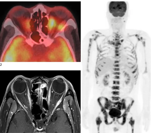
고용량 스테로이드 치료 이후에도 뚜렷한 증상 호전 없이 안 구통증이 동반되기 시작하고 안구운동제한으로 인한 복시가 악 화되어 스테로이드 치료 시작 25일 후 시행한 추적 안와자기공 명영상에서 우측 외직근 및 좌측 상직근을 제외한 외안근의 비 대가 악화되었을 뿐만 아니라 근육의 경계가 이전과 달리 뚜렷 한 다결절성 변화를 보이고 있었다(Fig. 1D - F). 같은 날 시 행한 18F-FDG PET/CT에서는 체간 및 사지골격에 미만성 골 전이 소견과 함께 양쪽 외안근에 미만성 FDG 섭취증가가 있었 다(Fig. 1G, H). 자기공명영상과 18F-FDG PET/CT 소견을 고려하여 전신 골격과 양쪽 외안근에 발생한 횡문근육종 전이로 진단하고 항암요법(vincristine, ifosfamide, dactinomycin) 을 추가적으로 시행하였다. 항암요법 후 2개월째 시행한 안와자 기공명영상에서 양쪽 외안근의 두께는 정상범위로 감소하였다
고 찰
성인에서 안와종양 중 전이성 종양은 2.5%에서 13%로 알려 져 있다(5, 6). 일반적인 안와 내 전이는 지방조직과 뼈에 발생 하지만, 외안근에 발생할 경우 단일 근육의 일측성 결절성 비대 (unilateral nodular enlargement)로 나타나는 것으로 알려 져 있다(1, 7). 외안근의 전이와 연관된 가장 흔한 원발성 종양 은 유방암, 악성흑색종, 위장관 악성종양, 그리고 폐암 순이다 (1). Lacey 등은 20명 환자에서의 외안근 전이 분석 결과 외안 근의 침범 빈도는 내직근(39%), 외직근(33%), 상직근(16%), 하직근(12%)의 순이었으며 대부분은 일측성 이었고 양측성 전 이는 17%에서 발생한 것으로 보고하였다(1).
g
i h
Fig. 1. (g-h)18F-FDG PET/CT performed on the same day with the 2nd follow-up orbit MRI. An axial 18F-FDG PET/CT im- age (g) shows intense hypermetabolism of the thickened bilateral medial and left lateral rectus muscles (SUVmax, 6.0 - 7.4 g/mL). The coronal maximum intensity projection image (h) shows multifocal hypermetabolic areas involving the axial and appendicular skeleton from disseminated bone metastasis. Contrast-enhanced axial T2-weighted image (i) after additional chemotherapy shows remarkable regression of the metastatic tumor involving the bilateral extraocular muscles.
외안근을 다발성으로 침범하여 미만성 비대의 형태로 발생한 경 우는 더욱 드물어 Medline 검색 결과 증례보고 두 예만 있을 뿐이다(3, 4). 이러한 양측 외안근의 미만성 비대는 영상학적 소견상 다양한 질환과 혼동 될 수 있으며 그 중에서 대표적인 질환이 갑상선 안병증이다. 갑상선 안병증은 CT나 MRI 상에 서 양쪽 외안근 특히 하직근과 내직근의 미만성 비후의 형태로 보인다(9). 근섬유만을 특이적으로 침범하고 힘줄과 근막은 침 범하지 않으므로 영상에서는 주변 조직과의 경계가 명확한 것이 특징이다(9). 또한 MRI에서는 주변 정상 근육보다 조영증강이 증가되고 T2강조영상에서 전반적인 고신호를 보일 수 있다(9).
대개의 경우 갑상선기능검사 상의 이상을 동반하지만 드물게 기 능검사 상의 이상보다 갑상선 안병증이 먼저 발생하는 경우도 20%라고 알려져 있다(10). 본 증례의 경우 처음 촬영한 MRI 상에서 미만성으로 비대된 외안근의 일부에서 전체적으로 부드 러운 방추형을 보이지 않고 건부와의 경계부위에서 결절 모양으 로 갑작스럽게 꺾이는 소견을 보이는 것과 조영증강이 현저히 증가되어 있지 않은 점 등은 흔히 보이는 갑상선 안병증의 영상 소견과의 차이점이라고 할 수 있다. 따라서 횡문근육종이나 기 타 악성종양의 병력이 있는 환자에서 외안근의 미만성 비대가 있을 때 앞서 언급한 것과 같은 소견들이 MRI에서 동반된 경우 갑상선 안병증 이외에 기저질환의 전이에 의한 소견일 가능성을 반드시 감별하여야 할 것으로 판단된다. 그 이외에 백혈병과 림 프종과 같은 침윤성 질환이나 안와 가성종양 등이 감별질환에 고려될 수 있으나 두 질환은 외안근을 침범했을 때 근육의 건부 를 흔히 침범하고 비대칭적인 침범 양상을 보이면서 주변 지방 조직으로의 침윤이 흔히 동반되는 점에서 본 질환과는 감별할 수 있겠다.
결론적으로 횡문근육종 환자에서 양쪽 외안근에 미만성으로 발생한 전이암은 매우 드물어 본 증례를 포함하여 세계적으로 모두 2예만 보고되어 있다. 미만성 외안근 비대의 형태로 발생 하여 갑상선 안병증과의 감별이 어려울 수 있으나 영상 소견에 서 비대된 외안근이 부드러운 방추형을 보이지 않고 건부와의 경계부위에서 결절 모양을 취하면서 갑작스럽게 꺾이는 모양을
보이거나 조영증강이 현저히 증가되어 있지 않는 경우 횡문근육 종과 같은 악성 종양의 전이를 감별질환에 포함해야 할 것으로 생각한다.
참 고 문 헌
1.Lacey B, Chang W, Rootman J. Nonthyroid causes of extraocu- lar muscle disease. Surv Ophthalmol 1999;44:187-213 2.Amato MM, Esmaeli B, Shore JW. Orbital rhabdomyosarcoma
metastatic to the contralateral orbit: a case report.
Ophthalmology 2002;109:753-756
3.Hatton MP, Green L, Boulos PR, Rubin PA. Rhabdomyosarco- ma metastases to all extraocular muscles. Ophthal Plast Reconstr Surg 2008;24:336-338
4.Gupta P, Singh U, Singh SK, Kapoor R, Gupta V, Das A.
Bilateral symmetrical metastasis to all extraocular muscles from distant rhabdomyosarcoma. Orbit 2010;29:146-148 5.Shields JA, Bakewell B, Augsburger JJ, Flanagan JC.
Classification and incidence of space-occupying lesions of the orbit. A survey of 645 biopsies. Arch Ophthalmol 1984;102:1606-1611
6.Lell M, Schulz-Wendtland R, Hafner A, Magener A, Bautz WA, Tomandl BF. Bilateral orbital tumour as the presentation of mammographically occult breast cancer. Neuroradiology 2004;46:682-685
7.Capone A, Slamovits TL. Discrete metastasis of solid tumors to extraocular muscles. Arch Ophthalmol 1990;108:237-243 8.Jones IS, Reese AB, Krout J. Orbital rhabdomyosarcoma: an
analysis of sixty-two cases. Trans Am Ophthalmol Soc 1965;63:223-255
9.Mafee MF. Orbit: embryology, anatomy, and pathology. In:
Som PM, ed. Head and neck imaging: 4th ed. Mosby, St. Louis.
2003:591-595
10.Burch HB, Wartofsky L. Graves’ ophthalmopathy: current con- cepts regarding pathogenesis and management. Endocr Rev 1993;14:747-793
Address reprint requests to : Jeong Hyun Lee, M.D., Ph.D., Head & Neck Imaging Department of Radiology, University of Ulsan College of Medicine, Asan Medical Center,
86 Asanbyeongwon-gil, Songpa-gu, Seoul 138-736, Korea.
Tel. 82-2-3010-4400 Fax. 82-2-476-0090 E-mail: [email protected]
Bilateral Extraocular Muscle Metastasis of Nasal Rhabdomyosarcoma Mimicking a Thyroid Associated Orbitopathy: A Case Report
Jin Ho Shin
1, Jeong Hyun Lee
1, Hyun Kyung Lim
1, Ha Young Lee
1, Jee Won Park
1, Hye Jin Baek
1, Young Jun Choi
1, Jin-Hee Ahn
2, Jung Hwan Baek
11Department of Radiology and Reasearch Institute of Radiology, University of Ulsan College of Medicine, Asan Medical Center
2Department of Oncology, University of Ulsan College of Medicine, Asan Medical Center
Metastases to the orbit usually affect the intraorbital fat and bone than the extraocular muscles.
Metastasis to the extracoular muscles commonly occurs unilaterally, and diffuse enlargement of the bilat- eral extraocular muscles due to metastasis is extremely rare. In this report, we will describe a case of dif- fuse metastasis to the bilateral extraocular muscles from nasal rhabdomyosarcoma masquerading as thy- roid associated orbitopathy. We will also discuss about the MR imaging findings helpful for differential di- agnosis from thyroid associated orbitopathy.
Index words :Rhabdomyosarcoma Alveolar
Extraocular muscle metastasis Magnetic resonance imaging (MRI) Thyroid associated orbitopathy
J. Korean Soc. Magn. Reson. Med. 15:176-180(2011)