This work has been supported by the Seoul National University Dental Hospital (SNUDH) research fund (#032014-0034). The authors reported no conflicts of interest related to this study.
Comparison of implant component fractures in external and internal type: A 12-year
retrospective study
Yuseung Yi1, Jai-Young Koak1, Seong-Kyun Kim1, Shin-Jae Lee2, Seong-Joo Heo1*
1Department of Prosthodontics & Dental Research Institute, School of Dentistry, Seoul National University, Seoul, Republic of Korea
2Department of Orthodontics, School of Dentistry, Seoul National University, Seoul, Republic of Korea
PURPOSE. The aim of this study was to compare the fracture of implant component behavior of external and internal type of implants to suggest directions for successful implant treatment. MATERIALS AND METHODS.
Data were collected from the clinical records of all patients who received WARANTEC implants at Seoul National University Dental Hospital from February 2002 to January 2014 for 12 years. Total number of implants was 1,289 and an average of 3.2 implants was installed per patient. Information about abutment connection type, implant locations, platform sizes was collected with presence of implant component fractures and their managements. SPSS statistics software (version 24.0, IBM) was used for the statistical analysis. RESULTS. Overall fracture was significantly more frequent in internal type. The most frequently fractured component was abutment in internal type implants, and screw fracture occurred most frequently in external type. Analyzing by fractured components, screw fracture was the most frequent in the maxillary anterior region and the most abutment fracture occurred in the maxillary posterior region and screw fractures occurred more frequently in NP (narrow platform) and abutment fractures occurred more frequently in RP (regular platform). CONCLUSION. In external type, screw fracture occurred most frequently, especially in the maxillary anterior region, and in internal type, abutment fracture occurred frequently in the posterior region. placement of an external type implant rather than an internal type is recommended for the posterior region where abutment fractures frequently occur. [J Adv Prosthodont 2018;10:155-62]
KEYWORDS: Implant fracture; Screw fracture; Abutment fracture; Fixture fracture
INTRODUCTION
Endosseous implants are reliable choice of treatment for the replacement of missing natural teeth. Although the
overall success rate of implant is relatively high, between 95 - 98%,1 they often encounter complications such as peri- implantitis and other technical problems. According to sev- eral previous studies, the most common cause of implant failure is peri-implantitis and technical complications are not uncommon.2-4 Technical problems of implant-supported restorations can be classified into two groups: those relating to the prosthesis, and those relating to the implant compo- nents.5-6 Technical problems relating to the prosthesis are such as veneering material or framework fractures and tech- nical problems relating to the implants components include screw loosening or screw fractures, abutment fracture and implant fixture fractures. If the problem is caused by the implant prosthesis itself, the problem can be overcome by refabricating the prosthesis. However, in the case of prob- lems associated with implant components, various solutions must be sought, simply to replace the implant components or, in some cases, remove the implants, which makes it diffi-
Corresponding author:
Seong-Joo Heo
Department of Prosthodontics, School of Dentistry, Seoul National University, 101, Daehak-ro, Jongno-gu, Seoul 03080, Republic of Korea Tel. +82220722661: e-mail, [email protected]
Received August 11, 2017 / Last Revision October 29, 2017 / Accepted October 30, 2017
© 2018 The Korean Academy of Prosthodontics
This is an Open Access article distributed under the terms of the Creative Commons Attribution Non-Commercial License (http://creativecommons.
org/licenses/by-nc/3.0) which permits unrestricted non-commercial use, distribution, and reproduction in any medium, provided the original work is properly cited.
cult to predict the solution. Adell et al.7 reported a 3.5%
implant fixture fracture in a 15-year study of Brånemark implant in 1981.Naert et al.8 reported a 0.53% implant frac- ture, 8.9% abutment screw fracture, and 1.2% occlusal screw fracture in a case study of implant supporting com- plete fixed prosthesis with Brånemark implant in 1992.As regards the fracture of implant, Rangert et al.9 said it was associated with bruxism or strong occlusal force, and it occurred more frequently in an single or double implant prosthesis of the posterior region. In a retrospective study of implant complications in 1997, Tolman and Laney report- ed10 that screw fractures occurred in 87 of 1,250 implants (7.0%). Although there have been several studies on fracture of the implant components, most studies report only failure of the osseointegration as failure of the implant and it is overlooked that implant failure can be caused by implant component fracture. Clinically, in the case of implant abut- ment fracture or screw fracture, it is recommended to remake the prosthesis after removing the fractured remnant.
However, it is difficult to remove the fragments and eventu- ally the implants must be removed, leading to implant failure.
The purpose of this study was to predict the prognosis of implants and to suggest directions for successful implant treatment by analyzing the factors affecting the fracture of implant components including abutment connection type, implant location and implant platform size.
MATERIALS AND METHODS
Data were collected from the clinical records of all patients who received one or more WARANTEC implants at Seoul National University Dental Hospital from February 2002 to January 2014 for 12 years and the following cases were excluded: i) implants failed in osseointegration, ii) implant placement after jaw resection and reconstruction, iii) the opposite arch was complete denture, iv) implant assisted over denture, v) insufficient clinical chart recording, vi) patients who have not visited since 2012. Data collection included 406 patients (205 males, 201 females), ranging in age from 21 to 94 years (mean 64.6 years, SD 11.5). Total number of implants was 1,289 and an average of 3.2 implants was installed per patient. Information about abut- ment connection type (internal or external), implant loca- tions, platform sizes was collected with presence of implant component fractures and their managements (Table 1). The information about implant component fractures was divid- ed into three types: screw fractures, abutment fractures, and fixture fractures. The management of fractures was classi- fied as screw replacement, prosthesis refabrication, and fix- ture removal.
SPSS statistics software (version 24.0, IBM, New York, NY, USA) was used for the statistical analysis. The Pearson chi-square test and Fisher’s exact test (P = .05) were used to evaluate the association between implant characteristics and implant components fracture.
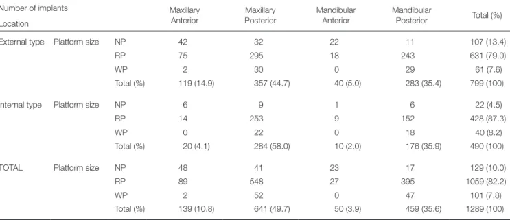
Table 1. Distribution of implants according to the location and platform size
Number of implants Maxillary
Anterior
Maxillary Posterior
Mandibular Anterior
Mandibular
Posterior Total (%) Location
External type Platform size NP 42 32 22 11 107 (13.4)
RP 75 295 18 243 631 (79.0)
WP 2 30 0 29 61 (7.6)
Total (%) 119 (14.9) 357 (44.7) 40 (5.0) 283 (35.4) 799 (100)
Internal type Platform size NP 6 9 1 6 22 (4.5)
RP 14 253 9 152 428 (87.3)
WP 0 22 0 18 40 (8.2)
Total (%) 20 (4.1) 284 (58.0) 10 (2.0) 176 (35.9) 490 (100)
TOTAL Platform size NP 48 41 23 17 129 (10.0)
RP 89 548 27 395 1059 (82.2)
WP 2 52 0 47 101 (7.8)
Total (%) 139 (10.8) 641 (49.7) 50 (3.9) 459 (35.6) 1289 (100)
NP = Narrow Platform, RP = Regular platform, WP = Wide platform
RESULTS
A total of 1,289 implants were placed in 406 patients during investigation period. 799 implants had external type abut- ment connection (62.0%) and 490 implants were internal type abutment connection (38.0%). 139 implants were placed in the maxillary anterior region (10.8%), 641 implants in the maxillary posterior region (49.7%), 50 implants in the man- dibular anterior region (3.9%), and 449 implants in the man- dibular posterior region (35.6%). According to the platform size, 129 implants (10.0%) with narrow platform (NP), 1059 implants (82.2%) with regular platform (RP), and 101 implants (7.8%) with wide platform (WP) were placed
(Table 1, Fig. 1).
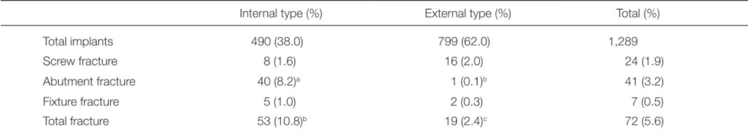
Of the total 1,289 implants, component fractures occurred in 72 implants (5.6%). In internal type implants, 53 components fractures occurred (10.8%): 8 screw fractures (1.6%), 40 abutment fractures (8.2%), and 5 fixture fracture (1.0%), and in external type implants, 19 components frac- tures occurred (2.4%): 16 screw fractures (2.0%), 1 abutment fracture (0.1%), and 2 fixture fractures (0.3%) (Table 2).
Overall fracture was significantly more frequent in inter- nal type (P < .001). The most frequently fractured compo- nent was abutment in internal type implants (8.2%), and screw fracture occurred most frequently (2.0%) in external type (Fig. 2).
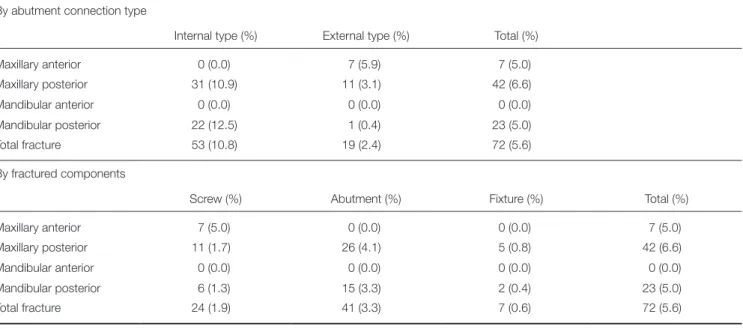
The implants were most placed in the maxillary posteri- or region (49.7%), followed by the mandibular posterior region (35.6%) (Table 1). Table 3 shows the most frequent location which implant component fractures occurred was the maxillary posterior region in internal type (10.8%), and maxillary anterior region in external type (5.9%).
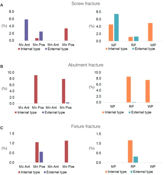
Analyzing by fractured components, screw fracture was the most frequent in the maxillary anterior region (5.0%) and the most abutment fracture occurred in the maxillary posterior region (4.1%) significantly (P = .013 < .050) (Table 3, Fig. 3A).
Table 2. Implant component fractures
Internal type (%) External type (%) Total (%)
Total implants 490 (38.0) 799 (62.0) 1,289
Screw fracture 8 (1.6) 16 (2.0) 24 (1.9)
Abutment fracture 40 (8.2)a 1 (0.1)b 41 (3.2)
Fixture fracture 5 (1.0) 2 (0.3) 7 (0.5)
Total fracture 53 (10.8)b 19 (2.4)c 72 (5.6)
Different letters mean significant difference (P < .05)
Fig. 2. Implant component fracture. Overall fracture was significantly more frequent in internal type. The most frequently fractured component was abutment in internal type implants, and screw fracture occurred most
frequently in external type.
(%) 10.0 8.0 6.0 4.0 2.0
0.0 Internal type External type Total All components Screw Abutment Fixture Platform size (%)
(%) 100.0 80.0 60.0 40.0 20.0 0.0
Fig. 1. Implant distribution according to the location and the platform sizes.
(%) 80.0 60.0 40.0 20.0 0.0
Implant location (%)
Mx Ant Mx Pos Mn Ant Mn Pos Internal type External type Total implant
Internal type External type Total implant NP RP WP
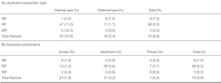
There was significant relationship between platform size and fractures. In external type, the number of fractures of NP was larger than that in other platform sizes, and more fractures occurred in the RP than other platform sizes in internal type.
Analyzing by fractured components, screw fractures occurred more frequently in NP (narrow platform) and abutment fractures occurred more frequently in RP (regular platform) (Table 4, Fig. 3B).
Table 3. Fractures depending on implant location By abutment connection type
Internal type (%) External type (%) Total (%)
Maxillary anterior 0 (0.0) 7 (5.9) 7 (5.0)
Maxillary posterior 31 (10.9) 11 (3.1) 42 (6.6)
Mandibular anterior 0 (0.0) 0 (0.0) 0 (0.0)
Mandibular posterior 22 (12.5) 1 (0.4) 23 (5.0)
Total fracture 53 (10.8) 19 (2.4) 72 (5.6)
By fractured components
Screw (%) Abutment (%) Fixture (%) Total (%)
Maxillary anterior 7 (5.0) 0 (0.0) 0 (0.0) 7 (5.0)
Maxillary posterior 11 (1.7) 26 (4.1) 5 (0.8) 42 (6.6)
Mandibular anterior 0 (0.0) 0 (0.0) 0 (0.0) 0 (0.0)
Mandibular posterior 6 (1.3) 15 (3.3) 2 (0.4) 23 (5.0)
Total fracture 24 (1.9) 41 (3.3) 7 (0.6) 72 (5.6)
Depending on implant location
Fig. 3. Implant component fracture. (A) Screw fracture was the most frequent in the maxillary anterior region and the most abutment fracture occurred in the maxillary posterior region significantly. (B) In external type, the number of fractures of NP was larger than that in other platform sizes, and more fractures occurred in the RP than other platform sizes in internal type. Screw fractures occurred more frequently in NP and abutment fractures occurred more frequently in RP. Mx = Maxillary, Ant = Anterior, Mn = Mandibular, Pos = Posterior, NP = Narrow platform, RP = Regular platform, WP = Wide platform.
A
B
Depending on implant platform size
(%)
(%)
(%)
(%)
DISCUSSION
The number of implants installed for 12 years from 2002 to 2014 was greater in the external type (62.0%) than in the internal type (38.0%). Depending on implant locations, implants were mostly placed at the maxillary posterior region (49.7%), followed by 35.6% at the mandibular poste- rior region, 10.8% at the maxillary anterior region and 3.9%
at the mandibular anterior region. Depending on implant Platform sizes, RP (Regular platform) was the most placed at all locations with 82.2%, NP (Narrow Platform) was placed in the anterior region with 10.0%, WP (Wide Platform) was placed in the posterior region mostly with 7.8% (Table 1, Fig. 1).
Of the total 1,289 implants, component fractures occurred in 72 implants (5.6%). In internal type implants, 53 components fractures occurred (10.8 %): 8 screw fractures (1.6%), 40 abutment fractures (8.2%), and 5 fixture fracture (1.0%), and in external type implants, 19 components frac- tures occurred (2.4%): 16 screw fractures (2.0%), 1 abut- ment fracture (0.1%), and 2 fixture fractures (0.3%). Table 5 shows the relationship between various factors and implant component fractures.
Comparing the overall implant component fracture of the internal and external types, the fracture rate in internal type was significantly higher than in external type (P <
.001). By implant components, there were no significant dif- ference between internal and external types in screw or fix- ture fractures, only in abutment fracture, internal type (8.2%) was significantly higher than external type (0.1%) (P
< .001).
Depending on the implant location, in internal types, there was no significant relationship between the implant location and each component fracture rates, however, in external types, there was a significant difference in total Table 4. Fractures depending on implant platform size
By abutment connection type
Internal type (%) External type (%) Total (%)
NP 1 (4.5) 8 (7.5) 9 (7.0)
RP 47 (11.0) 11 (1.7) 58 (5.5)
WP 5 (12.5) 0 (0.0) 5 (5.0)
Total fracture 53 (10.8) 19 (2.4) 72 (5.6)
By fractured components
Screw (%) Abutment (%) Fixture (%) Total (%)
NP 9 (7.0) 0 (0.0) 0 (0.0) 9 (7.0)
RP 13 (1.2) 38 (3.6) 7 (0.7) 58 (5.5)
WP 2 (2.0) 3 (3.0) 0 (0.0) 5 (5.0)
Total fracture 24 (1.9) 41 (3.2) 7 (5.2) 72 (5.6)
NP = Narrow Platform, RP = Regular Platform, WP = Wide Platform
Table 5. Implant component fractures
Screw Abutment Fixture Total
External type 2.00% 0.10% 0.30% 2.40%
Internal type 1.60% 8.20% 1.00% 10.80%
P value .402 < .001* > .999 < .001*
Component fractures depending on implant location
External type
Mx Ant 5.90% 0.00% 0.00% 5.90%
Mx Pos 2.50% 0.00% 0.60% 3.10%
Mn Ant 0.00% 0.00% 0.00% 0.00%
Mn Pos 0.00% 0.40% 0.00% 0.40%
PP value .003* .553 .683 .007*
Internal type
Mx Ant 0.00% 0.00% 0.00% 0.00%
Mx Pos 0.70% 9.20% 1.10% 10.90%
Mn Ant 0.00% 0.00% 0.00% 0.00%
Mn Pos 3.40% 8.00% 1.10% 12.50%
P value .186 .601 > .999 .327
Component fractures depending on implant platform size
External type
NP 7.50% 0.00% 0.00% 7.50%
RP 1.30% 0.20% 0.30% 1.70%
WP 0.00% 0.00% 0.00% 0.00%
P value .001* > .999 > .999 .003*
Internal type
NP 4.50% 0.00% 0.00% 4.50%
RP 1.20% 8.60% 1.20% 11.00%
WP 5.00% 7.50% 0.00% 12.50%
P value .092 .407 > .999 .662
*significant (P < .05).
Mx = Maxillary, Mn = Mandibular, Ant = Anterior, Pos = Posterior, NP = Narrow Platform, RP = Regular Platform, WP = Wide Platform
fracture rate (P = .007 < .05) and screw fracture (P = .003 <
.05) in maxillary anterior region was significantly higher (5.8%).
Analyzing according to implant platform size, NP (Narrow platform) showed significantly higher total fracture rate (P = .003 < .05) and higher screw fracture rates (P = .001 < .05) in external type and there was no significant dif- ference in internal type.
In most cases, screw loosening precedes screw fracture.
Screw loosening is a relatively frequent complication and it occurs more frequently in external type than internal type, in screw-retained type than in cemented type, in lower arch
than in upper arch, and in single tooth restoration than mul- tiple restorations.11,12
In this retrospective study, screw fracture occurred in internal type 1.6% and external type 2.0%, but there was no significant difference according to connection type. There was a significant difference according to implant position and platform size. The most frequent location was in the maxillary anterior region (5.9%), and most of them occurred in NP (narrow platform) according to the platform size (7.5%). In other words, screw fracture occurs most fre- quently in maxillary anterior region with NP (narrow plat- form) in external type (Fig. 4A). According to previous
Fig. 4. Implant component fractures (By implant component). (A) Screw fracture occurred in internal type 1.6% and external type 2.0%, but there was no significant difference according to connection type. There was a significant difference according to implant position and platform size. The most frequent location was in the maxillary anterior region (5.9%), and most of them occurred in NP. (B) Abutment fracture occurred more frequently in internal type (8.2%) than in external type (0.1%). There was no significant difference in fracture rate according to the implant location, but all fractures occurred in posterior region. (C) Fixture fracture occurred 1.0% in internal type, and 0.3% in external type, there was no significant difference according to implant connection type or implant location, however, all fixture fractures occurred at the posterior region in RP. Mx = Maxillary, Mn = Mandibular, Ant = Anterior, Pos = Posterior, NP = Narrow Platform, RP = Regular Platform, WP = Wide Platform.
B
C A
Fixture fracture Screw fracture
Abutment fracture
(%)
(%)
(%)
(%)
(%)
(%)
studies, screw fracture is caused by various factors such as excessive bite force, improper placement of implant, bone loss, inappropriate fit or design of prosthesis, accumulation of fatigue, defect in fabrication and type of implant.13-16 In internal type, the retention of the implant superstructure is obtained by the friction between the implant abutment and the fixture inner surface and screw. However, in external type, the contact surface between the implant abutment and the fixture is smaller than in internal type structurally, and the retentive force of the superstructure is achieved only by screws. Therefore, when excessive lateral force or other fac- tors are applied to the implant, the force is transmitted to the implant screw, and if the force exceeds the retention threshold of the screw, screw loosening will occur, or if it exceeds the fracture threshold, screw fracture will occur.
During eccentric movement, the implants in maxillary ante- rior region are affected by the lateral force and the bending force transmitted to the implant screw also increases, even- tually causes screw fracture. Since the fixation screw of NP (narrow platform) abutment has also narrow diameter, thus, the fracture resistance threshold is small and therefore sus- ceptible to fracture when the factors are applied.
Although many researches about implant screw or fix- ture fracture have been conducted, there is little analysis of abutment fracture. In this study, abutment fracture occurred more frequently in internal type (8.2%) than in external type (0.1%). There was no significant difference in fracture rate according to the implant location, but all fractures occurred in posterior region. (Fig. 4B) What is remarkable is all abut- ment fractures occurred in single implant restoration in pos- terior region using gold cast UCLA type with internal hex abutment connection. This type of implant prosthesis is a superstructure of a unit, so that when the lateral force applies, the force concentrates on the implant abutment neck area, causing a bending moment, which causes fracture of the abutment neck.
Implant fixture fracture is classified as late failure and have been reported to be caused by various factors. When the bone resorption occurs due to peri-implantitis, the bending stress from the masticatory force increases and the stress concentrates at the end of the abutment screw and acts as a starting point of the fracture.17,18
As a result of this study, fixture fracture occurred 1.0%
in internal type, and 0.3% in external type, there was no sig- nificant difference according to implant connection type or implant location, however, all fixture fractures occurred at the posterior region in RP (regular platform) (Fig. 4C).
Rangert et al. 17 reported that 90% of implant fractures are located in the molar and premolar regions of the mouth, where chewing forces and lateral movements associated with cusp inclination generate undesirable forces. When a implant is subjected to a force, external and internal types exhibit different force distribution structurally. In external type, when an external force is applied, the stress is trans- ferred to the screw, which is the most susceptible part, and the screw fracture occurs before the fixture is stressed beyond the fracture resistance, thereby preventing the frac-
ture of the fixture. In internal type, however, the mechanical interface between the abutment and the fixture is present, so that when the external force is applied, the stress trans- mitted to the abutment is transmitted to the fixture and if the stress exceeds the fracture resistance, fixture fracture is caused.
Clinically, when implant component fractures occur, the most important thing is whether the problem can be solved easily. When fixture fractures occur, it is considered as an implant failure since it can no longer function as an implant restoration and must be removed. However, in the case of screw or abutment fractures, the problem can be solved more easily if the fractured fragment can be removed. In other words, obtaining retrievability is the most important point to solve the problem. How to cope with fractures dur- ing this retrospective study was summarized in Table 6.
Only 12.5% of the fractured screws were not removed in internal type, and 6.2% in external type, in the case of abutment fracture, the abutment fragment removal was impossible in 27.5% and fixture were removed.
CONCLUSION
In external type, screw fracture occurred most frequently, especially in the maxillary anterior region, and in internal type, abutment fracture occurred frequently in the posterior region. The screw fracture seems to be easier to solve than the abutment fracture. Therefore, placement of an external type implant rather than an internal type is recommended for the posterior region where abutment fractures frequent- ly occur. If an internal type implant is inevitably installed in Table 6. How to cope with fractures
Fractured component Screw Abutment Fixture Internal type
Fixture removal 1 (12.5%) 10 (25%) 5 (100%) Prosthesis refabrication
(retrieve) 3 (37.5%) 29 (72.5%) 0 (0.0%)
Screw replacement
(retrieve) 4 (50.0%) 0 (0.0%) 0 (0.0%)
Maintain fracture status 0 (0.0%) 1* (2.5%) 0 (0.0%) External type
Fixture removal 0 (0.0%) 0 (0.0%) 2 (100%) Prosthesis refabrication
(retrieve) 0 (0.0%) 1 (100%) 0 (0.0%)
Screw replacement
(retrieve) 15 (93.8%) 0 (0.0%) 0 (0.0%)
Maintain fractured status 1** (6.2%) 0 (0.0%) 0 (0.0%)
* Fractured abutment could not be removed, being used.
** Fractured screw could not be removed, using new screw cut.
the posterior region, the SCRP type prosthesis or multi-unit abutment should be used instead of the one-unit UCLA type implant prosthesis for external force distribution.
ORCID
Seong Joo Heo https://orcid.org/0000-0003-0699-4141 REFERENCES
1. Laney WR, Jemt T, Harris D, Henry PJ, Krogh PH, Polizzi G, Zarb GA, Herrmann I. Osseointegrated implants for single- tooth replacement: progress report from a multicenter pro- spective study after 3 years. Int J Oral Maxillofac Implants 1994;9:49-54.
2. Wu PB, Yung WC. Factors contributing to implant failure.
Hong Kong Dent J 2005;2:8-12.
3. Carlson B, Carlsson GE. Prosthodontic complications in os- seointegrated dental implant treatment. Int J Oral Maxillofac Implants 1994;9:90-4.
4. Brägger U, Aeschlimann S, Bürgin W, Hämmerle CH, Lang NP. Biological and technical complications and failures with fixed partial dentures (FPD) on implants and teeth after four to five years of function. Clin Oral Implants Res 2001;12:26- 34.
5. Luterbacher S, Fourmousis I, Lang NP, Brägger U. Fractured prosthetic abutments in osseointegrated implants: a technical complication to cope with. Clin Oral Implants Res 2000;11:
163-70.
6. Binon PP. Implants and components: entering the new mil- lennium. Int J Oral Maxillofac Implants 2000;15:76-94.
7. Adell R, Lekholm U, Rockler B, Brånemark PI. A 15-year study of osseointegrated implants in the treatment of the edentulous jaw. Int J Oral Surg 1981;10:387-416.
8. Naert I, Quirynen M, van Steenberghe D, Darius P. A study of 589 consecutive implants supporting complete fixed pros- theses. Part II: Prosthetic aspects. J Prosthet Dent 1992;68:
949-56.
9. Rangert B, Krogh PH, Langer B, Van Roekel N. Bending overload and implant fracture: a retrospective clinical analysis.
Int J Oral Maxillofac Implants 1995;10:326-34.
10. Tolman DE, Laney WR. Tissue-integrated prosthesis compli- cations. Int J Oral Maxillofac Implants 1992;7:477-84.
11. Sailer I, Mühlemann S, Zwahlen M, Hämmerle CH, Schneider D. Cemented and screw-retained implant reconstructions: a systematic review of the survival and complication rates. Clin Oral Implants Res 2012;23:163-201.
12. Theoharidou A, Petridis HP, Tzannas K, Garefis P. Abutment screw loosening in single-implant restorations: a systematic review. Int J Oral Maxillofac Implants 2008;23:681-90.
13. Al Jabbari YS, Fournelle R, Ziebert G, Toth J, Iacopino AM.
Mechanical behavior and failure analysis of prosthetic retain- ing screws after long-term use in vivo. Part 1: Characterization of adhesive wear and structure of retaining screws. J Prosthodont 2008;17:168-80.
14. Al Jabbari Y, Fournelle R, Ziebert G, Toth J, Iacopino A.
Mechanical behavior and failure analysis of prosthetic retain-
ing screws after long-term use in vivo. Part 2: Metallurgical and microhardness analysis. J Prosthodont 2008;17:181-91.
15. Al Jabbari YS, Fournelle R, Ziebert G, Toth J, Iacopino AM.
Mechanical behavior and failure analysis of prosthetic retain- ing screws after long-term use in vivo. Part 3: Preload and tensile fracture load testing. J Prosthodont 2008;17:192-200.
16. Al Jabbari YS, Fournelle R, Ziebert G, Toth J, Iacopino AM.
Mechanical behavior and failure analysis of prosthetic retain- ing screws after long-term use in vivo. Part 4: Failure analysis of 10 fractured retaining screws retrieved from three patients.
J Prosthodont 2008;17:201-10.
17. Rangert B, Krogh PH, Langer B, Van Roekel N. Bending overload and implant fracture: a retrospective clinical analysis.
Int J Oral Maxillofac Implants 1995;10:326-34.
18. Morgan MJ, James DF, Pilliar RM. Fractures of the fixture component of an osseointegrated implant. Int J Oral Maxillofac Implants 1993;8:409-14.