In 2002, the World Health Organization defined osteo- chondroma as a cartilage-capped bony exostosis on the external surface of a bone containing a marrow cavity continuous with that of the underlying bone.1 Osteochon- droma accounts for approximately 35% to 50% of all be- nign bone tumors and 8% to 15% of all primary bone tu- mors.2 It can develop in any bone that is generated by en- dochondral ossification and is known to occur in almost every portion of the craniofacial skeleton, such as the skull base, maxillary sinus, zygomatic arch, nasal septum, and mandible.3 The mandibular condyle and the coronoid process are by far the most common sites of craniofacial osteochondroma.2 The clinical symptoms of mandibular condylar osteochondromas are facial asymmetry, cross- bite on the unaffected side, open bite on the affected side,
a deviated opening path, condylar motion limitation, disc displacement, and rarely, pain.4,5 While long-bone osteo- chondromas are usually asymptomatic and do not require surgery, resection is often appropriate for mandibular con- dylar osteochondromas because they cause functional and cosmetic problems6 and very rarely recur after treatment.
This report documents the case of a 21-year-old woman with mandibular condylar osteochondroma that recurred with a pattern similar to the original occurrence 3 years after the first excision and reshaping.
Case Report
A 21-year-old woman was referred to the Department of Oral and Maxillofacial Surgery, Kyungpook National University Dental Hospital, with facial asymmetry that was noticed 4 years ago. She had no history of facial trau- ma or ear infection. Crepitus and clicking were noted on the left and right temporomandibular joints, respectively, but pain was absent. The active range of motion was nor- mal at 40mm; however, the midline deviated 5.5mm to
Recurrent osteochondroma of the mandibular condyle: A case report
Young-Eun Kwon1, Karp-Shik Choi1, Chang-Hyeon An1, So-Young Choi2, Jae-Seo Lee3, Seo-Young An1,*
1Department of Oral and Maxillofacial Radiology, School of Dentistry, Kyungpook National University, Daegu, Korea
2Department of Oral and Maxillofacial Surgery, School of Dentistry, Kyungpook National University, Daegu, Korea
3Department of Oral and Maxillofacial Radiology, School of Dentistry, Chonnam National University, Gwangju, Korea
ABSTRACT
A 21-year-old woman presented with facial asymmetry. Crepitus and clicking of the temporomandibular joint were noted. The midline deviated 5.5mm to the left, and secondary malocclusion was observed. Panoramic and cone- beam computed tomographic images showed an irregular and exophytic bony mass on the anteromedial surface of the right mandibular condyle. A 3-phase bone scan revealed increased tracer uptake on the affected side. The lesion was treated with excision and reshaping under the diagnosis of osteochondroma confirmed by a histopathological examination. The lesion recurred after 3 years, and the patient underwent condylectomy. Mandibular condylar osteochondroma is often resected because it causes functional and aesthetic problems, but it rarely recurs. To the best of our knowledge, only 2 cases of recurrent osteochondromas of the mandibular condyle have been reported previously. Surgical treatment of the osteochondroma should be performed considering the possibility of recurrence, and long-term follow-up is recommended.(Imaging Sci Dent 2017; 47: 57-62)
KEY WORDS: Osteochondroma; Mandibular Condyle; Recurrence; Facial Asymmetry
Copyright ⓒ 2017 by Korean Academy of Oral and Maxillofacial Radiology
This is an Open Access article distributed under the terms of the Creative Commons Attribution Non-Commercial License(http://creativecommons.org/licenses/by-nc/3.0) which permits unrestricted non-commercial use, distribution, and reproduction in any medium, provided the original work is properly cited.
Imaging Science in Dentistry·pISSN 2233-7822 eISSN 2233-7830
*This study was supported by a National Research Foundation of Korea(NRF) grant funded by the Korean Government(NRF-2015R1C1A2A01055438).
Received September 21, 2016; Revised October 13, 2016; Accepted October 17, 2016
*Correspondence to : Prof. Seo-Young An
Department of Oral and Maxillofacial Radiology, School of Dentistry, Kyungpook National University, 2177 Dalgubeol-daero, Jung-gu, Daegu 41940, South Korea Tel) 82-53-600-7423, Fax) 82-53-425-6025, E-mail) [email protected]
the left and secondary malocclusion was observed during a physical examination(Fig. 1). A panoramic image(Or- thopantomograph OP 100D, Instrumentarium Imaging, Tuusula, Finland) revealed an irregular bony outgrowth on the anterior portion of the right condylar head(Fig. 2).
Cone-beam computed tomography(CBCT) images were acquired with Pax-Flex 3D(Vatech, Seoul, Korea), using a 120mm×85mm field of view at 90 kVp and 10 mA. A well-defined exophytic bony mass was observed on the anteromedial surface of the condylar neck, which present- ed as a bifid condylar head and caused a depression of the skull base(Fig. 3). A 3-phase bone scan was performed af- ter intravenously injecting 20-mCi 99mTc-hydroxydiphos- phonate(HDP); this revealed increased tracer uptake on the affected side(Fig. 4). Under the provisional diagnosis
of osteochondroma, the mass was resected by simple sur- gical excision and condylar reshaping was performed un- der general anesthesia using a preauricular approach. The removed mass had the following dimensions: 20mm×20 mm×18mm(Fig. 5). A histopathological examination showed the presence of a fibrous perichondrium, chond- roblasts, and chondroid matrix with chondrocytes in the lacuna. The endochondral ossification had matured into a cancellous bone with marrow, and the cartilaginous tissue was seen blending with the cancellous bone. The histo- pathological findings were consistent with osteochon- droma(Fig. 6). Further follow-up was scheduled after 6 months, but the patient did not present for the follow-up.
After 3 years, the patient visited again, complaining of a sudden deviation of the mandible to the left during ortho-
A
B
Fig. 1. A. Preoperative photograph shows facial asymmetry with chin deviation to the left side. B. Intraoral photograph reveals midline de- viation to the left(arrow) and secondary malocclusion in the closed position.
Fig. 2. Panoramic radiograph re- veals an irregular and large bony mass extending from the anterior portion of the right condylar head.
dontic treatment. A physical examination revealed a slight facial asymmetry and open bite in the anterior and left posterior areas. A similar pattern of bony outgrowth of the
right condyle was observed on the panoramic radiograph and contrast-enhanced computed tomography(CT)(Opti- ma CT660, GE Healthcare, Milwaukee, WI, USA) images (Fig. 7). Single-photon emission CT(SPECT)(Discovery NM/CT 670, GE Healthcare, Milwaukee, WI, USA) with
99mTc-HDP was used for examining the recurrence of the lesion. The 3-phase bone scan and axial-fused SPECT/CT images revealed intense uptake in the right condylar area (Fig. 8). More radical treatment was planned for the re- current lesion, and condylectomy was performed without reconstruction. A histological analysis revealed that the lesion was consistent with osteochondroma. Physical and radiographic examinations performed during the postop- erative follow-up at 6 months were uneventful.
Discussion
We report a case of recurrent osteochondroma of the mandibular condyle that was initially managed by con-
A B C
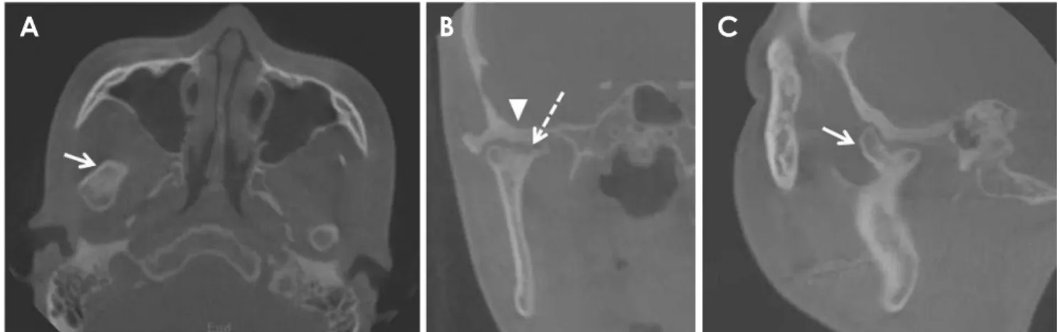
Fig. 3. A-C. Cone-beam computed tomography(CBCT) images demonstrate the well-defined margin of the radiopaque mass on the an- teromedial surface of the condylar neck(arrow) with a focal erosive change(dotted arrow). Depression and cortical thickenings of the skull base(arrowhead) can also be seen.
Fig. 4. A. 99mTc-hydroxydiphospho- nate bone scan, with a lateral spot view, showing an abnormal tracer uptake in the right condylar head. B.
No significant tracer avidity is seen on the left side.
A B
Fig. 5. A. A pre-auricular approach was used for exposing the condyle. B. The surgical specimen was approximately 20mm in length.
A B
servative surgical treatment considering the patient’s age.
Among the 235 cases of osteochondroma reviewed by Vezeau et al.6 and Peroz.7 in the English literature from 1927 to 2015, only 2 recurrent cases were reported. There- fore, to the best of our knowledge, only 3 recurrent cases of osteochondroma, including the present case, have been reported to date(Table 1). In one of the previously report- ed cases, the patient was surgically treated twice, and in the other, the patient was under observation for 15 years after the first operation. All lesions were located on the medial side of the condyle, and were on the anterior side in 2 cases. While mandibular condylar osteochondromas have a broad age distribution, ranging from 13 years to 76 years(mean, 41 years),7 recurrent cases are more likely to occur at a young age(recurrence occurred in the third decade of life in 2 of the 3 cases). Two of these 3 patients with recurrence were women; they presented with facial asymmetry as the main symptom and received simple resection initially. Two of the recurrent osteochondro- mas were detected by a routine radiographic examination without symptoms 1 year later, while the third was dis-
Fig. 6. Osteochondroma showing a fibrous perichondrium and chondroid matrix with chondrocytes in the lacuna. The cartilagi- nous tissue is seen blending with the cancellous bone(hematoxylin and eosin, original magnification: ×200).
Fig. 7. A. Panoramic radiograph of recurred osteochondroma shows the exophytic mass on the right condyle. B-C. An axial and coronal computed tomograph shows a sim- ilar pattern of bony outgrowth on the affected side.
A
B C
covered after 3 years and was accompanied by asymme- try. While the recurrence rate of osteochondroma in the long bones was 2%, the recurrence rate of mandibular condylar osteochondroma, including this case, was 1.3%
(3 out of 236 cases). However, perhaps due to the lack of long-term follow-up, we observed that the recurrent osteochondroma of the mandibular condyle had a border smaller than that of the original lesion. Further studies are needed on the recurrence of osteochondroma.
The cause of osteochondroma is presumed to be the protrusion of cartilaginous precursor cells through a de- fect in the epiphyseal plate, the migration of precursor cells from the epiphyseal to metaphyseal area, the hyper- plasia of cartilaginous cells due to tensional forces, and the differentiation of pluripotent cells in the periosteum into chondroblasts or osteoblasts.6 Hyperplasia of carti- laginous cells by tensional forces is the strongest hypoth- esis, because condylar osteochondromas occur commonly on the medial side(57%), followed by the anterior side (20%), and almost none occur in the lateral or superior aspects(<1%).8 This is supported by the theory of hy- perplastic changes in embryonic cartilaginous potential cells at the position of tendon insertions. Sequential forc-
es at the insertion site of the lateral pterygoid muscle may cause a local accumulation of these cells.2,5
Mandibular condylar osteochondroma appears as a mixed-density mass on panoramic radiographs because of the cartilage component and the calcified bone coexisting in the lesion, and the density increases with an increase in the calcified tissue.9 CT can reveal the calcified cartilag- inous cap and anatomical location of the lesion and may help in preoperative treatment planning.10 Magnetic res- onance imaging can help confirm the cartilaginous cap, which is characteristic of osteochondroma.9 Moreover, a bone scan can help identify abnormal metabolic bone activity through increased uptake of 99mTc-HDP in the affected condylar region, as seen in the present study. In most studies, the radiological appearance of condylar os- teochondroma is described as nodular or mushroom-like.9 However, adjoining structures such as the joint tuberos- ity, joint space, joint fossa, and joint capsule may affect the shape of the lesion by limiting the lesion growth.9 In the present case, the presence of an exophytic radiopaque mass protruding from the affected condylar neck to the anteromedial side suggested that its appearance was af- fected by the surrounding structure.
Fig. 8. A. Bone scan reveals in- creased tracer uptake in the recur- rence area. B. Fused single-photon emission computed tomography (SPECT)/CT shows intense tracer uptake in the affected condylar area.
A B
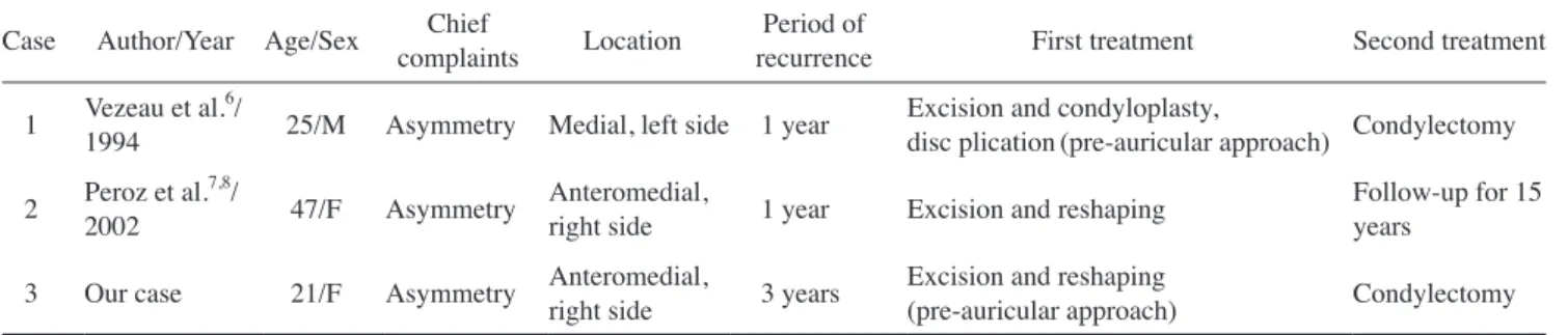
Table 1. Summary of cases of recurrent osteochondroma of the mandibular condyle Case Author/Year Age/Sex Chief
complaints Location Period of
recurrence First treatment Second treatment 1 Vezeau et al.6/
1994 25/M Asymmetry Medial, left side 1 year Excision and condyloplasty,
disc plication(pre-auricular approach) Condylectomy 2 Peroz et al.7,8/
2002 47/F Asymmetry Anteromedial, right side 1 year Excision and reshaping Follow-up for 15 years
3 Our case 21/F Asymmetry Anteromedial, right side 3 years Excision and reshaping
(pre-auricular approach) Condylectomy
Benign tumors that mainly develop in the condyle in- clude osteochondroma, osteoma, chondroma, condylar hyperplasia, condylar giant cell tumor, fibro-osseous le- sions, and vascular malformations.6 In the present case, osteochondroma, osteoma, and chondroma were included in the differential diagnosis. Osteoma could be exclud- ed because it usually appears as a pedunculated osseous mass on the mandibular condyle or neck, unlike the broad base seen in our case.4,9,11 Chondroma, a benign tumor that forms mature cartilage, is characterized radiological- ly by the presence of a mainly radiolucent and irregular mass, which can be distinguished from osteochondro-
ma.12,13 Osteochondromatosis was also ruled out because
no hot spots other than the mandibular condyle were ob- served on the bone scan.6
Condylar osteochondroma can be monitored without surgical intervention when it is asymptomatic and deter- mined to be stable by routine radiological and clinical ex- aminations.14 However, most cases are treated surgically.
Radical resection such as condylectomy is the traditional treatment with or without reconstruction.5 In contrast to this invasive surgical method, some authors recommend preserving as much of the condyle as possible,7 because the recurrence rate after surgical excision has been report- ed to be 2% in solitary osteochondroma cases of the long bone.3,5,15 Conservative methods have the advantage of preserving the vertical height and allowing stable occlu- sion, simplifying the procedure and eliminating the need for additional reconstruction.3,15 In the present case, the first surgical procedure was conservative, involving sur- gical excision and condylar reshaping using the pre-auric- ular approach, considering the patient’s age, although the lesion recurred 3 years later. This suggests that condylar osteochondroma treated conservatively can recur and that long-term follow-up is required for the surveillance of re- currence.
In conclusion, we reported a case of recurrent osteochon- droma of the mandibular condyle that was treated conser- vatively. Complete removal of the lesion and long-term follow-up are recommended for the prevention of recur- rence, despite the benign neoplastic nature of this osteo- chondroma.
References
1. Zhou Q, Yang C, Chen MJ. Osteochondroma of bilateral man- dibular condyle: a case report Int J Clin Exp Med 2015; 8:
2989-92.
2. Karras SC, Wolford LM, Cottrell DA. Concurrent osteochon- droma of the mandibular condyle and ipsilateral cranial base resulting in temporomandibular joint ankylosis: report of a case and review of the literature. J Oral Maxillofac Surg 1996;
54: 640-6.
3. Aydin MA, Küçükçelebi A, Sayilkan S, Celebioğlu S. Osteo- chondroma of the mandibular condyle: report of 2 cases treat- ed with conservative surgery. J Oral Maxillofac Surg 2001;
59: 1082-9.
4. Arora P, Deora SS, Kiran S, Bargale SD. Osteochondroma of condyle: case discussion and review of treatment modalities.
BMJ Case Rep 2014; 2014. pii: bcr2013200759.
5. Wolford LM, Mehra P, Franco P. Use of conservative con- dylectomy for treatment of osteochondroma of the mandibular condyle. J Oral Maxillofac Surg 2002; 60: 262-8.
6. Vezeau PJ, Fridrich KL, Vincent SD. Osteochondroma of the mandibular condyle: literature review and report of two atypi- cal cases. J Oral Maxillofac Surg 1995; 53: 954-63.
7. Peroz I. Osteochondroma of the condyle: case report with 15 years of follow-up. Int J Oral Maxillofac Surg 2016; 45: 1120- 8. Peroz I, Scholman H, Hell B. Osteochondroma of the mandib-2.
ular condyle: a case report. Int J Oral Maxillofac Surg 2002;
31: 455-6.
9. Zhang J, Wang H, Li X, Li W, Wu H, Miao J, et al. Osteo- chondromas of the mandibular condyle: variance in radio- graphic appearance on panoramic radiographs. Dentomaxillo- fac Radiol 2008; 37: 154-60.
10. Avinash KR, Rajagopal KV, Ramakrishnaiah RH, Carnelio S, Mahmood NS. Computed tomographic features of mandibular osteochondroma. Dentomaxillofac Radiol 2007; 36: 434-6.
11. Kondoh T, Seto K, Kobayashi K. Osteoma of the mandibular condyle: report of a case with a review of the literature. J Oral Maxillofac Surg 1998; 56: 972-9.
12. Chandu A, Spencer JA, Dyson DP. Chondroma of the mandib- ular condyle: an example of a rare tumour. Dentomaxillofac Radiol 1997; 26: 242-5.
13. Dhirawani RB, Anand K, Lalwani G, Pathak S, Thakkar B.
True chondroma of the mandibular condyle: a rare case. Ann Maxillofac Surg 2014; 4: 220-3.
14. Ward BB, Pires CA, Feinberg SE. Osteochondromas of the mandible: case reports and rationale for treatment. J Oral Maxillofac Surg 2005; 63: 1039-44.
15. Ortakoglu K, Akcam T, Sencimen M, Karakoc O, Ozyigit HA, Bengi O. Osteochondroma of the mandible causing severe facial asymmetry: a case report. Oral Surg Oral Med Oral Pathol Oral Radiol Endod 2007; 103: e21-8.