ISSN 0378-6471 (Print)⋅ISSN 2092-9374 (Online)
https://doi.org/10.3341/jkos.2019.60.3.223
Original Article
근시환자에서 당일 스마일수술과 계획 스마일수술의 결과 비교
Clinical Outcomes of One Day Small-incision Lenticule Extraction Compared with Scheduled Methods for Myopic Patients
김욱겸⋅류익희⋅김정섭⋅전가희⋅이인식⋅김희선⋅김진국
Wook Kyum Kim, MD, Ik Hee Ryu, MD, Jung Sub Kim, MD, Ga Hee Jeon, BS, In Sik Lee, MD, Hee Sun Kim, MD, Jin Kuk Kim, MD
비앤빛 강남밝은세상안과의원 B&VIIt Eye Center, Seoul, Korea
Purpose: To evaluate the clinical outcomes of one day small incision lenticule extraction (SMILE), which was performed on the same day as preoperative examinations in myopic patients.
Methods: This study included 163 patients (163 eyes) who underwent SMILE with Visumax from January to June in 2017. We divided the patients into two groups. The one day SMILE group underwent surgery on the same day as the preoperative exami- nations including dilated fundus examinations. The scheduled group underwent the surgery on a different day. Only the right eye was included in the analysis. Visual acuity and refractive results were compared between groups.
Results: The one day SMILE group included 83 patients and the scheduled SMILE group included 80 patients. At postoperative 1 month, the myopic errors were -0.07 ± 0.43 diopters (D) for the one day SMILE group and -0.09 ± 0.37 D for the scheduled SMILE group (p = 0.81); the astigmatic refractions were -0.48 ± 0.29 D and -0.46 ± 0.28 D, respectively (p = 0.57), the post- operative uncorrected visual acuities were -0.02 ± 0.11 logarithm of the minimum angle of resolution (logMAR) and -0.02 ± 0.11 logMAR, respectively (p = 0.79). At postoperative 6 months, the myopic errors were -0.06 ± 0.42 D for the one day SMILE group and -0.08 ± 0.34 D for the scheduled SMILE group (p = 0.81), and the astigmatic refractions were -0.48 ± 0.25 D and -0.48 ± 0.30 D, respectively (p = 0.99); the postoperative uncorrected visual acuities were -0.04 ± 0.09 logMAR and -0.05 ± 0.07 logMAR, re- spectively (p = 0.45).
Conclusions: The postoperative results of the one day SMILE patients, whose surgery was performed on the same day as the preoperative dilating fundus examination were not significantly different compared with patients treated with conventional sched- uled SMILE.
J Korean Ophthalmol Soc 2019;60(3):223-232
Keywords: Fundus exam, Laser refractive surgery, One day surgery, Pupil dilation, Small incision lenticule extraction
■Received: 2018. 7. 26. ■ Revised: 2018. 8. 27.
■Accepted: 2019. 2. 19.
■Address reprint requests to Wook Kyum Kim, MD B&VIIt Eye Center, #411 Seocho-daero, Seocho-gu, Seoul 06615, Korea
Tel: 82-2-501-6800, Fax: 82-2-590-2048 E-mail: [email protected]
*Conflicts of Interest: The authors have no conflicts to disclose.
ⓒ2019 The Korean Ophthalmological Society
This is an Open Access article distributed under the terms of the Creative Commons Attribution Non-Commercial License (http://creativecommons.org/licenses/by-nc/3.0/) which permits unrestricted non-commercial use, distribution, and reproduction in any medium, provided the original work is properly cited.
스마일(small-incision lenticule extraction, SMILE)은 펨 토초레이저를 사용하여 근시 및 난시를 교정하는 수술로서 결과의 안정성과 유효성이 입증되었다.1,2 스마일수술은 라 식, 라섹과 대등한 술 후 결과를 보이는 것으로 보고되었 다.3-5 스마일수술은 2-4 mm의 각막절개창을 통해 각막편 을 제거하고 보우만층을 포함한 각막 전면부를 보존하므로 수술 후 회복이 빠른 장점이 있다.4,6 스마일수술은 각막절 편을 만들지 않으므로 각막절편 주름, 유리절편 같은 합병
증이 없고 수술 후 충격이나 외상에 강하다.7,8 또한 각막조 직을 적게 약화시키므로 각막확장증의 위험이 낮고,9,10 각 막 신경 손상이 적어 안구건조증의 빈도를 낮출 수 있을 것 으로 기대할 수 있다.11-14
각막굴절교정수술 전에는 전안부 상태뿐만 아니라 망막 질환 여부를 확인하는 산동 후 검사가 필요하다.15-19 근시 정도가 심할수록 망막변성의 발생 빈도가 높아진다.20,21 라 식 수술 전 격자변성의 빈도는 6-10%로 보고되었으며,15-17 Lin and Tseng22은 치료가 필요한 망막 변성이 약 2%에서 발견되었다고 보고하였다. 라식 후 발생하는 망막박리의 빈도는 0.04-0.08%로 낮은 편이지만 발생하면 심각한 시력 장애를 유발할 수 있다.16,18,23 수술 전 망막박리 전구병변이 발견된 경우 예방적 레이저치료가 망막박리의 발생 빈도를 낮출 수 있다.18,22,24
굴절교정수술을 원하나 장기간 휴가를 낼 수 없는 환자 들은 회복이 빠른 수술을 원할 뿐만 아니라 수술 전 검사와 수술을 하루에 진행하기를 원한다. 이들의 요구에 부응하 여 최근 검사와 수술을 하루에 시행하는 당일 수술이 많아 졌으며, 당일 라식, 라섹뿐만 아니라 스마일수술도 당일 수 술이 많아지고 있다. 당일 수술은 산동제의 영향이 완전히 사라지지 않은 상태에서 수술을 진행하므로25 약간 산동된 상태가 수술에 미치는 영향에 대한 연구가 필요하다. 당일 라식, 라섹수술에 대해서는 기존 라식, 라섹수술과 비교하 여 대등한 결과를 보였음을 보고한 연구가 최근에 있었다.26 스마일수술은 안구추적장치를 사용하지 않고 각막을 약 한 음압으로 고정시켜 움직이지 않도록 한 상태에서 진행
하며,27,28 각막을 레이저의 환자 접속기(patient interface)에
고정하는 순간 제거될 각막편의 위치가 결정되기 때문에 각막고정 시 환자의 정확한 주시와 협조가 중요하다.29-32 산동된 동공 상태에서는 눈부심이 증가하고,33 동공 중심의 위치가 평소와 다르게 이동되어 동공 중심과 각막정점의 상대적 위치가 달라질 수 있다.34,35 Lee et al29은 동공 중심 과 각막정점과의 차이를 나타내는 카파각(angle kappa)이 큰 경우에 스마일수술 후 중심 이탈의 빈도가 더 높다고 하 였다.
당일 스마일수술이 늘어나고 있으나 수술 후 결과에 대 한 보고는 아직까지 없었다. 이에 본 저자들은 당일 스마일 수술과 기존의 스마일수술의 결과를 비교하여 망막검사로 동공이 조금 커져있는 상태에서 진행된 당일 스마일의 수 술 후 결과를 알아보고자 하였다.
대상과 방법
본 연구는 2017년 1월부터 6월까지 본원에서 스마일수
술을 받은 163명(163안)을 대상으로 하였으며 수술 전후의 의무기록을 후향적으로 분석하였다. 인증된 연구윤리심 의위원회(IRB)의 심의를 거쳐 연구계획을 승인(승인번호 P01-201806-21-015)받았다. 모든 환자에서 비쥬맥스 펨토 초레이저(VisumaxTM Femtosecond Laser, Carl Zeiss Meditec, Oberkochen, Germany)를 사용하여 스마일수술을 진행하였 다. 나이가 만 18세 이상, -8.00 D 이하의 근시, -4.00 D 이 하의 난시, 술 전 각막두께가 480 µm 이상, 술 후 잔여각막 두께가 270 µm 이상, 각막지형도에서 이상 소견이 없는 환 자를 대상으로 하였다.28,32 굴절수술 이외의 안과 수술 병력 이 있는 환자, 망막질환, 백내장, 시신경질환, 약시가 있는 환자, 동공모양에 이상이 있는 환자, 한눈보기(monovision) 수술을 받은 환자는 연구 대상에서 제외하였다.
스마일수술 방법
스마일수술은 0.5% Proparacaine hydrochloride (Alcain®, Alcon, Purrs, Belgium) 안약으로 마취한 후 시행하였다.
Visumax 레이저(VisumaxTM Femtosecond Laser, Carl Zeiss Meditec)를 각막에 고정한 후 레이저 조사를 시작하여 각 막편과 절개창을 만들었다. 환자에게 정면의 초록색 불빛 을 보게 한 상태에서 레이저 각막접촉부를 환자의 각막 가 운데에 닿게 하고 눈물에 의한 접촉면을 만들었다. 이 상태 에서 환자에게 초록색 불빛을 다시 한 번 정확히 주시하도 록 한 후, 음압을 걸어 각막을 레이저 각막접촉부에 고정하 였다. 각막편의 지름(lenticule diameter)은 6.0-6.5 mm로 하 였으며, 각막뚜껑의 직경(cap diameter)은 7.5 mm, 각막뚜 껑두께(cap thickness)는 120 µm로 하였다. 각막절개창은 145° 위치에 2.0 mm 크기로 만들었다. 레이저 세팅은 반복 속도(repetition rate) 500 kHz, 펄스에너지 130 nJ, 점간 간 격 4.0 µm으로 진행하였다.
레이저 조사가 이루어진 후 스마일용 주걱(spatula)을 사 용하여 절개창을 통해 각막편의 위층과 아래층을 순서대로 분리한 후 미세포셉을 사용하여 각막편을 제거하였다. 제 거된 각막편이 온전한지 확인한 후 각막실질낭을 평형용액 (BSS®, Alcon Laboratories, Inc. Fort Worth, TX, USA)으 로 세척하고 수술을 마무리하였다. 수술 후 0.5% 목시플록 사신(Vigamox®, Alcon, Fort Worth, TX, USA)과 로테맥스 점안액(Lotemax®, Bausch + Lomb, Inc., Bridgewater, NJ, USA)을 하루 3회씩 2주 동안 사용하도록 하였다.
당일 스마일수술의 진행 방법
당일 스마일군의 환자들은 당일 수술을 스스로 원하는 환자들이었다. 이들은 수술 전 검사 결과가 수술에 적합하 지 못할 경우에는 당일 수술이 불가할 수 있음을 안내받고
One day SMILE group Scheduled SMILE group p-value
Patients 83 80
Age (years) 25.52 ± 5.26 (20 to 47) 26.56 ± 6.09 (19 to 40) 0.24*
Gender (male:female) 25:58 38:42 0.66†
Preoperative refractive error
Spherical error (D) -4.29 ± 1.69 (-8.00 to -0.75) -4.09 ± 1.41 (-7.50 to -0.75) 0.41*
Cylinderic error (D) -0.75 ± 0.71 (-2.75 to 0) -0.64 ± 0.64 (-2.75 to 0) 0.29*
Preoperative spherical equivalent (D) -4.67 ± 1.76 (-8.38 to -1.38) -4.41 ± 1.5 (-8.25 to -0.88) 0.31*
Preoperative BCVA (logMAR) -0.03 ± 0.05 (-0.1 to 0.2) -0.04 ± 0.05 (-0.1 to 0) 0.36*
Scotopic pupil diameter (mm) 6.71 ± 0.64 (5.3 to 8.2) 6.69 ± 0.71 (4.6 to 8.5) 0.82*
Preoperative IOP (mmHg) 16.07 ± 2.36 (10 to 21) 15.44 ± 2.19 (11 to 20) 0.08*
Preoperative CCT (μm) 544.49 ± 29.53 (487 to 609) 548.13 ± 31.69 (492 to 634) 0.45*
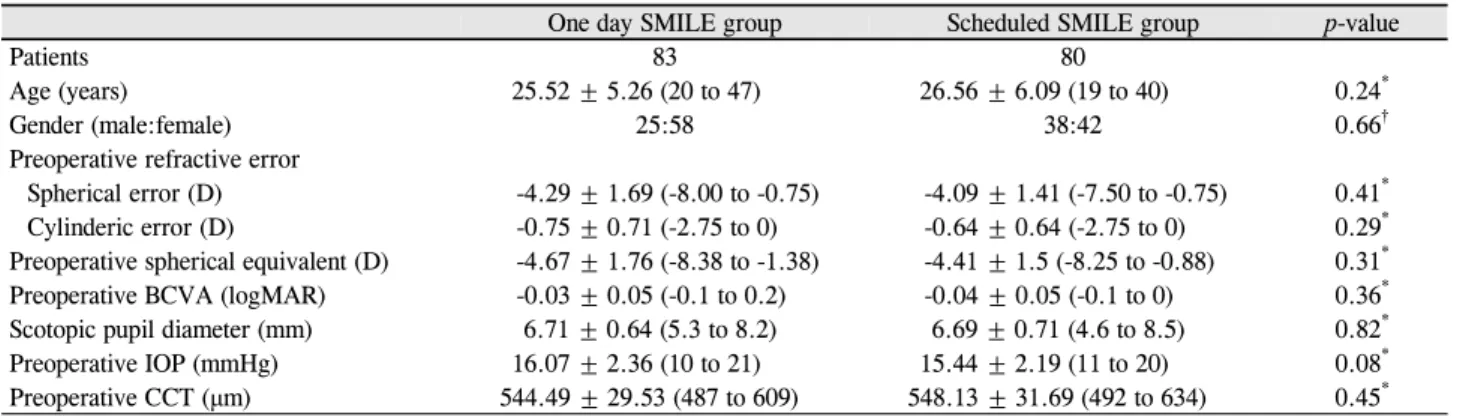
Values are presented as mean ± standard deviation (range) unless otherwise indicated.
SMILE = small incision lenticule extraction; D = diopters; BCVA = best corrected visual acuity; logMAR = logarithm of minimum angle of resolution; IOP = intraocular pressure; CCT = central cornea thickness.
*Independent t-test; †Chi-square test.
Table 1. Preoperative demographic characteristics of patients who underwent SMILE
사전에 동의하였다. 수술 당일 오전에 내원하여 전안부 상태, 굴절검사, 각막지형도, 각막두께 등을 검사하고 산동 후 망막검사를 진행하였다. 산동을 위하여 미드린 피(Mydrin P, Santen pharmaceutical Co., Ltd., Osaka, Japan)를 점안하 였으며 산동제 점안 후 약 30분 후 산동된 상태에서 간접 검안경(indirect ophthalmoscopy)을 사용하여 망막검사를 시행하였다. 술 전 검사에서 각막염, 결막염, 안검염, 심한 안구건조증, 녹내장, 망막변성이 발견된 경우는 당일 수술 을 취소하였으며, 특이 소견이 없는 경우 당일 스마일수술 을 진행하였다.
두 군에서의 수술 후 결과 비교
당일 스마일수술의 결과를 알아보기 위하여 당일 수술군 과 계획 수술군으로 나누어 수술 전후의 근시, 난시, 시력 을 포함한 자세한 안과적 검사를 시행하였다. 수술 전에 모 든 환자의 나안시력, 최대교정시력, 현성굴절검사, 세극등 현미경검사, 안압검사(NT-530P, Nidek, Gamagori, Japan), 산동 후 안저검사, 자동굴절검사계(ARK-530A, Nidek), 동 공크기검사(Keratograph 4, Oculus, Inc., Arlington, WA, USA), 각막내피세포검사(EM-4000, Tomey corp., Nagoya, Japan), 중심부 각막두께검사(NT-530P, Nidek), 각막지형도검사 등 을 시행하였다. 스마일수술 후 1개월, 6개월에 외래에 내원 하여 원거리나안시력(uncorrected distance visual acuity, UDVA), 최대교정시력, 자동굴절검사계에 의한 근시, 난시 정도, 안압검사, 세극등현미경검사를 시행하였다. 모든 환 자의 우안 결과값만을 분석에 사용하였다. 당일 수술 여부 외에 술 전 검사 방법, 수술 과정, 레이저 세팅 등은 두 군 에서 모두 동일하게 진행하였다. 계획 수술군 중 검사 후 수술까지 3개월 이상 경과한 경우에는 굴절이상의 변화를
확인하기 위하여 수술 전에 굴절검사를 한 번 더 하였다.
통계적 검정은 IBM SPSS ver. 18.0 프로그램(IBM Corp., Armonk, NY, USA)을 이용하였으며 두 군의 비교는 독립 t-test와 카이스퀘어(Chi-square) 방법을 이용하였으며, p값 이 0.05 미만인 경우 통계학적으로 유의하다고 정의하였다.
선형회귀분석의 설명력을 의미하는 결정계수(R2)값은 상관 계수(r)의 제곱으로서 이를 결과에 표시하였다.
결 과
스마일수술을 받은 환자 163명 중 당일 스마일군은 83명 이었고 계획 스마일군은 80명이었다. 두 군에서 나이, 술 전 근시, 난시, 교정시력, 동공크기, 각막두께는 차이가 없 었다(Table 1).
당일 스마일군과 계획 스마일군에서 술 후 1개월 시 구 면대응치는 각각 -0.31 ± 0.45디옵터, -0.32 ± 0.38디옵터였 고(p=0.97), 근시는 각각 -0.07 ± 0.43디옵터, -0.09 ± 0.37디 옵터였고(p=0.81), 난시는 각각 -0.48 ± 0.29디옵터, -0.46 ± 0.28디옵터였으며(p=0.57), 시력은 각각 -0.02 ± 0.11 logMAR, -0.02 ± 0.11 logMAR이었다(p=0.79) (Table 2). 수술 후 6개 월에 당일 스마일군과 계획 스마일군에서의 구면대응치는 각각 -0.30 ± 0.44디옵터, -0.32 ± 0.37디옵터였고(p=0.82), 근 시는 각각 -0.06 ± 0.42디옵터, -0.08 ± 0.34디옵터였고(p=0.81), 난시는 각각 -0.48 ± 0.25디옵터, -0.48 ± 0.30디옵터였으며 (p=0.99), 시력은 각각 -0.04 ± 0.09 logMAR, -0.05 ± 0.07 logMAR이었다(p=0.45) (Table 3).
스마일수술 후 구면대응치가 ±1.0 D 이내를 보인 환자는 수술 후 1개월에 당일 스마일군에서 94%, 계획 스마일군에 서 96%이었고, 수술 후 6개월에는 두 군에서 각각 96%,
One day SMILE group Scheduled SMILE group p-value*
Patients 83 80
UCVA at postop 1 month (logMAR) -0.02 ± 0.11 (-0.1 to 0.3) -0.02 ± 0.11 (-0.1 to 0.4) 0.79 Postoperative refractive error
Spherical error (D) -0.07 ± 0.43 (-1.00 to 1.00) -0.09 ± 0.37 (-1.00 to 0.75) 0.81
Cylinderic error (D) -0.48 ± 0.29 (-1.50 to 0) -0.46 ± 0.28 (-1.50 to 0) 0.57
Postoperative spherical equivalent (D) -0.31 ± 0.45 (-1.38 to 0.63) -0.32 ± 0.38 (-1.50 to 0.50) 0.97 Values are presented as mean ± standard deviation (range) unless otherwise indicated.
SMILE = small incision lenticule extraction; UCVA = uncorrected visual acuity; postop = postoperative; logMAR = logarithm of minimum angle of resolution; D = diopters.
*Independent t-test.
Table 2. Postoperative 1 month clinical outcomes of one day SMILE group and scheduled SMILE group
One day SMILE group Scheduled SMILE group p-value*
Patients 83 80
UCVA at postop 6 months (logMAR) -0.04 ± 0.09 (-0.1 to 0.3) -0.05 ± 0.07 (-0.1 to 0.25) 0.45 Postoperative refractive error
Spherical error (D) -0.06 ± 0.42 (-1.50 to 0.75) -0.08 ± 0.34 (-1.25 to 0.50) 0.81
Cylinderic error (D) -0.48 ± 0.25 (-1.00 to 0) -0.48 ± 0.30 (-1.50 to 0) 0.99
Postoperative spherical equivalent (D) -0.30 ± 0.44 (-1.88 to 0.50) -0.32 ± 0.37 (-2.00 to 0.38) 0.82 Values are presented as mean ± standard deviation (range) unless otherwise indicated.
SMILE = small incision lenticule extraction; UCVA = uncorrected visual acuity; postop = postoperative; logMAR = logarithm of minimum angle of resolution; D = diopters.
*Independent t-test.
Table 3. Postoperative 6 months clinical outcomes of one day SMILE group and scheduled SMILE group
A B
Figure 1. Refractive outcomes of the one day SMILE group and scheduled SMILE group. The spherical equivalent of one day
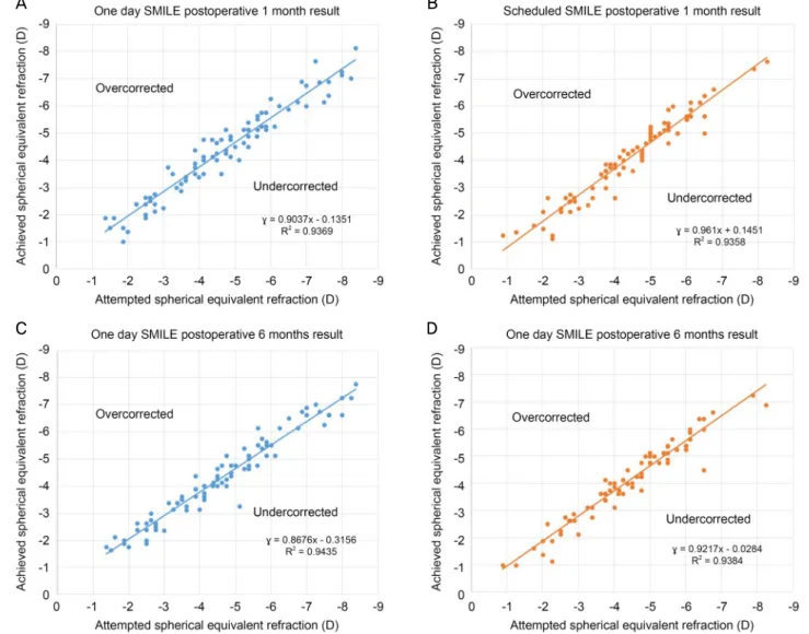
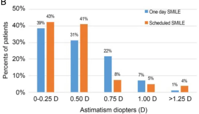
SMILE and scheduled SMILE at postoperative 1 month (A). The spherical equivalent of one day SMILE and scheduled SMILE at postoperative 6 months (B). SMILE = small incision lenticule extraction; D = diopters.96%였으며(Fig. 1), 목표 구면대응치와 잔여 구면대응치의 차이인 잔여 구면대응치는 두 군에서 유의한 차이는 보이 지 않았다. 결정계수(R2)는 수술 후 1개월에 당일 스마일군 에서 0.9369, 계획 스마일군에서 0.9358이었고 수술 후 6개월 에 각각 0.9435, 0.9384였다(Fig. 2). 스마일수술 후 난시가 0.75디옵터 이내인 환자는 수술 후 1개월에 당일 스마일군 에서 92%, 계획 스마일군에서 91%였고, 수술 후 6개월에는 각각 93%, 89%였다(Fig. 3). 수술 후 나안시력을 수술 전 교정시력으로 나눈 값인 효용성지수는 스마일수술 후 1개 월에 당일 스마일군에서 0.95 ± 0.18, 계획 스마일군에서
0.95 ± 0.17로 두 군 간의 차이가 없었으며(p=0.84), 수술 후 6개월에는 각각 0.99 ± 0.13, 계획 스마일군에서 1.00 ± 0.12로 차이가 없었다(p=0.53) (Table 4).
고 찰
스마일은 시력 회복이 빠른 수술이지만 초기 뿌연 증상 으로 라식보다 시력이 완전히 회복되기까지의 시간이 더
소요된다.36,37 스마일수술 초기의 뿌연 증상의 원인은 수술
부위에서 산란되는 빛의 강도가 높고 각막뚜껑(cap)과 각막
A B
C D
Figure 2. Predictability of spherical equivalent correction of the one day SMILE group and scheduled SMILE group. The results of
one day SMILE (A) and scheduled SMILE (B) at postoperative 1 month. The results of one day SMILE (C) and scheduled SMILE (D) at postoperative 6 months. SMILE = small incision lenticule extraction; D = diopters.실질 사이의 미세변형이 원인이다.8,38-41 수술 1-2주 후 대비 감도 저하와 빛번짐이 대부분 호전되지만40,42 완전한 회복 을 위해서는 수술 후 6개월까지 경과 관찰이 필요하다.8,37,41 이러한 이유로 본 연구에서는 두 군의 수술 후 1개월과 6개 월의 검사값들을 비교 분석하였다. 본 연구의 당일 스마일 군과 계획 스마일군의 평균 logMAR 나안시력은 수술 후 1개 월에 -0.02, 6개월에 -0.04로 나타났으며 두 군의 차이는 보 이지 않았다. 이러한 결과는 스마일수술 후 logMAR 평균 나안시력이 0.02에서 -0.17로 보고한 기존 연구들43,44과 유 사한 결과이며 수술 후 1개월보다 수술 후 6개월에 나안시 력이 호전된 것은 스마일수술 후 시력이 3-6개월까지 향상 될 수 있다는 기존 연구들과 일치한다.8,45
각막굴절교정수술 후 결과를 좋게 하기 위해서 수술 중
광학부 중심을 각막정점에 정확히 맞추는 것이 필요하
다.46-48 Bueeler et al46은 각막굴절수술 후 3 mm 동공 기준
으로 레이저 조사 중심이 0.2 mm 이상 벗어나면 시력에 영 향을 줄 수 있다고 하였다. Piñero and Teus8은 스마일수술 시 광학부가 중심에서 벗어나면 코마와 같은 고위수차가 증가한다고 하였다. Lee et al29은 스마일수술 후 중심이탈 이 0.335 mm 이상이면 그 이하인 군에 비하여 코마와 구면 수차가 증가하였다고 보고하였다.
광원을 향해서 정확히 보고 있는 상태의 안구에 맺힌 각 막 반사점인 제 1 퍼킨지 반사(first Purkinje image)를 각막 정점(vertex)이라고 하며, 이는 스마일수술 시 각막 중심으 로 받아들여지고 있다.29 레이저 조사의 중심을 각막정점으 로 수술하는 것이 동공 중심으로 수술하는 것에 비하여 우
Figure 3. Preoperative and postoperative astigmatic refraction
distribution of the one day SMILE group and scheduled SMILE group. Preoperative astigmatic diopters of two groups (A).Astigmatic diopters after the one day SMILE and scheduled SMILE at postoperative 1 month (B). Astigmatic diopters after the one day SMILE and scheduled SMILE at postoperative 6 months (C). SMILE = small incision lenticule extraction; D = diopters.
A B
C
One day SMILE group Scheduled SMILE group p-value*
SMILE (1 month) 0.95 ± 0.18 0.95 ± 0.17 0.84
SMILE (6 months) 0.99 ± 0.13 1.00 ± 0.12 0.53
Values are presented as mean ± standard deviation.
UDVA = uncorrected visual acuity; CDVA = corrected distace visual acuity; SMILE = small incision lenticule extraction.
*Independent t-test.
Table 4. The efficacy indices (postoperative UDVA/ preoperative CDVA) of both groups at postoperative 1 month and 6 months
수한 수술 후 결과를 보였다.46-51 환자가 초록색 불빛을 정 확히 보고 있을 때 반사되는 초록색 불빛이 각막정점이므 로 스마일수술 시 환자의 협조가 필요하다.28,31,32,52 각막이 고정되는 순간 각막이 미세하게 움직여 발생하는 오차는 각막의 두 정점인 각막정점(vertex)과 각막정점(apex)의 위 치 차이가 클수록 발생 빈도가 높다.53 또한 이러한 과정이 술자의 주관적 판단에 따라 수동으로 진행되므로 술자의 경험이 수술 후 결과에 영향을 미친다.27,30
각막을 고정하기 직전에 환자가 초록색 불빛을 잘 주시 하는지 술자가 확인할 수 있는 방법은 두 가지다. 첫째, 각 막이 접촉되었을 때 생긴 눈물층에 의한 각막접촉면이 대 칭인지 확인하는 것이다. 둘째, 동공 중심과 각막정점의 상 대적인 위치를 각막지형도와 비교하는 것이다. 동공이 평 상시보다 산동된 상태에서는 둘째 방법을 사용하는 것이 부정확할 수 있다. 동공이 커지면 동공 중심과 각막정점의 상대적 위치가 변할 수 있기 때문이다.32,34,35,54-57
스마일수술의 광학부 중심이탈이 라식, 라섹 수술과 비 교하여 더 나쁘다는 보고는 아직까지 없다.58 라식, 라섹은 수술 중 안구추적장치를 사용할 수 있는 장점이 있으나 엑 시머레이저 조사가 각막절편이 젖혀져 있거나 각막상피가 제거된 상태에서 진행되므로 환자가 초록색 불빛을 선명하 게 볼 수 없다. 이에 비하여, Visumax 레이저(VisumaxTM Femtosecond Laser, Carl Zeiss Meditec)를 사용한 스마일 수술은 각막상피가 온전하여 환자가 초록색 불빛을 선명하 게 볼 수 있는 상태에서 각막 고정이 이루어지고, 안구가 고정된 상태로 레이저 조사가 이루어진다는 장점이 있다. 스마일수술에 사용되는 Visumax 펨토초레이저(VisumaxTM Femtosecond Laser, Carl Zeiss Meditec)는 환자가 초록색 불빛을 정확히 볼 수 있도록 환자의 근시, 난시 정도에 따 라서 불빛이 조절되며, 낮은 음압을 사용하여 고정하므로 수술 중 시야가 캄캄해지는 현상이 발생하지 않아서 환자 의 공포심을 줄여 협조도를 높일 수 있다.36,59
스마일수술 후 난시가 저교정되는 현상이 보고되었으나 난시축을 정확히 맞추고 노모그램을 조정하여 극복될 수 있었다고 하였다.8,60,61 굴절교정 시 난시축이 5° 틀어지면 난시의 교정 효과가 1.5% 감소하고, 15°가 틀어지면 13.4%
감소하고, 30° 틀어지면 50% 감소한다.30 스마일수술은 안 구가 고정된 상태에서 진행되므로 동적 회선 보정은 필요 가 없으나 정적 회선 보정은 필요하다. Alpin62은 레이저수 술환자의 38%가 앉았을 때에 비해 누운 자세에서 5° 이상 의 안구가 회선되었다고 하며, Ganesh et al63은 스마일수술 중 5° 이상의 회선을 보인 환자가 약 20%였다고 보고하였 다. 스마일수술 시 안구회선을 예방하고 난시교정 효과를 높이기 위하여 각막에 마킹을 한 후 각막고정을 시행하는 방법들이 시도되고 있다.32,63 최근 Pedersen et al60은 스마 일수술 후 잔여 난시가 0.50디옵터 이내인 경우가 70%, 1.0디 옵터 이내인 경우가 94%라고 보고하였다. 본 연구에서는 각막에 마킹을 하는 방법은 사용하지 않았으며, 난시교정 결과는 수술 후 6개월에 당일 스마일군에서 0.50디옵터 이 내가 72%, 1.0디옵터 이내가 100%, 계획 스마일군에서는 각각 70%, 97%로 이전 연구와 유사한 결과를 보였다.60,64
스마일수술 전 망막검사를 위하여 사용되는 산동제는 부 교감신경 길항제와 교감신경 작용제로서 일정 시간 동공근 에 작용하여 빛에 대한 동공 반응을 억제한다. 산동검사 시 주로 사용되는 미드린 피(Mydrin P, Santen pharmaceutical Co., Ltd.)의 주성분은 트로피카마이드 0.5-1%와 페닐에프 린 2.5%로서 동공 확장의 최대 효과는 30분 후에 나타나며 회복에는 4-10시간이 걸린다.18 당일 스마일수술을 하는 환 자의 일부는 동공이 커져 있는 상태로 수술을 진행한다. 스 마일수술 중 동공 크기가 수술에 미치는 영향은 두 가지 측 면을 생각할 수 있다. 첫째, 눈부심이 증가하여 환자가 초 록색 불빛을 주시하는데 영향을 받을 수 있다는 점이고 둘 째, 술자가 각막정점을 레이저의 환자 접속기에 고정시킬 때 커져 있는 동공에 영향을 받을 수 있다는 점이다. 당일 스마일군과 계획 스마일군환자들의 수술 후 결과가 대등하 다는 것은 산동된 상태가 스마일수술의 결과에 미치는 영 향이 크지 않음을 시사한다.
본 연구는 당일 스마일수술의 결과를 근시, 난시, 시력으 로만 비교하였다는 제한점이 있지만 당일 스마일수술의 결 과를 처음으로 보고하였다는 점에서 의미가 있다. 술 후 고 위수차를 비교하기 위한 파면분석검사, 중심이탈 정도를 확인하기 위한 각막지형도검사, 시력의 질을 비교하기 위 한 optical quality analysis system (OQAS)검사를 포함된 추가 연구가 앞으로 필요할 것으로 생각된다.65
결론적으로 Visumax 레이저(VisumaxTM Femtosecond Laser, Carl Zeiss Meditec)를 사용한 당일 스마일수술은 산동 검
사 후 완전히 회복되지 않은 동공 상태에서 수술을 진행하 였음에도 기존 방법과 비교하여 수술 후 결과에 차이가 없 었다. 이러한 결과는 당일 스마일수술을 원하는 환자와 집 도하는 술자 모두가 안심하고 수술을 할 수 있는 근거가 될 것이다.
REFERENCES
1) Sekundo W, Gertnere J, Bertelmann T, Solomatin I. One-year re- fractive results, contrast sensitivity, high-order aberrations and complications after myopic small-incision lenticule extraction (ReLEx SMILE). Graefes Arch Clin Exp Ophthalmol 2014;252:
837-43.
2) Pedersen IB, Ivarsen A, Hjortdal J. Three-year results of small in- cision lenticule extraction for high myopia: refractive outcomes and aberrations. J Refract Surg 2015;31:719-24.
3) Kobashi H, Kamiya K, Igarashi A, et al. Two-years results of small-incision lenticule extraction and wavefront-guided laser in situ keratomileusis for myopia. Acta Ophthalmol 2018;96:e119-26.
4) Reinstein DZ, Archer TJ, Gobbe M. Small incision lenticule ex- traction (SMILE) history, fundamentals of a new refractive surgery technique and clinical outcomes. Eye Vis (Lond) 2014;1:3.
5) Vestergaard A, Ivarsen A, Asp S, Hjortdal JØ. Femtosecond (FS) laser vision correction procedure for moderate to high myopia: a prospective study of ReLEx (R) flex and comparison with a retro- spective study of FS-laser in situ keratomileusis. Acta Ophthalmol 2013;91:355-62.
6) Ganesh S, Brar S, Arra RR. Refractive lenticule extraction small incision lenticule extraction: a new refractive surgery paradigm.
Indian J Ophthalmol 2018;66:10-9.
7) Ağca A, Demirok A, Yildirim Y, et al. Refractive lenticule ex- traction (ReLEx) through a small incision (SMILE) for correction of myopia and myopic astigmatism: current perspectives. Clin Ophthalmol (Auckland, NZ) 2016;10:1905-12.
8) Piñero DP, Teus MA. Clinical outcomes of small-incision lenticule extraction and femtosecond laser-assisted wavefront-guided laser in situ keratomileusis. J Cataract Refract Surg 2016;42:1078-93.
9) Seven I, Vahdati A, Pedersen IB, et al. Contralateral eye compar- ison of SMILE and flap-based corneal refractive surgery: computa- tional analysis of biomechanical impact. J Refract Surg 2017;33:
444-53.
10) Shetty R, Francis M, Shroff R, et al. Corneal biomechanical changes and tissue remodeling after SMILE and LASIK. Invest Ophthalmol Vis Sci 2017;58:5703-12.
11) Denoyer A, Landman E, Trinh L, et al. Dry eye disease after re- fractive surgery comparative outcomes of small incision lenticule extraction versus LASIK. Ophthalmology 2015;122:669-76.
12) kobashi H, Kamiya K, Shimizu K. Dry eye after small incision len- ticule extraction and femtosecond laser-assisted LASIK: meta-analysis.
Cornea 2017;36:85-91.
13) Xia LK, Ma J, Liu HN, et al. Three-year results of small incision lenticule extraction and wavefront-guided femtosecond laser-as- sisted laser in situ keratomileusis for correction of high myopia and myopic astigmatism. Int J Ophthalmol 2018;11:470-7.
14) Shen Z, Shi K, Yu Y, et al. Small incision lenticule extraction
(SMILE) versus femtosecond laser-assisted in situ keratomileusis (FS-LASIK) for myopia: a systematic review and meta-analysis.
PLoS ONE 2016;11:e0158176.
15) Lam DS, Fan DS, Chan WM, et al. Prevalence and characteristics of peripheral retinal degeneration in chinese adults with high my- opia: a cross-sectional prevalence survey. Optom Vis Sci 2005;82:
235-8.
16) Arevalo JF. Retinal complications after laser-assisted in situ kera- tomileusis (LASIK). Curr Opin Ophthalmol 2004;15:184-91.
17) Lewis H. Peripheral retinal degenerations and the risk of retinal detachment. Am J Ophthalmol 2003;136:155-60.
18) Lin J, Xie X, Du X, et al. Incidence of vitreoretinal pathologic con- ditions in myopic eyes after laser in situ keratomileusis. Zhonghua Yan Ke Za Zhi 2002;38:546-9.
19) Ghosh S, Couper TA, Lamoureux E, et al. Evaluation of iris recog- nition system for wavefront-guided laser in situ keratomileusis for myopic astigmatism. J Cataract Refract Surg 2008;34:215-21.
20) Pierro L, Camesasca FI, Mischi M, Brancato R. Peripheral retinal changes and axial myopia. Retina 1992;12:12-7.
21) Kirker GE, McDonald DJ. Peripheral retinal degeneration in high myopia. Can J Ophthalmol 1971;6:58-61.
22) Lin SC, Tseng SH. Prophylactic laser photocoagulation for retinal breaks before laser in situ keratomileusis. J Refract Surg 2003;
19:661-5.
23) Wilkes SR, Beard CN, Kurland LT, et al. The incidence of retinal detachment in Rochester, Minnesota, 1970–1978. Am J Ophthalmol 1982;94:670-3.
24) Arevalo JF, Ramirez E, Suarez E, et al. Rhegmatogenous retinal detachment in myopic eyes after laser in situ keratomileusis.
Frequency, characteristics, and mechanism. J Cataract Refract Surg 2001;27:674-80.
25) Wright KW. Strube YN. Pediatric ophthalmology and strabismus, 3rd ed. New York: Oxford University Press, 2012;92.
26) Kim WK, Ryu IH, Lee IS, et al. Comparison of postoperative re- sults of one day laser-assisted in-situ keratomileusis, laser-assisted sub-epithelial keratectomy surgery, and conventional surgery. J Korean Ophthalmol 2018;59:410-8.
27) Chan TCY, Ng ALK, Cheng GPM, et al. Effect of the learning curve on visual and refractive outcomes of small-incision lenticule extraction. Cornea 2017;36:1044-50.
28) Kang DSY, Lee H, Reinstein DZ, et al. Comparison of the dis- tribution of lenticule decentration following SMILE by subjective patient fixation or triple marking centration. J Refract Surg 2018;
34:446-52.
29) Lee H, Roberts CJ, Arba-Mosquera S, et al. Relationship between decentration and induced corneal higher-order aberrations follow- ing small-incision lenticule extraction procedure. Invest Ophthalmol Vis Sci 2018;59:2316-24.
30) Titiyal JS, Kaur M, Rathi A, et al. Learning curve of small incision lenticule extraction: challenges and complications. Cornea 2017;
36:1377-82.
31) Steinwender G, Shajari M, Mayer WJ, et al. Impact of a displaced corneal apex in small incision lenticule extraction. J Refract Surg 2018;34:460-5.
32) Jun I, Kang DSY, Reinstein DZ, et al. Clinical outcomes of SMILE with a triple centration technique and corneal wavefront-guided transepithelial PRK in high astigmatism. J Refract Surg 2018;34:
156-63.
33) Zhao F, Han T, Chen X, et al. Minimum pupil in pupillary response to light and myopia affect disk halo size: a cross-sectional study.
BMJ Open 2018 10;8:e019914.
34) De Ortueta D, Arba-Mosquera S. Laser in situ keratomileusis for high hyperopia with corneal vertex centration and asymmetric offset. Eur J Ophthalmol 2017;27:141-52.
35) Narváez J, Brucks M, Zimmerman G, et al. Intraoperative cyclo- rotation and pupil centroid shift during LASIK and PRK. J Refract Surg 2012;28:353-7.
36) Damgaard IB, Ang M, Farook M, et al. Intraoperative patient expe- rience and postoperative visual quality after smile and lasik in a randomized, paired-eye, controlled study. J Refract Surg 2018;34:
92-9.
37) Ganesh S, Gupta R. Comparison of visual and refractive outcomes following femtosecond laser-assisted LASIK with SMILE in pa- tients with myopia or myopic astigmatism. J Refract Surg 2014;
30:590-6.
38) Riau AK, Angunawela RI, Chaurasia SS, et al. Effect of different femtosecond laser-firing patterns on collagen disruption during re- fractive lenticule extraction. J Cataract Refract Surg 2012;38:1467-75.
39) Wei S, Wang Y, Wu D, et al. Ultrastructural changes and corneal wound healing after SMILE and PRK procedures. Curr Eye Res 2016;41:1316-25.
40) Han T, Zhao J, Shen Y, et al. A three-year observation of corneal backscatter after small incision lenticule extraction (SMILE). J Refract Surg 2017;33:377-82.
41) Agca A, Ozgurhan EB, Yildirim Y, et al. Corneal backscatter anal- ysis by in vivo confocal microscopy: fellow eye comparison of small incision lenticule extraction and femtosecond laser-assisted LASIK. J Ophthalmol 2014;2014:265012.
42) Yao P, Zhao J, Li M, et al. Microdistortions in bowman’s layer fol- lowing femtosecond laser small incision lenticule extraction ob- served by fourier-domain OCT. J Refract Surg 2013;29:668-74.
43) Agca A, Ozgurhan EB, Demirok A, et al. Comparison of corneal hysteresis and corneal resistance factor after small incision lentic- ule extraction and femtosecond laser-assisted LASIK: a pro- spective fellow eye study. Cont Lens Anterior Eye 2014;37:77-80.
44) Kamiya K, Shimizu K, Igarashi A, Kobashi H. Visual and re- fractive outcomes of femtosecond lenticule extraction and small incision lenticule extraction for myopia. Am J Ophthalmol 2014;
157:128-34.e2.
45) Son GS, Lee SC, Lim TH. Clinical outcomes of small incision len- ticule extraction including visual qaulity analysis. J Korean Opthalmol 2016;57:562-7.
46) Bueeler M, Mrochen M, Seiler T. Maximum permissible lateral de- centration in aberration-sensing and wavefront-guided corneal ablation. J Cataract Refract Surg 2003;29:257-63.
47) Mrochen M, Kaemmerer M, Mierdel P, Seiler T. Increased high- er-order optical aberrations after laser refractive surgery: a problem of subclinical decentration. J Cataract Refract Surg 2001;27:362-9.
48) Wang L, Koch DD. Residual higher-order aberrations caused by clinically measured cyclotorsional misalignment or decentration during wavefront-guided excimer laser corneal ablation. J Cataract Refract Surg 2008;34:2057-62.
49) Okamoto S, Kimura K, Funakura M, et al. Comparison of myopic LASIK centered on the coaxially sighted corneal light reflex or line of sight. J Refract surg 2009;25(10 Suppl):S944-50.
50) Reinstein DZ, Archer TJ, Gobbe M. Is topography-guided ablation
profile centered on the corneal vertex better than wavefront-guided ablation profile centered on the entrance pupil? J Refract Surg 2012;28:139-43.
51) Arbelaez MC, Vidal C, Arba-Mosquera S. Clinical outcomes of corneal vertex versus central pupil references with aberration-free ablation strategies and LASIK. Invest Ophthalmol Vis Sci 2008;
49:5287-94.
52) Reinstein DZ, Gobbe M, Gobbe L, et al. Optical zone centration ac- curacy using corneal fixation-based SMILE compared to eye track- er-based femtosecond laser-assisted LASIK for myopia. J Refract Surg 2015;31:586-92.
53) Steinwender G, Shajari M, Mayer WJ, et al. Impact of a displaced corneal apex in small incision lenticule extraction. J Refract Surg 2018;34:460-5.
54) Uozato H, Guyton DL. Centering corneal surgical procedures. Am J Ophthalmol 1987;103(3 Pt 1):264-75.
55) Yang Y, Thompson K, Burns SA. Pupil location under mesopic, photopic, and pharmacologically dilated conditions. Invest Ophthalmol Vis Sci 2002;43:2508-12.
56) Erdem U, Muftuoglu O, Gundogan FC, et al. Pupil center shift rela- tive to the coaxially sighted corneal light reflex under natural and pharmacologically dilated conditions. J Refract Surg 2008;24:530-8.
57) Schruender SA, Fuchs H, Spasovski S, Dankert A. Intraoperative corneal topography for image registration. J Refract Surg 2002;18:
S624-9.
58) Alió del Barrio JL, Vargas V, Al-Shymali O, Alió JL. Small in- cision lenticule extraction (SMILE) in the correction of myopic as- tigmatism: outcomes and limitations - an update. Eye Vis 2017;4:26.
59) Hall RC, Rosman M, Chan C, et al. Patient and surgeon experience during laser in situ keratomileusis using 2 femtosecond laser systems. J Cataract Refract Surg 2014;40:423-9.
60) Pedersen IB, Ivarsen A, Hjortdal J. Changes in astigmatism, densi- tometry, and aberrations after SMILE for low to high myopic astig- matism: a 12-month prospective study. J Refract Surg 2017;
33:11-7.
61) Swami AU, Steinert RF, Osborne WE, White AA. Rotational mal- position during laser in situ keratomileusis. Am J Ophthalmol 2002;133:561-2.
62) Alpins N. Astigmatism analysis by the Alpins method. J Cataract Refract Surg 2001;27:31-49.
63) Ganesh S, Brar S, Pawar A. Results of intraoperative manual cy- clotorsion compensation for myopic astigmatism in patients under- going small incision lenticule extraction (SMILE). J Refract Surg 2017;33:506-12.
64) Ivarsen A, Hjortdal J. Correction of myopic astigmatism with small incision lenticule extraction. J Refract Surg 2014;30:240-7.
65) Kanellopoulos AJ, Asimellis G. LASIK ablation centration: an ob- jective digitized assessment and comparison between two gen- erations of an excimer laser. J Refract Surg 2015;31:164-9.
= 국문초록 =
근시환자에서 당일 스마일수술과 계획 스마일수술의 결과 비교
목적: 근시환자에서 산동 후 망막검사를 포함한 수술 전 검사와 수술을 하루에 진행하는 당일 스마일수술의 결과를 알아보고자 하였 다.
대상과 방법: 2017년 1월부터 6월까지 본원에서 스마일수술을 받은 환자 163명(163안)을 대상으로 하였다. 수술 전 검사와 수술을 같 은 날에 진행한 당일 스마일군과 검사 후 다른 날에 수술을 진행한 계획 스마일군으로 나누어 수술 후 1, 6개월의 근시, 난시 및 시력 을 비교하였다.
결과: 당일 스마일군은 83명, 계획 스마일군은 80명이었으며, 수술 후 1개월에 근시는 각각 -0.07 ± 0.43디옵터, -0.09 ± 0.37디옵 터였고(p=0.81), 난시는 각각 -0.48 ± 0.29디옵터, -0.46 ± 0.28디옵터였으며(p=0.57), 시력은 각각 -0.02 ± 0.11 logMAR, -0.02
± 0.11 logMAR이었다(p=0.79). 수술 후 6개월에 당일 스마일군과 계획 스마일군의 근시는 각각 -0.06 ± 0.42디옵터, -0.08 ± 0.34 디옵터였고(p=0.81), 난시는 각각 -0.48 ± 0.25디옵터, -0.48 ± 0.30디옵터였으며(p=0.99), 시력은 각각 -0.04 ± 0.09 logMAR, -0.05 ± 0.07 logMAR이었다(p=0.45).
결론: 산동제로 동공이 약간 커진 상태에서 진행한 당일 스마일수술은 검사와 수술을 다른 날에 진행한 스마일수술법에 비하여 술 후 근시, 난시, 나안시력에서 유의한 차이를 보이지 않았다.
<대한안과학회지 2019;60(3):223-232>
김욱겸 / Wook Kyum Kim
비앤빛 강남밝은세상안과의원 B&VIIt Eye Center