Sacral insufficiency fractures (SIF) are a common, but often underdiagnosed source of lower back pain without apparent trauma. This report presents the clinical outcome of a 75-year-old female with SIF, and an underlying medical history of osteoporosis and rheumatoid arthritis. She was treated non-operatively, in-hospital, with Korean medicine. Patient progress was assessed using the numerical rating scale and self- reported symptoms. Post-treatment, the numerical rating scale score for pain in her hip decreased from 7 to 2. At admission, the patient was unable to sit, and could only walk 3 m with assistance. At discharge, she could sit for longer than 1 hour and walk further than 200 m unassisted. On the follow-up visit, the patient was asymptomatic, and x-ray scans showed ossification of the sacral and pubic fractures. These results suggest that, Korean medicine can effectively reduce pain and aid rehabilitation in patients with SIF, without the need for surgery.
©2020 Korean Acupuncture & Moxibustion Medicine Society. This is an open access article under the CC BY- NC-ND license (http://creativecommons.org/licenses/by-nc-nd/4.0/).
Article history:
Submitted: April 25, 2020 Revised: June 02, 2020 Accepted: June 08, 2020 Keywords:
conservative treatment, pelvis, spinal stenosis, stress fracture
https://doi.org/10.13045/jar.2020.00129 pISSN 2586-288X eISSN 2586-2898
Case Report
Effect of Complex Korean Medicine Treatment on Sacral Insufficiency Fracture: A Case Report
Ga Hyeon Jung, Hyun Lee, Hwa Yeon Ryu, Jae Hui Kang *
Department of Acupuncture and Moxibustion Medicine, College of Korean Medicine, Daejeon University, Cheonan, Korea
ABSTRACT
Journal of Acupuncture Research
Journal homepage: http://www.e-jar.org
Introduction
Sacral insufficiency fractures (SIF) have been described as a common, but often underdiagnosed cause of lower back pain and mobility, especially in elderly patients with osteoporosis [1-3].
Patients with osteoporosis have low bone mineral density, poor bone quality, and are susceptible to falling due to muscle weakness [4].
Patients with SIF present with vague symptoms, including lower back pain and buttock pain [5], while some may present with neurological deficits [1]. Additionally, SIF are difficult to diagnose using x-ray scans alone. These factors make a definitive diagnosis of SIF difficult to achieve [6,7].
Past medical history
Conservative treatment options should be offered first to patients with SIF [6]. Contrary to previous studies, recent research suggests that early mobilization improves outcomes and reduces complications [8]. Several studies have reported the effectiveness of conservative treatment for fractures, including Korean medicine
treatment such as acupuncture, moxibustion, herbal medication, bee venom, and pharmacopuncture [9]. In Korea, there have been reports of pelvic fractures caused by apparent trauma or traffic accidents [9-12]. This case report describes the effectiveness of Korean medicine treatment for SIF in an elderly patient with osteoporosis.
Case Report Patient
○○○ (F/75) Chief complaint
Pain in both hips and inner thighs.
Past medical history
Hospitalization on July 24
th, 2019, due to systemic arthralgia, for 1 month, during which she was diagnosed with rheumatoid arthritis and osteoporosis, and steroid medication was prescribed.
* Corresponding author. Jae Hui Kang
Department of Acupuncture and Moxibustion Medicine, Cheonan Korean Medicine Hospital of Daejeon University, 4, Notaesan-ro, Seobuk-gu, Cheonan-si, Chungcheongnam- do, 331-958, Korea
E-mail: [email protected]
ORCID: Ga Hyeon Jung https://orcid.org/ 0000-0002-7759-0972, Jae Hui Kang https://orcid.org/ 0000-0003-4812-0557
©2020 Korean Acupuncture & Moxibustion Medicine Society. This is an open access article under the CC BY-NC-ND license (http://creativecommons.org/licenses/by-nc-
nd/4.0/).
Family history None.
Present illness
Early in November 2019 the patient started to experience spontaneous pain in her right hip whilst squatting. On November 15
th, 2019 she had a magnetic resonance imaging (MRI) scan of the lumbar spine which showed spinal stenosis affecting L2-5. A nerve block injection was administered twice. She was then hospitalized in a Western hospital from November 25
thuntil December 3
rd, 2019, during which time, a bone scan, X-rays of the lumbar spine, pelvis, and hips, in addition to an MRI scan of the lumbar spine and sacrum were performed. She was diagnosed with SIF and spinal stenosis. After discharge, she visited the Korean Medicine hospital for treatment.
Duration of treatment
December 3
rd, 2019 to January 22
nd, 2020 (51 days of hospitalization)
Clinical chemistry
Laboratory investigations, including total blood count, routine biochemical tests, erythrocyte sedimentation rate, and C-reactive protein levels fell within normal limits. Urinalysis demonstrated the presence of 5-7 red blood cells per high power field, 7-10 white blood cells, 10-15 epithelial cells, and a few bacteria. With respect to the urinalysis, the patient was asymptomatic.
Radiology
A bone scan, pelvic and lumbar MRI scans, and anteroposterior x-ray scans of the pelvis were taken (Figs. 1-4).
Patient protection policy on patient information use
To protect the patient’s personal information, medical records were obtained from the Institutional Review Board (no: DJUMC- 2020-BM-02).
Treatment Acupuncture
The acupuncture needles used were 0.25×30 mm and 30×40 mm stainless steel standardized, and disposable (Eastern acupuncture
Fig. 1. A bone scan of the pelvis. The bone scan showed a pattern of vertical uptake in both alae with a horizontal component in the body of the sacrum, indicating an insufficiency fracture of the right ala. Increased bone production was also evident in the left pubic area (November 15
th, 2019).
Fig. 1. A bone scan of the pelvis showed a pattern of vertical up take in both alae with a horizontal component in the body of the sacrum, indicating an insufficiency fracture of the right ala. Incr eased bone activity was also evident in the left pubic area (Nove mber 15, 2019).
Fig. 2. Magnetic resonance images of the hip (15
thNovember 2019). Axial (fat-saturated T2-weighted; left), and coronal (fat-saturated T2-weighted; right) images show a low- energy intensity lesion on the right sacrum and left pubis. A diffuse bone marrow edema
involving the right sacral ala with a non-displaced fracture line was evident. Fig. 2. Magnetic resonance images of the hip (15 November 201 9). Axial (fat-saturated T2-weighted) (left), coronal fat-saturated T2-weighted (right) images show a low-energy intensity lesion on the right sacrum and left pubis. Diffuse bone marrow edema i nvolving the right sacral ala with a non-displaced fracture line w as evident.
Fig. 4. Anteroposterior radiology scans of the pelvis (25
thNovember 2019). Sacral and pubic fractures were hardly visible on the X-ray image. Low bone density was observed
in the left superior ramus of the pubic symphysis due to the fracture. Fig. 4. Anteroposterior radiograph of the pelvis (25 November 2 019). Sacral and pubic fractures were hardly visible on the X-ra y image. Low bone density in the left superior ramus of the pubi c symphysis was seen due to fracture.
Fig. 3. Magnetic resonance images of the lumbar spine (15
thNovember 2019). There were no new lesions observed except for underlying central spinal stenosis in L2-3, L3-4, and L4-5.
Fig. 3. Magnetic resonance images of the lumbar spine (15 Nov
ember 2019). There was not any new lesion shown except for un
derlying central spinal stenosis in L2-3, L3-4, and L4-5.
equipment manufacturer, Boryung, Korea). Acupuncture was administered at BL27, BL28, BL29, BL30, BL31, BL32, BL33, BL34, BL35, BL36, BL37, BL53, BL54, GB29, GB30, LR9, and the tender points on tissues, including the gluteus muscle, piriformis, iliopsoas, adductor, and hamstrings, for 15 minutes twice daily.
Pharmacopuncture
Pharmacopuncture was performed, once daily, on each day of hospitalization. Acupoints BL27, BL28, BL29, BL30, BL53, BL54, or trigger points around the hip were used. Aconitum ciliare decaisne pharmacopuncture (ADP) (Korean Pharmacopuncture Institute, Seoul, Korea) was administered just before acupuncture therapy. Doses of 0.05 to 0.1 mL were injected at each acupoint at a depth of between 1 cm to 1.2 cm. The maximum, total dose per treatment was 1.0 mL This was administered using a 1.0 mL disposable syringe with needle (Jungrim Medical, Seoul, Korea, 30 G ×1.27cm, 12.7 mm needle).
Bee venom (BV)
Due to the potential risks associated with the use of bee venom (delayed hypersensitivity reaction or anaphylaxis), informed consent was obtained from the patient prior to treatment.
A hypersensitivity skin test to BV (1:20,000, 0.1 mL; Korean Pharmacopuncture Institute, Seoul, Korea) was performed, and
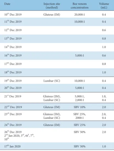
was negative. The concentration of BV administered was serially increased in the order of 1:20,000, 1:10,000, 1:5,000, 1:2,000, and sweet bee venom (SBV) 25% and SBV 50% were also used for intramuscular (IM) injections. The method of injection, either subcutaneous (SC) or IM injection, was chosen according to the site of the lesion. The volume injected also increased with each treatment if no hypersensitivity reaction occurred (Table 1). BV was substituted with SBV, if the patient felt pain from the IM injection of BV in the gluteus area. BV was injected using 30 G, 1 mL syringes.
Herbal medicine
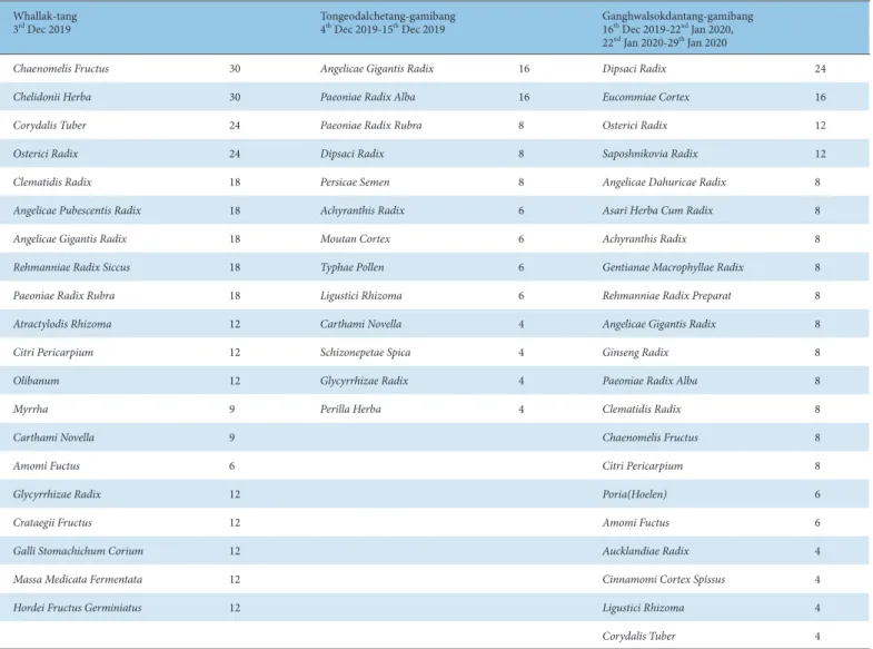
The patient took herbal medication 3 times daily. Whallak- tang, Tongeodalchetang-gamibang (TDT-gamibang) and Ganghwalsokdantang-gamibang (GST-gamibang) were prescribed (Table 2).
Physiotherapy
Transcutaneous electrical nerve stimulation (TENS) was applied once daily for 20 minutes in the region of BL37 to reduce pain and relieve tight muscles.
Moxibustion treatment
Indirect electric moxibustion (Technoscience, Seoul, Korea) therapy was applied once daily at BL25, BL26, and BL36.
Evaluation
Numerical rating scale
The patient’s pain was assessed daily using the NRS. A score of 0 indicated no pain and 10 the most severe pain.
Subjective state of the patient
The patient’s electronic medical records were reviewed to evaluate changes in the symptoms.
Radiological findings
X-ray scans of both hips were taken weekly during hospitalization, and on April 2
nd, 2020 at the outpatient follow-up consultation.
Progress of the patient under treatment Numeric rating scale
On admission, the patient complained of pain and rated her pain score as a NRS 7 in both her hips and inner hamstrings. Her pain was most severe in the supine position, causing sleep disturbances at night. From the 3
rdday of treatment and onwards, the patient’s hip pain started to decline (Fig. 5).
Walking and sitting positions
At admission, the patient was only able to use a wheelchair or walk 1 or 2 steps with assistance. Due to the intensity of pain, she could not remain in a seated position. On Day 3 after admission, she could walk 3 m using a walker, and on Day 9, she could remain seated for more than 10 minutes. On Day 25, she could walk unassisted for 3 m, and for 150 m using a walker. On Day 27, she walked 30 m, unassisted. On Day 30, she could sit for 20 minutes, but she suffered left posterior thigh pain. On Day 41, because her hip pain was greatly reduced, she could remain seated for 30 minutes. On Day 42, she tried to walk unassisted for 300 m, resting after every 100 m. On Day 48, she remained seated for 1 hour. At discharge, she could walk unassisted for more than 200 m. On the
Date Injection site
(method) Bee venom
concentration Volume (mL)
10
thDec 2019 Gluteus (IM) 20,000:1 0.4
11
thDec 2019 10,000:1 0.4
12
thDec 2019 0.6
13
thDec 2019 0.8
14
thDec 2019 1.0
16
thDec 2019 5,000:1 0.6
17
thDec 2019 0.8
18
thDec 2019 1.0
19
thDec 2019 Lumbar (SC) 10,000:1 0.4
20
thDec 2019 5,000:1 0.4
21
stDec 2019 Gluteus (IM),
Lumbar (SC) 5,000:1,
2,000:1 1.0,
0.4
22
ndDec 2019 Gluteus (IM) SBV 10% 2.0
23
rdDec 2019 Gluteus (IM),
Lumbar (SC) SBV 25%,
2000:1 2.0,
0.4
24
thDec 2019 Gluteus (IM) SBV 25% 2.0
26
thDec 2019 2
ndJan 2020, 3
rd, 6t
h, 7
th, 10
thSBV 50% 2.0
17
thJan 2020 SBV 50% 1.0
IM, intramuscular; SBV, sweet bee venom; SC, subcutaneous.
Table 1. Bee Venom Injection Timetable.
Whallak-tang
3
rdDec 2019 Tongeodalchetang-gamibang
4
thDec 2019-15
thDec 2019 Ganghwalsokdantang-gamibang 16
thDec 2019-22
ndJan 2020, 22
ndJan 2020-29
thJan 2020
Chaenomelis Fructus 30 Angelicae Gigantis Radix 16 Dipsaci Radix 24
Chelidonii Herba 30 Paeoniae Radix Alba 16 Eucommiae Cortex 16
Corydalis Tuber 24 Paeoniae Radix Rubra 8 Osterici Radix 12
Osterici Radix 24 Dipsaci Radix 8 Saposhnikovia Radix 12
Clematidis Radix 18 Persicae Semen 8 Angelicae Dahuricae Radix 8
Angelicae Pubescentis Radix 18 Achyranthis Radix 6 Asari Herba Cum Radix 8
Angelicae Gigantis Radix 18 Moutan Cortex 6 Achyranthis Radix 8
Rehmanniae Radix Siccus 18 Typhae Pollen 6 Gentianae Macrophyllae Radix 8
Paeoniae Radix Rubra 18 Ligustici Rhizoma 6 Rehmanniae Radix Preparat 8
Atractylodis Rhizoma 12 Carthami Novella 4 Angelicae Gigantis Radix 8
Citri Pericarpium 12 Schizonepetae Spica 4 Ginseng Radix 8
Olibanum 12 Glycyrrhizae Radix 4 Paeoniae Radix Alba 8
Myrrha 9 Perilla Herba 4 Clematidis Radix 8
Carthami Novella 9 Chaenomelis Fructus 8
Amomi Fuctus 6 Citri Pericarpium 8
Glycyrrhizae Radix 12 Poria(Hoelen) 6
Crataegii Fructus 12 Amomi Fuctus 6
Galli Stomachichum Corium 12 Aucklandiae Radix 4
Massa Medicata Fermentata 12 Cinnamomi Cortex Spissus 4
Hordei Fructus Germiniatus 12 Ligustici Rhizoma 4
Corydalis Tuber 4
Table 2. Herbal Composition of 3 Herbal Medications for Daily Dosage.
Fig. 5. Changes in the NRS Score for Pain. From Day 3, the patient’s hip pain started to decline (NRS 6), and on Day 6 she could sleep without night pain. On Day 14, narcotic analgesics were stopped, the dosage of which had been tapered off since Day 8. Even though the analgesic effect ended, both the hip and leg pain had decreased to a NRS score of 5.5. On Day 24, her subjective pain during sitting was greatly improved. On Day 50, the pain of both hips had nearly disappeared.
Adm, admission date; D/C, discharge date
0 1 2 3 4 5 6 7 8
NRS (Numerical Rating Scale)
NRS