Korean J Gastroenterol Vol. 68 No. 1, 54-56 http://dx.doi.org/10.4166/kjg.2016.68.1.54 pISSN 1598-9992 eISSN 2233-6869
IMAGE OF THE MONTH
Korean J Gastroenterol, Vol. 68 No. 1, July 2016 www.kjg.or.kr
다발성 용종 형태를 보인 진행 위암의 대장 전이
이석종, 정우철
가톨릭대학교 의과대학 성빈센트병원 내과학교실
Colorectal Metastasis of Advanced Gastric Cancer Presenting as Multiple Colorectal Polyps
Seok Jong Lee and Woo Chul Chung
Department of Internal Medicine, St. Vincent’s Hospital, College of Medicine, The Catholic University of Korea, Seoul, Korea
CC This is an open access article distributed under the terms of the Creative Commons Attribution Non-Commercial License (http://creativecommons.org/licenses/
by-nc/4.0) which permits unrestricted non-commercial use, distribution, and reproduction in any medium, provided the original work is properly cited.
Copyright © 2016. Korean Society of Gastroenterology.
교신저자: 정우철, 16247, 수원시 팔달구 중부대로 93-6, 가톨릭대학교 성빈센트병원 소화기내과
Correspondence to: Woo Chul Chung, Department of Internal Medicine, The Catholic University of Korea, St. Vincent’s Hospital, 93-6 Jungbu-daero, Paldal-gu, Suwon 16247, Korea. Tel: +82-31-249-7138, Fax: +82-31-253-8898, E-mail: [email protected]
Financial support: None. Conflict of interest: None.
Fig. 2. Esophagogastroduodenoscopic finding. A 1.8 cm depressed lesion with mucosal convergence is seen in the great curvature side of the mid-body.
Fig. 1. Initial abdominal CT finding. The bilateral masses (right ovary, 22.3×21.4×16.9 cm; left ovary, 6.2×6.2×5.5 cm) and large amount of ascites suggested advanced ovary cancer.
증례: 46세 여자 환자가 내원 2개월 전부터 발생한 복부 팽만감을 주소로 내원하였다. 내원 당시 활력징후는 혈압 100/60 mmHg, 맥박 78회/분, 호흡수 20회/분, 체온 36.8oC 였다. 복부 진찰에서 특정 부위에 국한된 압통이나 반발통은 없었고, 종괴는 촉지되지 않았다. 말초혈액검사에서 백혈구는 4,700/mm3, 혈색소 11.7 g/dL, 혈소판 146,000/mm3였으며
생화학검사에서 총 단백 5.1 g/dL, 알부민 3.2 g/dL, 총 빌리 루빈 0.7 mg/dL, AST 26 IU/L, ALT 14 IU/L, 알칼리인산분 해효소 2,118 IU/L, 아밀라아제 28 U/L, Na 138 mmol/L, K 4.1 mmol/L였다. 흉부 방사선검사 및 심전도에서는 특이한 소견이 없었으며, 복부 컴퓨터단층촬영검사에서 많은 양의 복 수와 양측 난소의 낭종성 종대 소견이 관찰되었다(우측
Lee SJ and Chung WC. Colorectal Metastasis of Advanced Gastric Cancer
55
Vol. 68 No. 1, July 2016
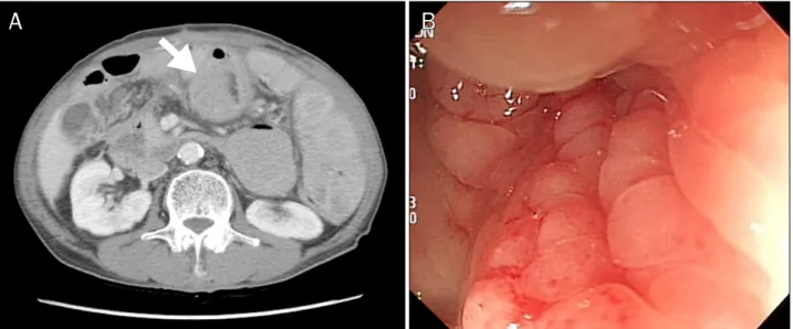
Fig. 5. Another case of colonic metastasis of gastric adenocarcinoma. (A) Segmental wall thickening at the mid transverse colon was observed at abdominal CT (arrow). (B) Metastatic colon cancer from gastric adenocarcinoma shows typical linea platisca pattern at mid transverse colon.
Fig. 4. Microscopic finding of colonic lesion. Poorly differentiated ade- nocarcinoma with signet ring cell component is seen (H&E, ×200).
Fig. 3. Colonoscopic finding. Raised erosion (0.5 cm) is revealed in the mid descending colon.
22.3×21.4×16.9 cm, 좌측 6.2×6.2×5.5 cm; Fig. 1). 또한 흉부와 요추의 전이 소견이 관찰되어 악성 난소 종양의 골 전이를 의심하였다. 내원 3일째에 검사한 CEA는 2.71 ng/mL (정상, 5 ng/mL)였고, CA 125는 655.1 U/mL (정상, 35 U/mL)였다. 전이성 병소를 완전히 배제하기 위해 상부위장관 내시경과 대장 내시경을 실시하였다. 상부위장관 내시경검사 에서 위 체부 대만부에 궤양성 병변과 함께 많은 양의 삼출물 과 주름 집중상, 주름 단절 소견을 관찰하였고(Fig. 2), 대장 내시경에서는 하행 결장과 직장에 다발성 융기형 미란 소견이 있었다(Fig. 3). 조직생검을 시행하였고, 이후 결과에서 분화 도가 나쁜 반지고리 세포암의 악성 종양으로 판정하였다. 특히
대장의 생검 결과에서도 반지고리 세포암 형태로 관찰되었고 (Fig. 4), 최종으로 위암의 복막, 난소, 골, 대장 전이로 진단하 였다. 이후 환자에게 항암화학요법을 권하였으나 거부하고 대 증요법을 실시하다가 2개월 후 사망하였다.
진단: 위암의 대장 전이
위암의 전이는 주로 혈행성 전파(hematogenous spread), 림프계(lymphatic spread), 주위 장기를 통한 직접침윤(direct invasion), 또는 복막이나 복강(transcoelomic spread)을 통 해서 이루어진다.1 장기로는 주로 간, 폐, 뼈로 전이되며 대장 으로의 전이는 비교적 드물다고 알려져 있다.2 위암 이외에도 대장 병변의 원발 병소로는 유방, 피부(melanoma), 신장, 전 립선 혹은 난소의 악성 종양이 있다.3 이런 경우에는 혈행성
56
이석종, 정우철. 진행 위암의 대장 전이The Korean Journal of Gastroenterology
전파가 가장 흔하고, 점막하 림프계에서 전이성 병소가 쌓이 게 된다. 형태학적으로는 주로 linea plastisca나 annular stricture로 나타나는데(Fig. 5),4 단발성 혹은 다발성 용종의 형태를 보이는 위암의 대장 전이는 매우 드물어서 현재까지 약 10개 정도의 증례가 있다.3,5-9 증례 중에서 위암과 대장으 로 전이된 소견이 동시에 발견된 경우가 3차례였으며, 2개의 증례에서는 위전절제술 후 대장 전이를 확인하였고, 나머지는 사후 부검을 통해서 전이를 확인하였다.
이번 증례는 복수를 주소로 내원한 환자에서 상부위장관 내시경과 대장 내시경을 통하여 분화도가 불량한 반지고리 세 포암이 확인되었다. 이 환자는 원발성 위암 및 원발성 대장암 이 각각 나타났을 가능성은 높지 않으나, 원발성 대장암의 위 전이 또는 원발성 위암의 대장 전이를 각각 감별할 필요는 있다. 일반적으로 원발 장소를 확인하고자 CK7, CK20에 대 한 면역화학염색을 시행하는데, CK20이 양성인 경우 대장에 서 기인한 원발성 선암을 시사하고, CK7이 양성일 경우 위에 서 기인한 선암일 가능성이 높다. 이때 면역화학염색법에 의 한 진단의 민감도와 특이도는 각각 72%와 96%로, 상당히 높 은 가능성으로 원발 장소를 구분할 수 있는 것으로 보고하였 다.10 이번 증례는 육안으로 보았을 때 전형적인 원발성 진행 위암의 모양을 보였고, 대장 이외에 난소, 복막, 골 전이가 확 인되어 면역학적 검사까지 진행할 필요가 없었던 경우이다.
하지만, 수술 치료 후에 대장 검사를 시행하게 된다면 검사자 가 전이성 병변에 대해 인식해야 할 필요가 있으며, 이번 증례 를 통해 단발 혹은 다발성 대장의 용종성 병변이 진행 위암의 전이성 병변이 될 수 있다는 것을 고려해야 한다.
REFERENCES
1. Batson OV. The function of the vertebral veins and their role in the spread of metastases. 1940. Clin Orthop Relat Res 1995;
(312):4-9.
2. Rodríguez Salas N, González Paz C, Rivera T, López Alfonso A, Martín Marino A, Lara Alvarez MA. Colonic anastomosis and co- lonic polyp mucosal metastasis of signet ring cell gastric adenocarcinoma. Clin Transl Oncol 2010;12:238-239.
3. Ogiwara H, Konno H, Kitayama Y, Kino I, Baba S. Metastases from gastric adenocarcinoma presenting as multiple colonic polyps:
report of a case. Surg Today 1994;24:473-475.
4. Katon RM, Brendler SJ, Ireland K. Gastric linitis plastica with metastases to the colon: a mimic of Crohn's disease. J Clin Gastroenterol 1989;11:555-560.
5. Gao B, Xue X, Tai W, et al. Polypoid colonic metastases from gas- tric stump carcinoma: a case report. Oncol Lett 2014;8:1119- 1122.
6. Metayer P, Antonietti M, Oumrani M, Hemet J, Lemoine F, Basuyau J. Metastases of a gastric adenocarcinoma presenting as colonic polyposis. Report of a case. Dis Colon Rectum 1991;
34:622-623.
7. Lee IH, Lee JE, Byeon SW, et al. A case of advanced gastric cancer presenting as multiple colonic lymphoid hyperplasia. Korean J Gastroenterol 2015;66:221-226.
8. Tiszlavicz L. Stomach cancer metastasizing into a solitary ad- enomatous colonic polyp. Orv Hetil 1990;131:1259-1261.
9. Tiszlavicz L. Metastasis of a stomach carcinoma in a solitary ad- enomatous cecal polyp. Zentralbl Allg Pathol 1990;136:277- 282.
10. Park SY, Kim HS, Hong EK, Kim WH. Expression of cytokeratins 7 and 20 in primary carcinomas of the stomach and colorectum and their value in the differential diagnosis of metastatic carci- nomas to the ovary. Hum Pathol 2002;33:1078-1085.