ISSN 2234-3806 • eISSN 2234-3814
466 www.annlabmed.org https://doi.org/10.3343/alm.2020.40.6.466 https://doi.org/10.3343/alm.2020.40.6.466
Proenkephalin Predicts Organ Failure, Renal
Replacement Therapy, and Mortality in Patients With Sepsis
Hanah Kim , M.D., Ph.D.1, Mina Hur , M.D., Ph.D.1, Joachim Struck , Ph.D.2, Andreas Bergmann , Ph.D.2, and Salvatore Di Somma , M.D., Ph.D.3; on behalf of GREAT Network
1Department of Laboratory Medicine, Konkuk University School of Medicine, Seoul, Korea; 2Sphingotec GmbH, Hennigsdorf, Germany; 3Department of Medical-Surgery Sciences and Translational Medicine, School of Medicine and Psychology, Sapienza–University, Sant’ Andrea Hospital, Rome, Italy Background: Kidney failure occurs frequently and is associated with high mortality during
sepsis. Proenkephalin (PENK) is an emerging biomarker of kidney function. We explored whether PENK levels could predict severity, organ failure, and mortality in septic patients.
Methods: We measured the PENK level in the plasma of 215 septic patients using the sphingotest penKid assay (Sphingotec GmbH, Hennigsdorf, Germany). This was analyzed in terms of sepsis severity, vasopressor use, 30-day mortality, sequential organ failure as- sessment (SOFA) renal subscore, the Chronic Kidney Disease Epidemiology Collaboration estimated glomerular filtration rate (CKD-EPI eGFR) categories, and renal replacement therapy (RRT) requirement.
Results: The PENK levels were significantly higher in patients with septic shock, vasopres- sor use, and non-survivors than in patients with solitary sepsis, no vasopressor use, and survivors, respectively (P =0.02, P =0.007, P <0.001, respectively). The PENK levels were significantly associated with SOFA renal subscore and CKD-EPI eGFR categories (both P <0.001). The distribution of lower eGFR ( <60 mL/min/1.73 m2), RRT requirement, SOFA renal subscore, and the number of organ failures differed significantly according to the PENK quartile (P for trend<0.001 or 0.017). The 30-day mortality rate also differed significantly according to the PENK quartile (P for trend<0.001).
Conclusions: PENK could be an objective and reliable marker to predict severity, organ failure, and 30-day mortality in septic patients.
Key Words: Proenkephalin, Sepsis, Organ failure, SOFA score, Kidney function, Mortality
Received: December 25, 2019 Revision received: April 9, 2020 Accepted: May 11, 2020 Corresponding author:
Mina Hur, M.D., Ph.D.
Department of Laboratory Medicine, Konkuk University School of Medicine, Konkuk University Medical Center, 120-1 Neungdong-ro, Gwangjin-gu,
Seoul 05030, Korea Tel: +82-2-2030-5581 E-mail: [email protected]
© Korean Society for Laboratory Medicine This is an Open Access article distributed under the terms of the Creative Commons Attribution Non-Commercial License (https://creativecom- mons.org/licenses/by-nc/4.0) which permits unrestricted non-commercial use, distribution, and reproduction in any medium, provided the original work is properly cited.
INTRODUCTION
Organ failure is a hallmark of sepsis [1]. Kidney failure frequently occurs in sepsis, and sepsis is the most common etiology of acute kidney injury (AKI) [2]. AKI is not a single disease but a loose collection of syndromes as diverse as sepsis, cardiorenal syndrome, and urinary tract obstruction [3]. Sepsis encompass a spectrum of disorders involving both the heart and kidneys, in
which acute or chronic dysfunction in one organ can induce acute or chronic dysfunction in another [4]. Even though the entire phenomenon of sepsis-associated AKI is not fully under- stood, its long-term adverse outcomes are related to multiple or- gan failures [5, 6]. Because both sepsis and AKI are indepen- dently associated with adverse outcomes, the early detection of AKI is critical to provide opportunities for successful intervention and resuscitation in septic patients.
The early detection of kidney failure by monitoring kidney func- tion using injury and/or stress biomarkers would be crucial to decrease hospital mortality and the duration of hospital stays [7].
However, the current clinical diagnosis of kidney failure relies on markers such as glomerular filtration rate (GFR) based on in- creased serum creatinine (sCr) levels and decreased urine out- put [8, 9]. In sepsis, the sequential (sepsis-related) organ failure assessment (SOFA) scoring system has been used to assess or- gan functions, and the sCr level is used to evaluate kidney func- tion [10]. Changes in sCr levels or urine output are neither sen- sitive nor specific for AKI; the sCr level is also affected by nu- merous factors, and its solitary use might not reflect the actual degree of kidney function [3]. Moreover, the inverse relationship between GFR and sCr levels is nonlinear in patients with near- normal kidney function [8]. Therefore, early and reliable bio- markers are required for critically ill septic patients at risk of de- veloping AKI or likely to need clinical treatments, such as renal replacement therapy (RRT) [11].
Proenkephalin A 119-159 (PENK) is a stable surrogate marker for endogenous enkephalins, and it has been suggested to be a functional kidney marker closely related to the iohexol-determined GFR, which is considered the gold standard [12-14]. PENK has been investigated as a novel biomarker for AKI in various clini- cal settings, including sepsis and heart failure [14-22]. Further, few recent studies showed that high PENK levels are associated with the deterioration of kidney function and adverse clinical outcomes for critically ill patients. However, no study has ex- plored whether PENK levels can substitute for or augment the current role of the SOFA scoring system in septic patients. We explored the clinical utility of PENK for sepsis patients in terms of sepsis severity, organ failure, and mortality. We hypothesized that PENK levels would be related to the renal subscore of the SOFA score, estimated GFR (eGFR), RRT requirement, and short-term mortality in sepsis.
METHODS
Study population and design
A total of 215 patients were enrolled at Konkuk University Medi- cal Center (KUMC), Seoul, Korea, from August 2016 to August 2017. They were diagnosed as having sepsis (N=109, 50.7%) or septic shock (N=106, 49.3%) according to the Sepsis-3 cri- teria [1]. For all patients, standard-of-care biomarkers, including white blood cells, C-reactive protein, lactate, procalcitonin, and sCr levels, were measured on the day of diagnosis; on the same day, the SOFA score was assessed in six different organ systems
with an assigned value from 0 (normal) to 4 (high degree of fail- ure) [10]. All patients received proper standard-of-care treatments.
Their medical records were reviewed retrospectively to retrieve demographic, clinical, and laboratory data; the baseline charac- teristics of the study population are summarized in Table 1.
This study protocol was reviewed and exempt from approval by the Institution Review Board of KUMC (KUH1200085). This registry study required neither study-intended blood sampling nor additional interventions; therefore, the requirement to obtain written informed consent from the patients was waived. Resid- ual EDTA plasma samples that were available on the same day of sepsis diagnosis were collected, split into small aliquots, and then stored at -70°C until use.
Assay
Samples that had been collected from August 2016 to August 2017 were analyzed in September 2017. PENK is known to be stable for at least 2.5 years at -70°C (unpublished data) and for up to three freeze–thaw cycles [23]. The long-term stability of PENK has been tested in a set of patients EDTA plasma sam- ples covering the measurement interval; frozen samples were thawed at room temperature and gently mixed immediately be- fore measuring PENK levels. PENK levels were measured using the sphingotest penKid assay (Sphingotec GmbH, Hennigsdorf, Germany). This is a chemiluminescence sandwich immunoas- say using two monoclonal antibodies directed against the mid- dle portion of PENK (anti-PENK 129-144 mAb as a tracer anti- body) and the C-terminus of PENK (anti-PENK 152-159 mAb as a capture antibody) [23]. The assay was calibrated using di- lutions of synthetic PENK 119-159. Samples/calibrators (50 μL) were pipetted into white polystyrene coated 96-well microtiter plates (Greiner Bio-One International AG, Austria). After adding labeled anti-PENK 129-144 mAb (150 μL), the microtiter plates were incubated for 18 hours at 22°C without agitation. Unbound tracer was removed using washing solution (350 μL per well, five times), and remaining chemiluminescence was measured for 1 second per well using the Centro LB 960 microtiter plate lu- minescence reader (Berthold Technologies GmbH & Co. KG, Germany). The PENK level was determined using a five-point calibration curve (27.4–2,223 pmol/L). Calibrators and samples were run in duplicate with a required <20% coefficient of varia- tion (CV) between the duplicates. The mean value of duplicates of each sample was used for statistical analysis. The analytical sensitivity (limit of detection) was 17.3 pmol/L (CV=2.7%), and the measurable range was 17.3–2,223 pmol/L. The manufac- turer’s recommended reference range at the 99th percentile is
468 www.annlabmed.org https://doi.org/10.3343/alm.2020.40.6.466 Table 1. Characteristics of the study population
Variable All patients (N=215) Sepsis (N=109) Septic shock (N=106) P
Patient enrollment
ICU* 92 (42.8) 29 (26.6) 63 (59.4) <0.001
Emergency room 123 (57.2) 80 (73.4) 43 (40.6) <0.001
Age (yr) 71 (58–79) 70 (58–79) 72 (59–79) >0.9
Males 127 (59.1) 65 (59.6) 62 (58.5) 0.9
Clinical outcomes
Hospital stay (day) 15 (6–31) 15 (7–28) 16 (5–43) 0.7
Vasopressor use† 123 (57.2) 17 (15.6) 106 (100.0) <0.001
Renal replacement therapy 22 (10.2) 7 (6.4) 15 (14.2) 0.07
30-day all-cause mortality (day) 66 (30.7) 18 (16.5) 48 (45.3) <0.001
Comorbidities
Cardiovascular 116 (54.0) 68 (62.4) 48 (45.3) 0.07
Cerebrovascular 97 (45.1) 48 (44.0) 49 (46.2) 0.8
Renal and genitourinary 60 (27.9) 35 (32.1) 25 (23.6) 0.5
GI & hepatobiliary 20 (9.3) 8 (7.3) 12 (11.3) 0.8
Respiratory 20 (9.3) 11 (10.1) 9 (8.5) >0.9
Hemato-oncological 8 (3.7) 5 (4.6) 3 (2.8) >0.9
Others 7 (3.3) 4 (3.7) 3 (2.8) >0.9
Type of infections
Bacteremia 214 (99.5) 108 (99.1) 106 (100.0) 0.3
Respiratory 98 (45.6) 39 (35.8) 59 (55.7) 0.06
Urinary 63 (29.3) 41 (37.6) 22 (20.8) 0.2
GI & hepatobiliary 56 (26.0) 21 (19.3) 35 (33.0) 0.3
Soft tissue 10 (4.7) 6 (5.5) 4 (3.8) >0.9
Others‡ 7 (3.3) 4 (3.7) 3 (2.8) >0.9
SOFA score 7 (4 –10) 5 (3–8) 13 (10–15) <0.001
Cardiovascular 3 (0–4) 0 (0–1) 4 (4–4) <0.001
Central nervous system 0 (0–2) 0 (0–1) 2 (0–3) <0.001
Coagulation 1 (0–2) 1 (0–2) 2 (0–2) 0.02
Liver 0 (0–1) 0 (0–1) 1 (0–2) 0.03
Renal 1 (0–2) 1 (0–2) 1 (1–2) 0.05
Respiratory 3 (1–4) 1 (0–3) 4 (2–4) <0.001
Laboratory parameters
WBC (×109/L) 12.8 (6.8–16.9) 11.7 (6.7–15.1) 14.6 (7.2–20.1) 0.02
CRP (mg/L) 162 (102–254) 157 (92–225) 184 (115–270) 0.08
Lactate (mmol/L) 3.56 (2.00–6.04) 2.03 (1.38–3.34) 4.89 (3.71–9.55) <0.001
Procalcitonin (µg/L) 17.7 (6.5–44.4) 13.7 (5.1–24.4) 26.7 (8.7–68.1) <0.001
Creatinine (µmol/L) 139.7 (84.0–249.3) 114.1 (75.2–245.8) 163.6 (102.6–253.8) 0.04
eGFR (mL/min/kg/1.73 m2) 42.3 (22.0–82.8) 54.4 (23.3–90.5) 37.0 (21.5–69.0) 0.06
Proenkephalin (pmol/L) 103.0 (52.5–207.5) 75.7 (44.4–183.9) 118.7 (71.7–245.3) 0.02
Data are expressed as number (percentage) or median (interquartile range).
P values were derived using the Mann–Whitney test or Chi-squared test to compare sepsis and septic shock patients.
*The 92 ICU patients were enrolled from medical (N=60, 65.2%), surgical (N=24, 26.1%), and neurological (N=8, 8.7%) ICUs; †Vasopressors were used alone (N=95) or in combination (N=28), using norepinephrine (N=115), dopamine (N=30), dobutamine (N=7, 3.3%), and epinephrine (N=6, 2.8%);
‡Others included catheters (N=4), central nervous system (N=2), and foreign body (N=1).
Abbreviations: ICU, intensive care unit; GI, gastrointestinal; SOFA, sequential organ failure assessment; WBC, white blood cells; CRP, C-reactive protein; eGFR, estimated glomerular filtration rate.
https://doi.org/10.3343/alm.2020.40.6.466 www.annlabmed.org 469 24.6–80 pmol/L, and 80 pmol/L is suggested as a clinical cut-
off [24].
Statistical analysis
All continuous variables showed non-parametric distribution;
accordingly, the data were expressed as median (interquartile range) or number (percentage). Mann–Whitney test or chi-squared test was used to compare clinical and laboratory variables be- tween the two groups, namely sepsis and septic shock. Krus- kal–Wallis test with a post-hoc test was used to compare PENK levels among the groups according to the SOFA renal score (from 0 to 4), the chronic kidney disease epidemiology collaboration (CKD-EPI) eGFR categories (from G1 to G5), and the RRT re- quirement (no RRT, RRT on day 1, RRT later during hospitaliza- tion) [25].
The PENK levels were divided into quartiles, and a chi-squared test or Fisher’s exact test was used to compare the eGFR (eGFR
<60 mL/min/kg/1.73 m2), RRT requirement, and the number of organ failures based on the SOFA scoring system (≥2) in each PENK quartile and SOFA renal subscore group; the Cochran- Armitage test for trends was used for trend analysis. Kaplan–Meier survival curves and hazard ratios (HRs) with the 95% confidence
interval (CI) were used to compare 30-day mortality in each PENK quartile and for each SOFA renal subscore group; the log-rank test for trends was used for trend analysis. The HR was used to estimate the relative risk of the survival event (survival vs non- survival) in each group and was considered significant when the CI did not include the value 1. A chi-squared test or Fisher’s ex- act test was also used to compare survival events. These analy- ses were conducted for all patients, as well as in each group (sep- sis and septic shock). MedCalc Statistical Software version 19.1.7 (MedCalc Software Bvba, Ostend, Belgium) and Analyse-it for Microsoft Excel 5.30.2 (Analyse-It Software, Ltd., Leeds, United Kingdom) were used for statistical analyses. Rounding rules were applied to summary statistics, and two-sided P <0.05 were con- sidered statistically significant [26].
RESULTS
Distribution of PENK levels
The PENK levels were significantly higher in the septic shock group than in the sepsis group (118.7 pmol/L vs 75.7 pmol/L, P =0.02; Table 1). The PENK levels were also significantly higher in patients with vasopressor use than in those without vasopres-
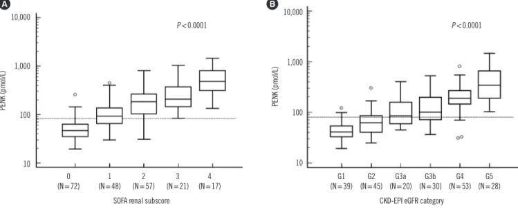
Fig. 1. Comparison of PENK levels according to the sequential organ failure assessment (SOFA) renal subscore and the CKD-EPI estimated GFR (eGFR) categories. (A) PENK levels (median and IQR) increased significantly according to the increased SOFA renal subscores (from 0 to 4) as follows: 46.9 pmol/L (351–62.9) in 0; 92.4 pmol/L (64.8–136.1) in 1; 182.9 pmol/L (103.7–264.0) in 2; 208.3 pmol/L (145.5–
370.2) in 3; 482.3 pmol/L (312.2–819.4) in 4. (B) PENK levels (median and IQR) increased significantly according to the increased CKD- EPI eGFR categories as follows: 40.7 pmol/L (32.7–54.0) in G1; 61.9 pmol/L (40.4–85.8) in G2; 84.8 pmol/L (58.4–158.0) in G3a; 100.8 pmol/L (71.7–195.6) in G3b; 188.2 pmol/L (144.1–264.0) in G4; 341.0 pmol/L (188.4–650.0) in G5. In each figure, the Y-axis is presented as a logarithmic scale.
Abbreviations: PENK, proenkephalin; SOFA, sequential organ failure assessment; CKD-EPI eGFR, the Chronic Kidney Disease Epidemiology Collaboration estimated glomerular filtration rate; IQR, interquartile range.
352
Fig. 1. Comparison of PENK levels according to the sequential organ failure assessment 353
(SOFA) renal subscore and the CKD-EPI estimated GFR (eGFR) categories. (A) PENK levels 354
(median and IQR) increased significantly according to the increased SOFA renal subscores 355
(from 0 to 4) as follows: 46.9 pmol/L (351 – 62.9) in 0; 92.4 pmol/L (64.8 – 136.1) in 1; 182.9 356
19
352
Fig. 1. Comparison of PENK levels according to the sequential organ failure assessment 353
(SOFA) renal subscore and the CKD-EPI estimated GFR (eGFR) categories. (A) PENK levels 354
(median and IQR) increased significantly according to the increased SOFA renal subscores 355
(from 0 to 4) as follows: 46.9 pmol/L (351 – 62.9) in 0; 92.4 pmol/L (64.8 – 136.1) in 1; 182.9 356
10,000
1,000
100
10
0
(N=72) 1
(N=48) 2 (N=57) 3
(N=21) 4 (N=17) SOFA renal subscore
PENK (pmol/L)
P <0.0001
A 10,000
1,000
100
10
G1 (N=39)
CKD-EPI eGFR category
PENK (pmol/L)
P <0.0001 B
G2 (N=45) G3a
(N=20) G3b (N=30) G4
(N=53) G5 (N=28)
470 www.annlabmed.org https://doi.org/10.3343/alm.2020.40.6.466 sor use (116.9 pmol/L vs 72.7 pmol/L, P =0.007) and in the non-
survivors than in the survivors (171.5 pmol/L vs 79.8 pmol/L, P <0.001; data not shown). PENK levels gradually increased according to the increased SOFA renal subscores and CKD-EPI eGFR categories (Fig. 1A and 1B, both P <0.001); this signifi- cance was constantly observed across each group (P <0.05, post-hoc test), except for eGFR categories G3a and G3b. Re- garding RRT requirements, the PENK levels were significantly higher in patients who required RRT on day 1 (N =3) than in those who did not require RRT (N=193) or required RRT later during hospitalization (N=19) (1,026.8 pmol/L vs. 284.3 pmol/L vs. 88.4 pmol/L, P <0.001, P for trend <0.001); a similar trend was noted for sCr, CKD-EPI eGFR, and SOFA renal scores (all P <0.001; data not shown).
PENK quartiles and SOFA renal subscores for predicting clinical outcomes
The PENK levels were divided into quartiles as follows: Q1<52.5 pmol/L (N =54); 52.5 pmol/L ≤Q2 <103.0 pmol/L (N =53);
103.0 pmol/L≤Q3<207.5 pmol/L (N=54); Q4 ≥ 207.5 pmol/L (N=54). Tables 2 and 3 show the PENK quartiles and SOFA re- nal subscores, respectively, to predict clinical outcomes.
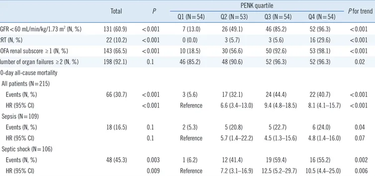
The distribution of lower eGFR (<60 mL/min/1.73 m2), RRT requirement, SOFA renal subscore, and the number of organ
failures differed significantly according to the PENK quartile. The 30-day mortality rate also differed significantly according to the PENK quartile (P for trend <0.001). Compared with the PENK Q1, higher PENK groups (Q2–Q4) were associated with poor survival outcomes [HR (95% CI) ranging from 4.5 (1.3–15.6) to 12.5 (5.2–29.7)]; this significant finding was consistently ob- served in each group, namely sepsis and septic shock, as well as in total patients (Table 2). When the clinical cut-off for PENK (80 pmol/L) was applied, the patients with increased PENK lev- els showed poor survival outcomes than the patients with nor- mal PENK levels [HR (95% CI)=3.0 (1.8–4.9), P <0.001; data not shown].
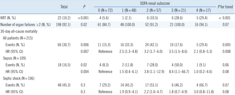
Regarding SOFA renal subscores, although RRT requirements were significantly different according to each subscore, the num- ber of organ failures did not reach statistical significance. Although no patient received RRT during hospitalization in the PENK Q1 group, four of the 72 patients (5.6%) with a SOFA renal subscore of 0 received RRT during hospitalization. The 30-day mortality rate differed significantly according to the SOFA renal subscore (P for trend=0.005, for event). Compared with the SOFA renal subscore 0 group, the other subscore groups (1–4) were associ- ated with poor survival outcomes based on total patients [HR (95% CI) ranging from 2.1 (0.8–5.3) to 3.5 (1.5–8.6)]. When the patients were divided into sepsis and septic shock groups,
Table 2. PENK quartiles for eGFR, RRT, SOFA renal subscore, number of organ failures, and 30-day all-cause mortality
Total P PENK quartile
P for trend Q1 (N=54) Q2 (N=53) Q3 (N=54) Q4 (N=54)
eGFR<60 mL/min/kg/1.73 m2 (N, %) 131 (60.9) <0.001 7 (13.0) 26 (49.1) 46 (85.2) 52 (96.3) <0.001
RRT (N, %) 22 (10.2) <0.001 0 (0.0) 3 (5.7) 3 (5.6) 16 (29.6) <0.001
SOFA renal subscore ≥1 (N, %) 143 (66.5) <0.001 10 (18.5) 30 (56.6) 50 (92.6) 53 (98.1) <0.001
Number of organ failures ≥2 (N, %) 198 (92.1) 0.1 46 (85.2) 48 (90.6) 52 (96.3) 52 (96.3) 0.02
30-day all-cause mortality All patients (N=215)
Events (N, %) 66 (30.7) <0.001 3 (5.6) 17 (32.1) 24 (44.4) 22 (40.7) <0.001
HR (95% CI) <0.001 Reference 6.6 (3.4–13.0) 9.4 (4.8–18.5) 8.1 (4.1–15.7) <0.001
Sepsis (N=109)
Events (N, %) 18 (16.5) 0.1 2 (5.3) 5 (20.8) 5 (22.7) 6 (24.0) 0.04
HR (95% CI) 0.1 Reference 5.7 (1.4–22.2) 4.5 (1.3–15.6) 4.8 (1.4–16.0) 0.07
Septic shock (N=106)
Events (N, %) 48 (45.3) 0.003 1 (6.2) 12 (41.4) 19 (59.4) 16 (55.2) 0.002
HR (95% CI) 0.009 Reference 7.2 (3.1–16.9) 12.5 (5.2–29.7) 10.5 (4.4–25.0) 0.006
P was derived using a Chi-squared test, Fisher’s exact test, or Kaplan–Meier survival analysis. The P for trend was derived using a Cochran–Armitage test for trends or log-rank test for trends.
Abbreviations: eGFR, estimated glomerular filtration rate; HR, hazard ratio; N, number; PENK, proenkephalin; Q, quartile; RRT, renal replacement therapy;
SOFA, sequential organ failure assessment.
Table 3. SOFA renal subscores for RRT, number of organ failures, and 30-day all-cause mortality
Total P SOFA renal subscore
P for trend 0 (N=72) 1 (N=48) 2 (N=57) 3 (N=21) 4 (N=17)
RRT (N, %) 22 (10.2) <0.001 4 (5.6) 1 (2.1) 6 (10.5) 6 (28.6) 5 (29.4) < 0.001
Number of organ failures ≥2 (N, %) 198 (92.1) 0.02 61 (84.7) 48 (100.0) 52 (91.2) 21 (100.0) 16 (94.1) 0.07 30-day all-cause mortality
All patients (N=215)
Events (N, %) 66 (30.7) 0.006 11 (15.3) 16 (33.3) 24 (42.1) 10 (17.6) 5 (29.4) 0.005
HR (95% CI) 0.007 Reference 2.5 (1.3–4.8) 3.2 (1.7–6.0) 3.5 (1.5–8.6) 2.1 (0.8–5.3) 0.008
Sepsis (N=109)
Events (N, %) 18 (16.5) 0.02 4 (8.3) 2 (11.8) 7 (28.0) 4 (50.0) 1 (9.1) 0.06
HR (95% CI) 0.004 Reference 1.5 (0.4–6.1) 3.8 (1.1–12.9) 8.4 (1.1–66.7) 1.0 (0.2–4.6) 0.08
Septic shock (N=106)
Events (N, %) 48 (45.3) 0.3 7 (29.2) 14 (45.2) 17 (53.1) 6 (46.2) 4 (66.7) 0.07
HR (95% CI) 0.3 Reference 1.9 (0.9–4.1) 2.2 (1.0–4.7) 1.8 (0.7–4.9) 3.0 (0.8–11.8) 0.08
P was derived using a chi-squared test, Fisher’s exact test, or Kaplan–Meier survival analysis. The P for trend was derived using a Cochran–Armitage test for trends or log-rank test for trends.
Abbreviations: HR, hazard ratio; N, number; PENK, proenkephalin; RRT, renal replacement therapy; SOFA, sequential organ failure assessment.
however, this significance was observed only in septic patients with SOFA renal subscores of 2 and 3 [HR (95% CI), 3.8 (1.1–
12.9) and 8.4 (1.1–66.7)] (Table 3).
DISCUSSION
We demonstrated the clinical utility of PENK to assess organ failure and predict mortality in septic patients. The PENK level was significantly associated with septic shock, vasopressor use, and 30-day mortality; these findings are in line with those of some recent studies [16, 19, 27]. In our data, PENK levels showed significant associations with SOFA renal subscores and CKD-EPI eGFR categories (Fig. 1). It was noteworthy that the PENK level showed more prognostic efficacy than the SOFA renal subscore for predicting the RRT requirement; RRT was required for some patients even with a SOFA renal subscore of 0, although RRT was not required for patients in the PENK Q1 group. A recent study also showed that PENK is an effective predictor of AKI de- velopment particularly in septic patients presenting to the emer- gency department with normal sCr levels [28]. PENK quartiles were also associated with an increasing trend in the number of organ failures (Table 2); this implies that the PENK level might reflect sepsis-induced organ failure simply and objectively and has the clinical potential to substitute for or augment SOFA scores.
Another noticeable finding in our study was the significant as- sociation between the PENK quartile and 30-day mortality rate.
In all patients, both the PENK quartile and SOFA renal subscore
showed significant associations with the 30-day mortality rate (Tables 2 and 3). However, when patients were divided into sep- sis and septic shock groups, only the PENK quartile predicted and stratified the 30-day mortality rate consistently, whereas the SOFA renal subscore showed limitations. Compared with PENK Q1, high levels of PENK (Q2–Q4) were related to a higher mor- tality risk. Additionally, high levels of PENK (Q2–Q4) during sep- sis identified patients with a high mortality risk (despite the fact that they did not present with shock), whereas very low levels of PENK (Q1) during septic shock identified patients with a low mor- tality risk. This finding implies that PENK levels could be useful to stratify patients further even in the same stage of sepsis or septic shock.
The present data also support the use of a clinical cut-off for PENK (80 pmol/L) [15-17]. In the groups with poor clinical sta- tus or outcomes, including septic shock, vasopressor use, and non-survivors, the median value of PENK levels was higher than the clinical cut-off. This clinical cut-off was useful to identify pa- tients with increased mortality risk, and likewise for risk stratifi- cation by PENK quartile (Q1 vs Q2–Q4). Although its prognostic validity should be supported by further studies, applying a single clinical cut-off value for PENK or an array of such values, includ- ing PENK quartiles, would be one of important and interesting topic.
This study has several limitations. This was a small, single- center registry study; accordingly, the severity and mortality of sepsis in this study might be different from those of other popu-
472 www.annlabmed.org https://doi.org/10.3343/alm.2020.40.6.466 lation cohorts. Second, we could not keep a strict sampling time
and could not define the exact time delay from sepsis diagnosis to sample collection, although it was performed within 24 hours.
The in vivo half-life and clearance of PENK are still unknown, and a recent study showed that 24-hour changes in PENK lev- els are associated with subsequent AKI at 48 hours and seven days [17]. In contrast, sCr cannot reflect real-time GFR. Specifi- cally, sCr peaks on day 4 with a rise of approximately 115% af- ter the AKI episode, showing a considerable time lag between the changes in GFR and the subsequent increase in sCr and recov- ery [29]. As a biomarker of kidney function in the acute stage, PENK could show significant changes during the first 24 hours after sepsis diagnosis; therefore, observations of delta changes in PENK levels would increase its clinical significance.
In conclusion, we demonstrated that the PENK level is associ- ated with sepsis severity, organ failure, RRT requirement, and short-term mortality in patients with sepsis and septic shock.
The PENK quartile seems to be superior to the SOFA renal sub- score for predicting and stratifying 30-day mortality in each sep- sis and septic shock patient group. PENK could thus be an ob- jective and reliable marker that has the potential to substitute for or augment the current role of the SOFA scoring system in criti- cally ill septic patients.
ACKNOWLEDGEMENTS
None.
AUTHOR CONTRIBUTIONS
HK designed the study, analyzed the data, and wrote the draft;
MH conceived the study, analyzed the data, and finalized the draft; JS and AB discussed the data; SDS discussed the data and reviewed the manuscript. All authors read and approved the final manuscript.
CONFLICTS OF INTEREST
AB and JS are employed by Sphingotec GmbH, the company that provided the PENK assays for this study. The other authors declare no conflicts of interest. The authors alone are responsi- ble for the content and writing of the paper.
RESEARCH FUNDING
Not applicable.
ORCID
Hanah Kim https://orcid.org/0000-0002-3266-638X Mina Hur https://orcid.org/0000-0002-4429-9978 Joachim Struck https://orcid.org/0000-0002-9793-7160 Andreas Bergmann https://orcid.org/0000-0002-4466-1682 Salvatore Di Somma https://orcid.org/0000-0002-1717-6585
REFERENCES
1. Singer M, Deutschman CS, Seymour CW, Shankar-Hari M, Annane D, Bauer M, et al. The third international consensus definitions for sepsis and septic shock (Sepsis-3). JAMA 2016;315:801-10.
2. Bellomo R, Kellum JA, Ronco C, Wald R, Martensson J, Maiden M, et al. Acute kidney injury in sepsis. Intensive Care Med 2017;43:816-28.
3. Ronco C, Bellomo R, Kellum JA. Acute kidney injury. Lancet 2019;394:
1949-64.
4. Rangaswami J, Bhalla V, Blair JEA, Chang TI, Costa S, Lentine KL, et al.
Cardiorenal syndrome: classification, pathophysiology, diagnosis, and treatment strategies: a scientific statement from the American Heart As- sociation. Circulation 2019;139:e840-78.
5. Lee SA, Cozzi M, Bush EL, Rabb H. Distant organ dysfunction in acute kidney injury: a review. Am J Kidney Dis 2018;72:846-56.
6. Poston JT and Koyner JL. Sepsis associated acute kidney injury. BMJ 2019;364:k4891.
7. Kellum JA, Bellomo R, Ronco C. Progress in prevention and treatment of acute kidney injury: moving beyond kidney attack. JAMA 2018;320:
437-8.
8. KDIGO clinical practice guideline for acute kidney injury. Kidney Int Sup- pl 2012;2:1-141.
9. Palevsky PM, Liu KD, Brophy PD, Chawla LS, Parikh CR, Thakar CV, et al. KDOQI US commentary on the 2012 KDIGO clinical practice guide- line for acute kidney injury. Am J Kidney Dis 2013;61:649-72.
10. Vincent JL, Moreno R, Takala J, Willatts S, De Mendonça A, Bruining H, et al. The SOFA (Sepsis-related Organ Failure Assessment) score to de- scribe organ dysfunction/failure. On behalf of the working group on sep- sis-related problems of the European Society of Intensive Care Medicine.
Intensive Care Med 1996;22:707-10.
11. Joannidis M and Forni LG. Clinical review: timing of renal replacement therapy. Crit Care 2011;15:223.
12. Ernst A, Köhrle J, Bergmann A. Proenkephalin A 119-159, a stable pro- enkephalin A precursor fragment identified in human circulation. Pep- tides 2006;27:1835-40.
13. Beunders R, Struck J, Wu AHB, Zarbock A, Di Somma S, Mehta RL, et al. Proenkephalin (PENK) as a novel biomarker for kidney function. J Appl Lab Med 2017;2:400-12.
14. Beunders R, van Groenendael R, Leijte G, Kox M, Pickkers P. Proen- kephalin compared to conventional methods to assess kidney function in critically ill sepsis patients. Shock 2020 Jan 21. doi: 10.1097/SHK.
0000000000001510.
15. Marino R, Struck J, Hartmann O, Maisel AS, Rehfeldt M, Magrini L, et al. Diagnostic and short-term prognostic utility of plasma pro-enkepha- lin (pro-ENK) for acute kidney injury in patients admitted with sepsis in the emergency department. J Nephrol 2015;28:717-24.
16. Kim H, Hur M, Lee S, Marino R, Magrini L, Cardelli P, et al. Proenkeph- alin, neutrophil gelatinase-associated lipocalin, and estimated glomeru- lar filtration rates in patients with sepsis. Ann Lab Med 2017;37:388-
97.
17. Caironi P, Latini R, Struck J, Hartmann O, Bergmann A, Bellato V, et al.
Circulating proenkephalin, acute kidney injury, and its improvement in patients with severe sepsis or shock. Clin Chem 2018;64:1361-9.
18. Hollinger A, Wittebole X, François B, Pickkers P, Antonelli M, Gayat E, et al. Proenkephalin A 119-159 (Penkid) is an early biomarker of septic acute kidney injury: the Kidney in Sepsis and Septic Shock (Kid-SSS) Study. Kidney Int Rep 2018;3:1424-33.
19. Moledina DG. Penkid: a novel biomarker of reduced GFR in sepsis. Kid- ney Int Rep 2018;4:17-9.
20. Shah KS, Taub P, Patel M, Rehfeldt M, Struck J, Clopton P, et al. Proen- kephalin predicts acute kidney injury in cardiac surgery patients. Clin Nephrol 2015;83:29-35.
21. Emmens JE, Ter Maaten JM, Damman K, van Veldhuisen DJ, de Boer RA, Struck J, et al. Proenkephalin, an opioid system surrogate, as a nov- el comprehensive renal marker in heart failure. Circ Heart Fail 2019;12:
e005544.
22. Kanagala P, Squire IB, Jones DJL, Cao TH, Chan DCS, McCann G, et al. Proenkephalin and prognosis in heart failure with preserved ejection fraction: a GREAT network study. Clin Res Cardiol 2019;108:940-9.
23. Donato LJ, Meeusen JW, Lieske JC, Bergmann D, Sparwaßer A, Jaffe
AS. Analytical performance of an immunoassay to measure proenkeph- alin. Clin Biochem 2018;58:72-7.
24. Wu AHB and Anand I. The biological variation of plasma proenkepha- lin: data from a stable heart failure cohort. Clin Chem Lab Med 2019;
57:e105-7.
25. Levey AS, Stevens LA, Schmid CH, Zhang YL, Castro AF 3rd, Feldman HI, et al. A new equation to estimate glomerular filtration rate. Ann In- tern Med 2009;150:604-12.
26. Cole TJ. Too many digits: the presentation of numerical data. Arch Dis Child 2015;100:608-9.
27. Legrand M, Hollinger A, Vieillard-Baron A, Dépret F, Cariou A, Deye N, et al. One-year prognosis of kidney injury at discharge from the ICU: a multicenter observational study. Crit Care Med 2019;47:e953-61.
28. Rosenqvist M, Bronton K, Hartmann O, Bergmann A, Struck J, Meland- er O. Proenkephalin a 119-159 (penKid)–a novel biomarker for acute kidney injury in sepsis: an observational study. BMC Emerg Med 2019;
19:75.
29. Thomas ME, Blaine C, Dawnay A, Devonald MA, Ftouh S, Laing C, et al. The definition of acute kidney injury and its use in practice. Kidney Int 2015;87:62-73.